
This series of cases highights an under-utilized method of dural fistula cure. The main two approaches are transarterial and transvenous by venous sinus sacrifice. The former frequently results in embolizaiton of feedsrs remote from fistula site, while latter often needlessly sacrifices a perfectly salvageable sinus. An observation made long ago by Mironov and since then confirmed by others is that many fistulas are reducible to a single channel parallel to the sinus upon which myriad arterial feeders converge. This channel then opens into the sinus proper. Closing this channel while keeping the sinus open cures the fistula. Here is one example, courtesy of Drs. Eytan Raz and Peter Kim Nelson
Patient presents with pulsatile tinnitus
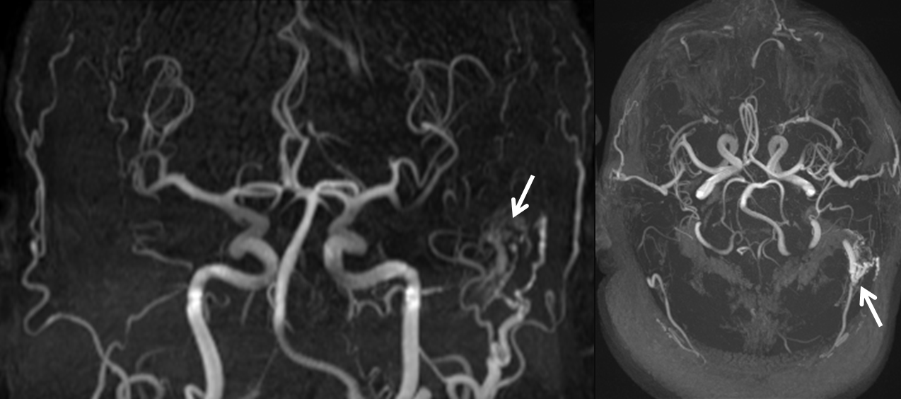
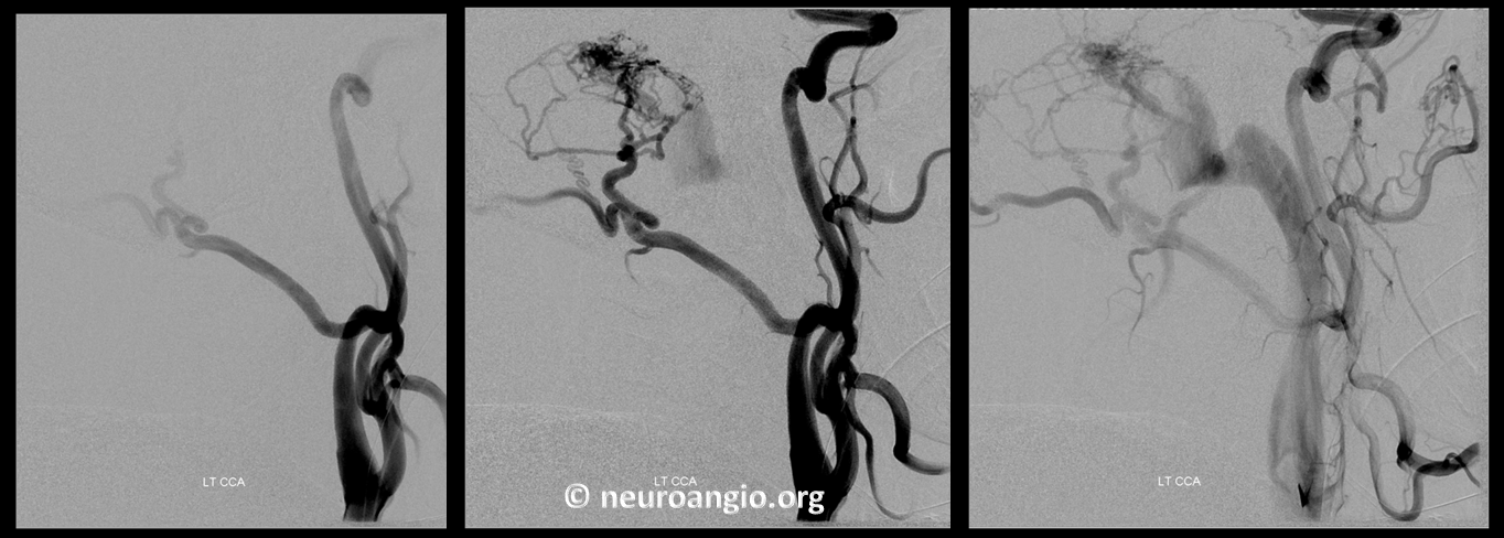
No ICA contribution. Sinus is open

Typical ECA supply of grade 1 fistula
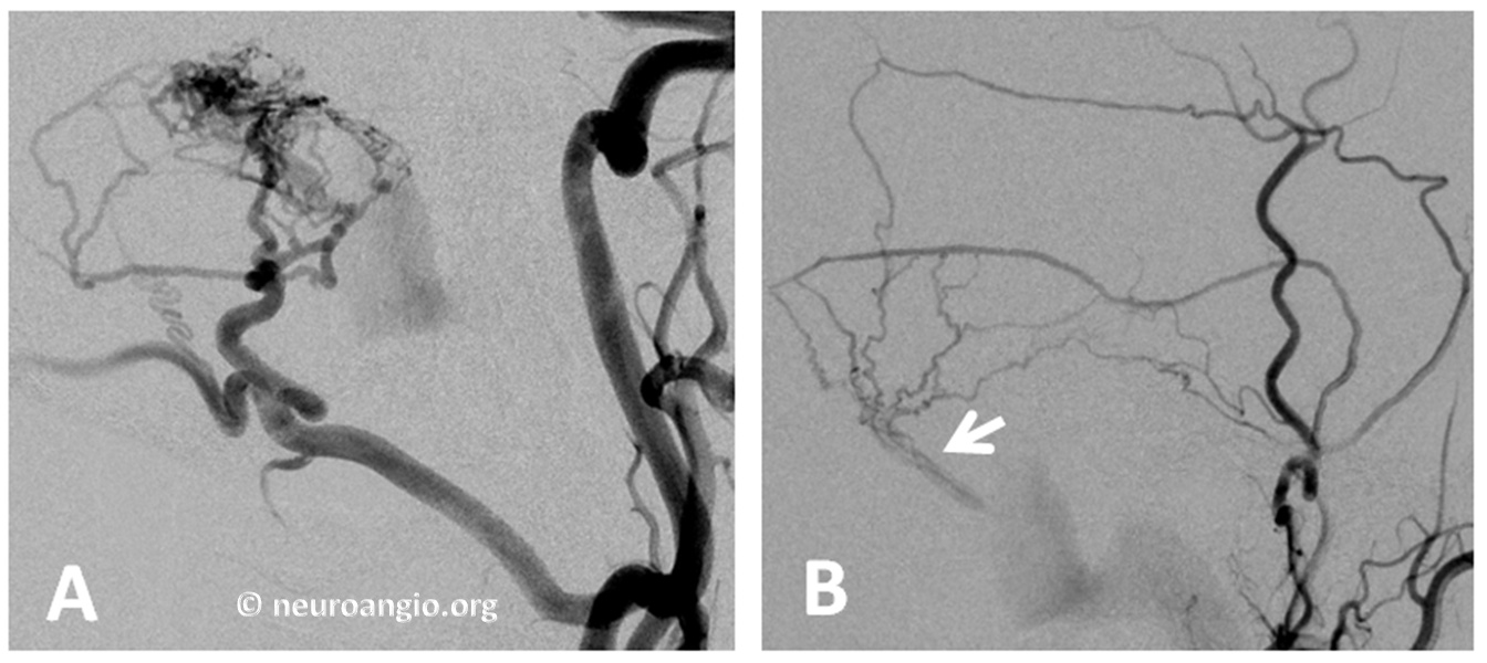
Distal ECA injection (B) beautifully shows common channel in wall of sinus (arrow)
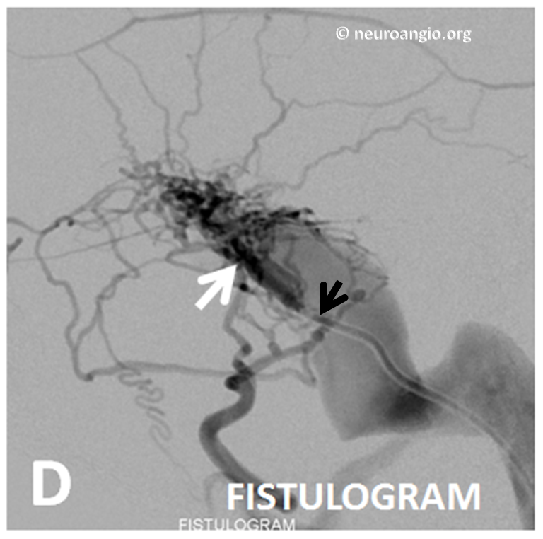
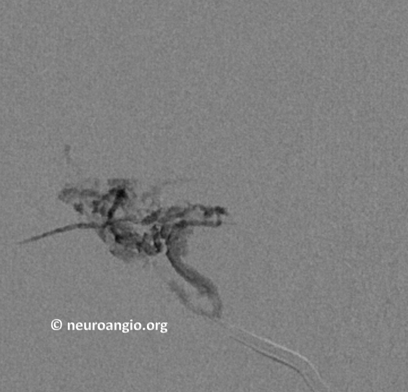
A 5F vert (black arrow) is placed transvenously into the common channel (white arrow) and an absolutely beautiful fistulogram is obtained, retrogradely opacifying the entire arterial network feeding the fistula. What can be better than this to show fistula anatomy?
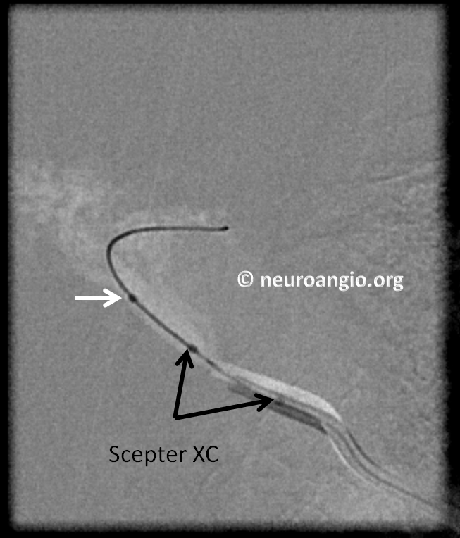
Next,a Scepter is placed into the common channel to allow for flow control
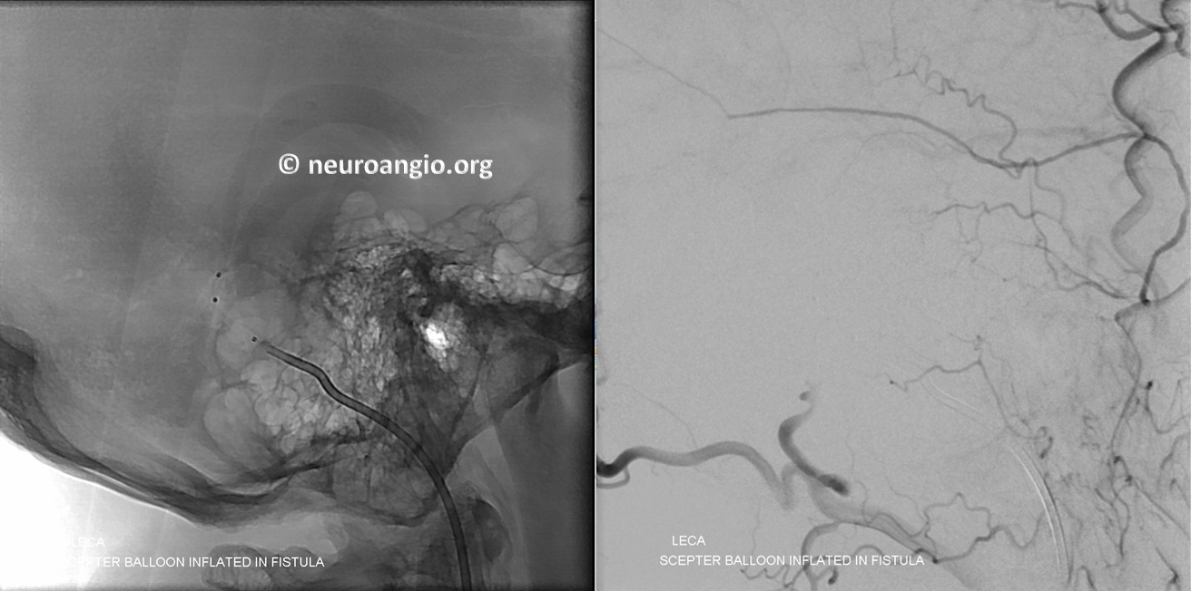
Injection of ECA with scepter in place shows fistula occlusion, confirming that this single channel is the common vessel upon which all fistula supply converges.
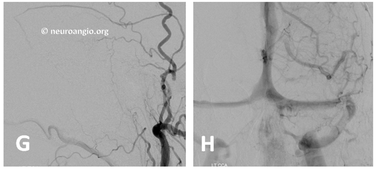
nBCA embo cast
Post
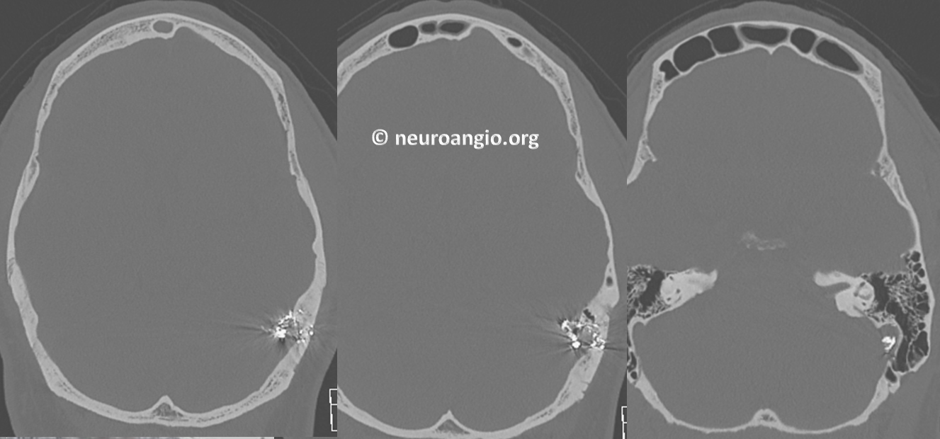
CT with glue cast in place
The common channel is medial to the sinus
See another common channel case here
See literature on superselective transvenous embolization — there are many more of these than meet the eye. It is a safe and effective treatment method, which avoids unnecessary permeation of vessels distal from fistula site and occlusion of a perfectly salvageable sinus
Further reading:
Mironov A. Selective transvenous embolization of dural fistulas without occlusion of the dural sinus. AJNR American journal of neuroradiology 1998;19:389-391
Piske RL, Campos CM, Chaves JB, et al. Dural sinus compartment in dural arteriovenous shunts: a new angioarchitectural feature allowing superselective transvenous dural sinus occlusion treatment.
Caragine LP, Halbach VV, Dowd CF, et al. Parallel venous channel as the recipient pouch in transverse/sigmoid sinus dural fistulae. Neurosurgery 2003;53:1261-1266; discussion 1266-1267