
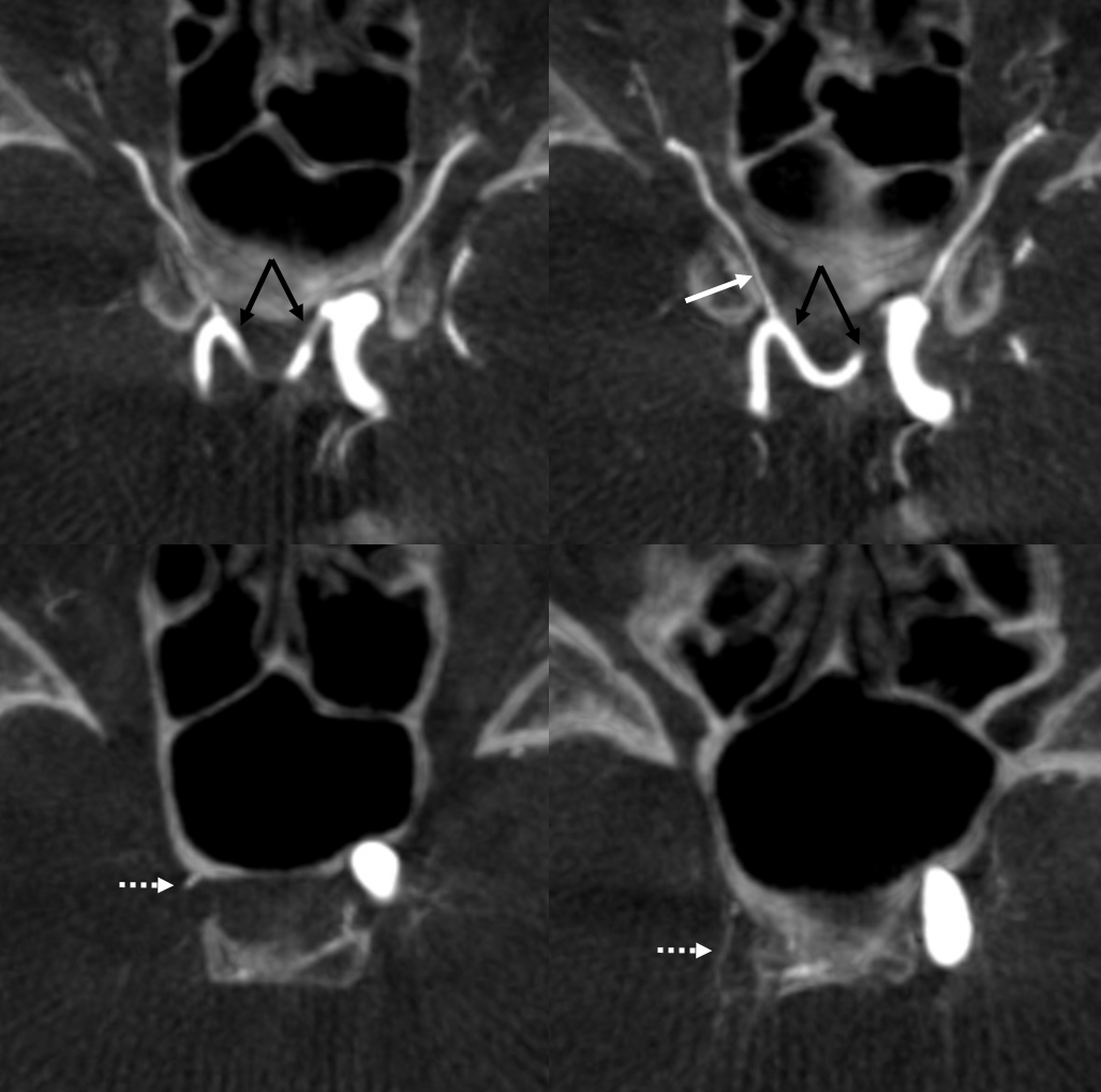
A very rare find — looks the same every time — a vessel traversing the anterior sella turcica, connecting the two carotids. In every instance we are aware of, it is associated with carotid agenesis — as a means of reconstitution. Supposedly it is a remnant of the inferior hypophyseal artery — but most likely this idea an error since the inferior hypophyseal artery is located posterior to the posterior clinoid — thus outside of the back part of sella turcica — whereas this vessels is inside the front part of it. Lasjaunias suggested that this segmental carotid agenesis leads to reconstitution of the carotid distal to the agenesis segment. The primitive maxillary artery is supposed to reconstitute the ICA when proximal petrous segment is absent — beyond the more common “aberrant carotid” reconstitution by the inferior tympanic-caroticotympanic circuit. However, given what we said above, if the Lasjaunias theory holds, then the agenesis segment reconstituted by the primitive maxillary artery must be cavernous. We actually do have some support for this in the case below. Others believe current vascular neuroembryology is a branch of metaphysics, or divination… You decide what you believe in… Regardless of belief, primitive maxillary artery is very much a fact, and a very interesting one, as we can see below.
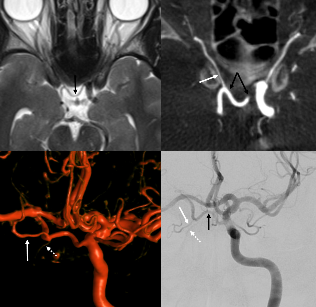
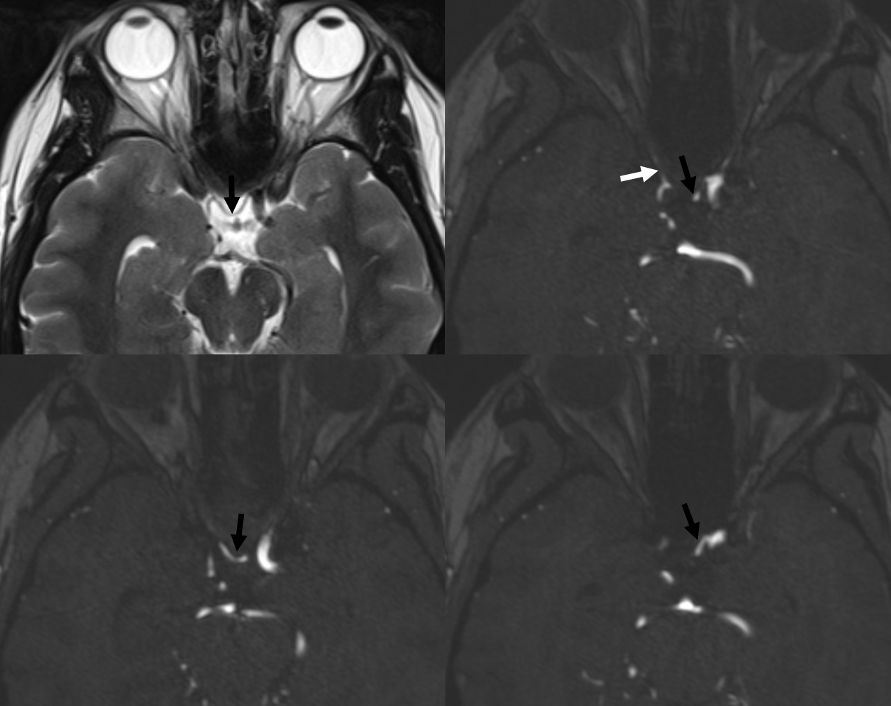
MRI — here and everywhere, the Primitive Maxillary Artery (PMA) is black, right ophthalmic is white, and anteromedial branch of the ILT (retrogradely supplying cavernous sinus ILT area in this cool case) is dashed white. Note that the PMA is located in the anterior part of the sella, and at the level of the ophthalmic arteries (which here arise from their usual proximal intradural locations)
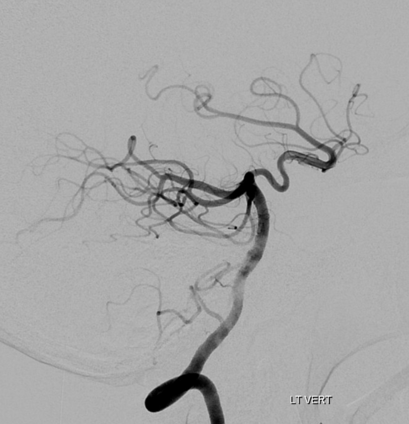
Carotid agenesis. PCOM helps also
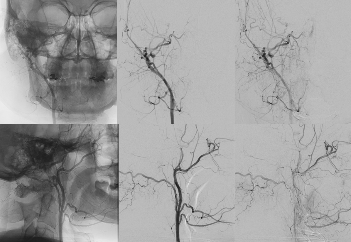
Right carotid agenesis. Notice a tiny pituitary blush from the accessory meningeal artery (not labeled — look for it)
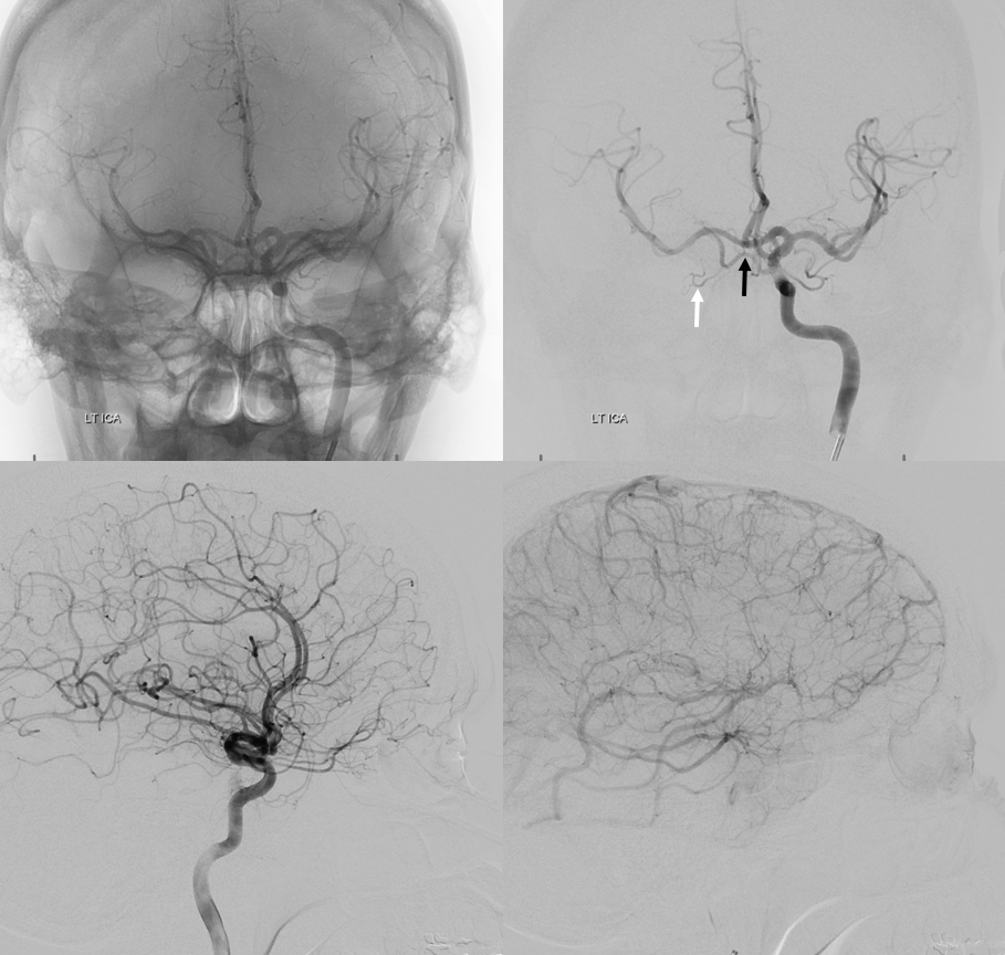
Angio. The PMA (black arrow) is what really supplies the right ophthalmic (white arrow). Lateral images show two choroidal blushes
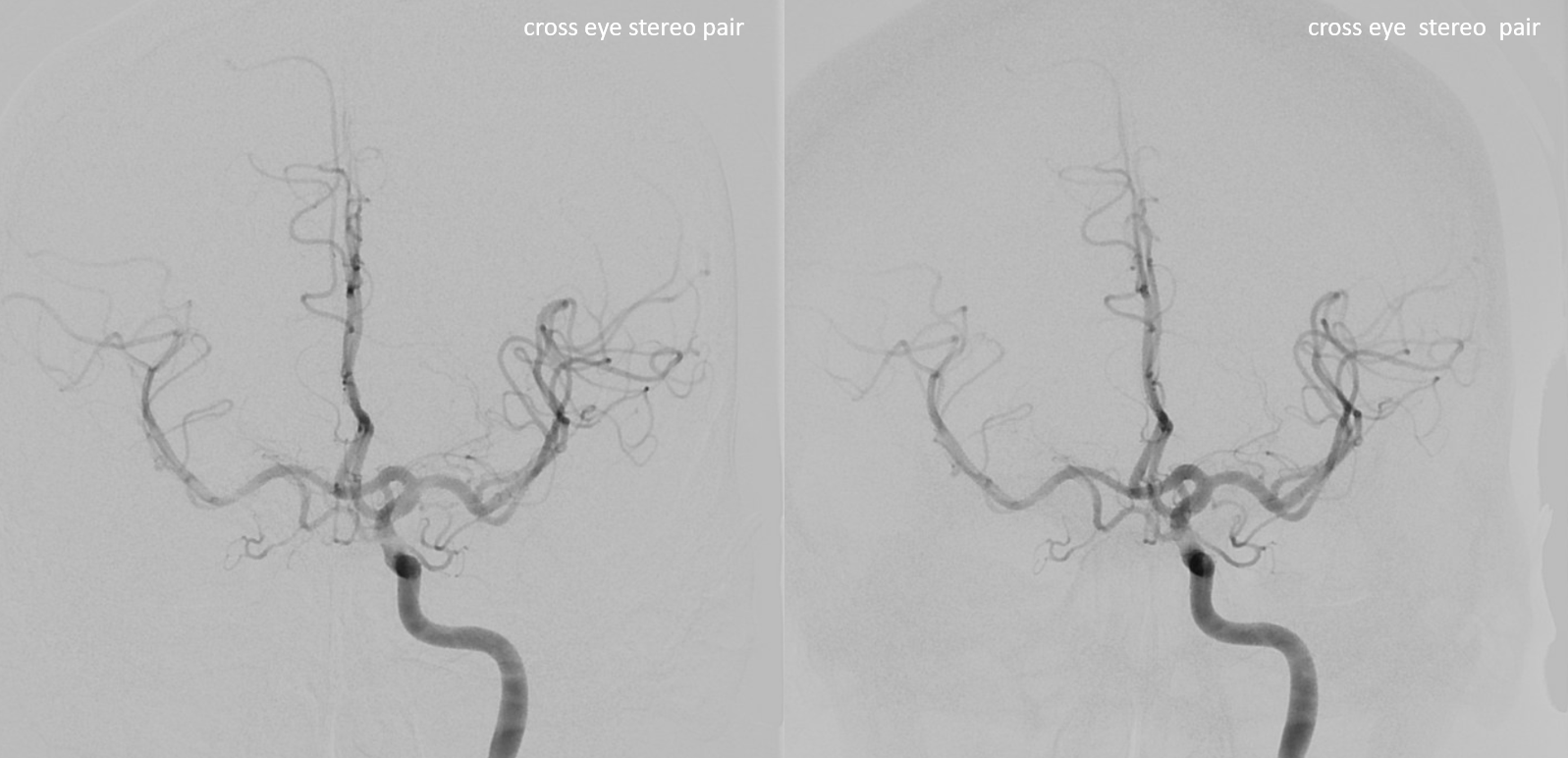
Stereo frontal views
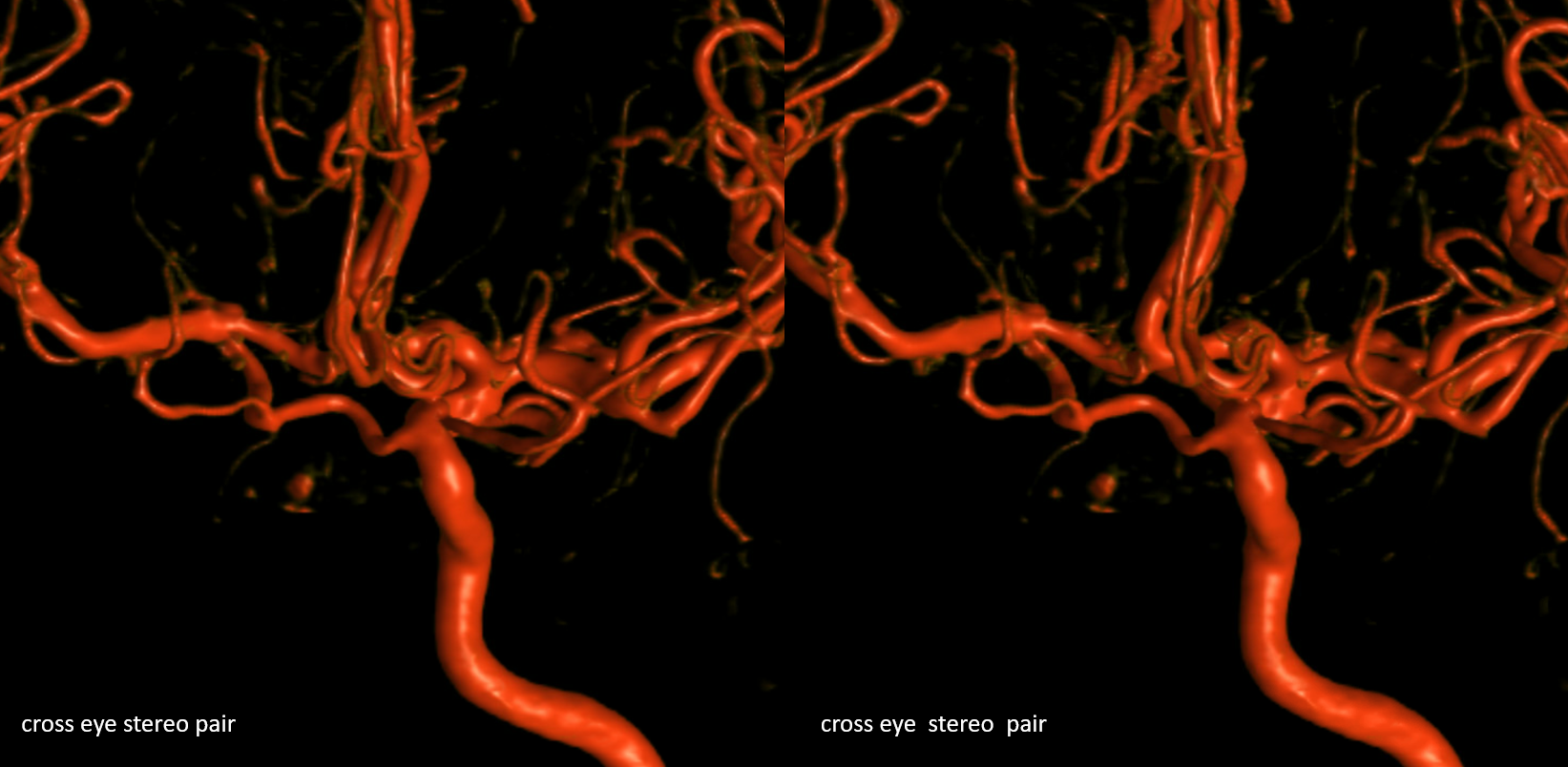
Stereo Volume Rendered images
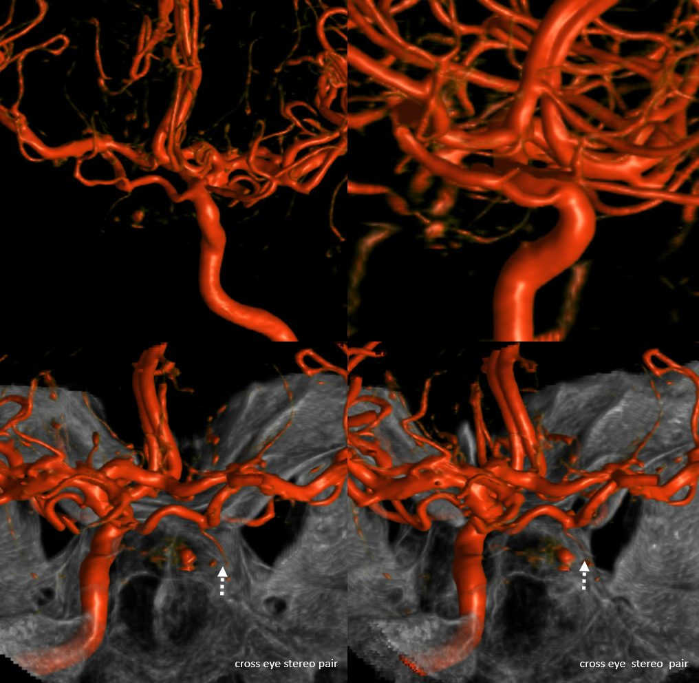
More VR images — bottom pair is cross-eye stereos. The anteromedial branch (or its equivalent, for the purists) is dashed white arrows
Rotational angio axials
Our conclusion is that the Primitive Maxillary Artery is more related to the superior hypophyseal artery than the inferior one. Makes a lot more sense — and Lasjaunias theory of segmental agenesis and reconstitution still holds in general