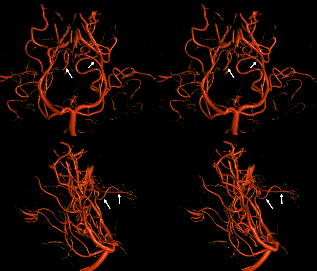
Stereo Lateral; Red=posterior lateral choroidal; Orange=posterior Medial choroidal (bilateral are seen on the left image)
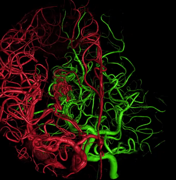
Posterior choroidal arteries with anastomosis near foramen of Monro, and associated thalamoperforating branches
Dominant posterior medial choroidal artery. The hemodynamic balance of the posterior medial and lateral choroidal arteries can be shifted in either direction. The dominant vessel traverses the foramen of Monro to supply the choroid plexus of the correspondingly hypoplastic feeder. In this example, the medial choroidal (red) is dominant and extends superolaterally across the foramen of Monro point (yellow) to supply the lateral ventricular territory (orange). A hypoplastic lateral choroidal artery (black) is present. The splenial arteries are labeled in purple.
Choroid Plexus Blush
Variable — sometimes quite “vascular” — tends to stay late into venous phase. Typical curvilinear look. In the image below, right side has a large posterior lateral choroidal (arrows) and associated choroid plexus blush.
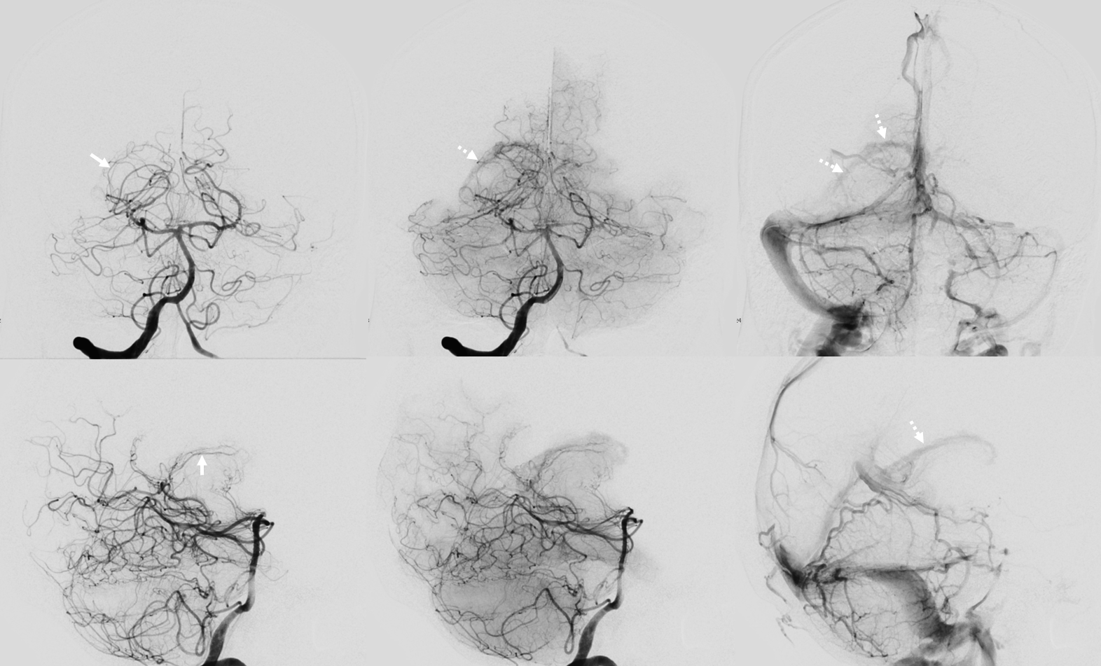
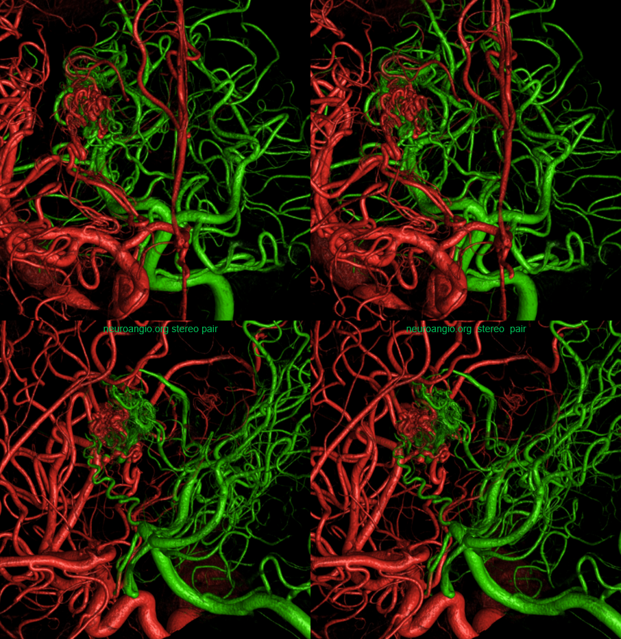
Posterior Lateral Choroidal Artery and Thalamoperforating arteries supplying a left thalamic AVM
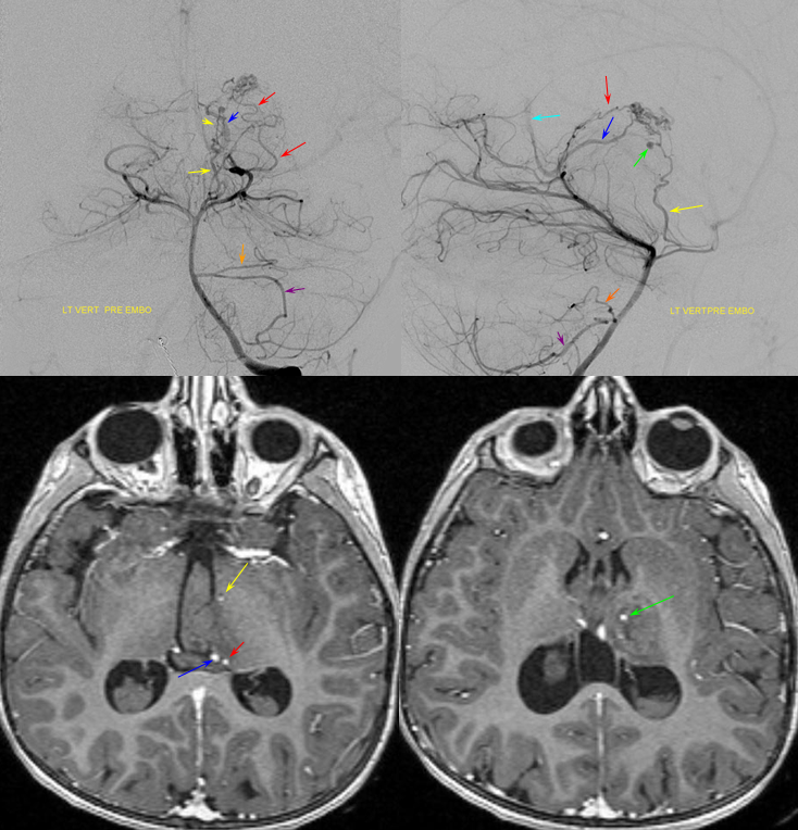
The anterior thalamus near foramen of Monro is usually supplied by an anteromedial perforator coming off the posterior communicating artery, as is seen in this case. A ruptured AVM with a pseudoaneurysm (green) arising from an anterior thalamic perforator (yellow) supplies the lesion together with the posterior medial choroidal artery (blue) and posterior thalamic artery (red). These arteries and pseudoaneurysm can be identified with high degree of certainty on a pre-angiographic post-contrast MRI T1 gradient echo sequence (MP-RAGE in this case).
Lateral Thalamic AVM
Another example of the balance between choroidal arteries. This lateral thalamic AVM is supplied by anterior choroidal, posterior lateral choroidal, anterior thalamic, and lateral thalamic arteries.
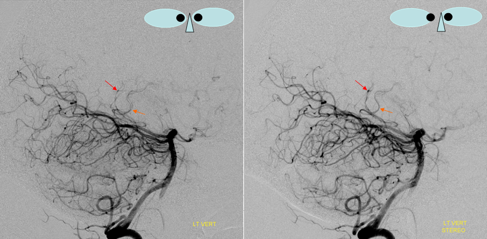
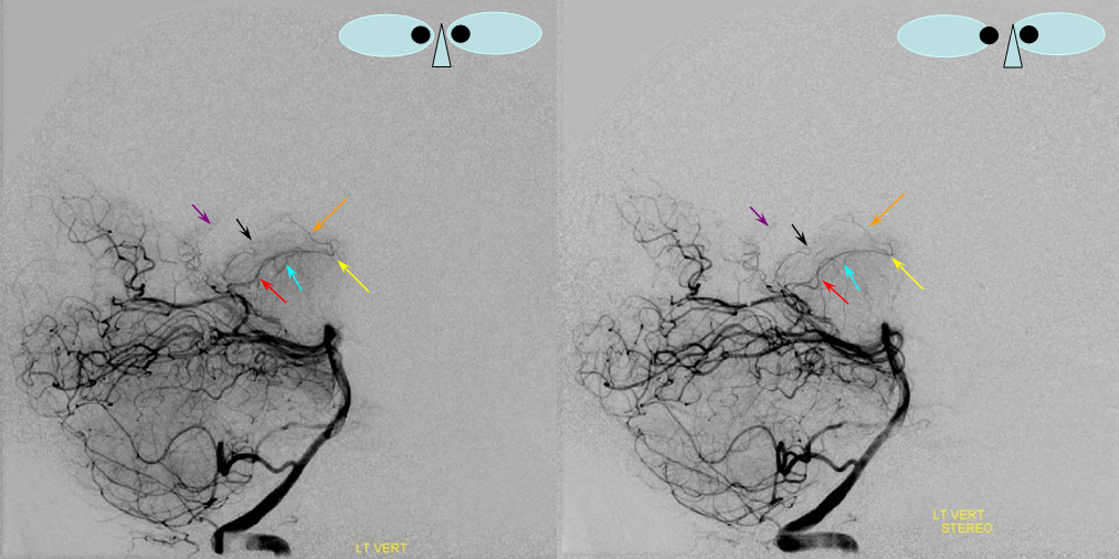
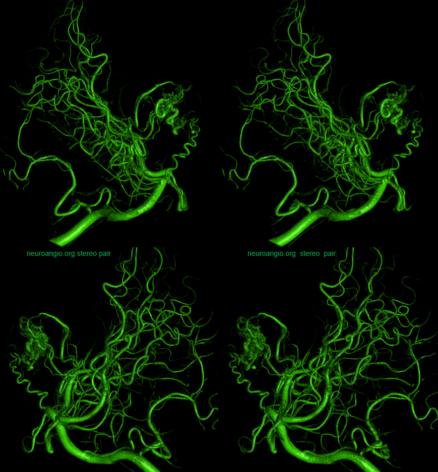
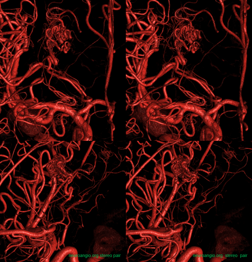
DYNA stereo vert injections
DYNA stereo ICA injection
Fusion DYNA
Movie
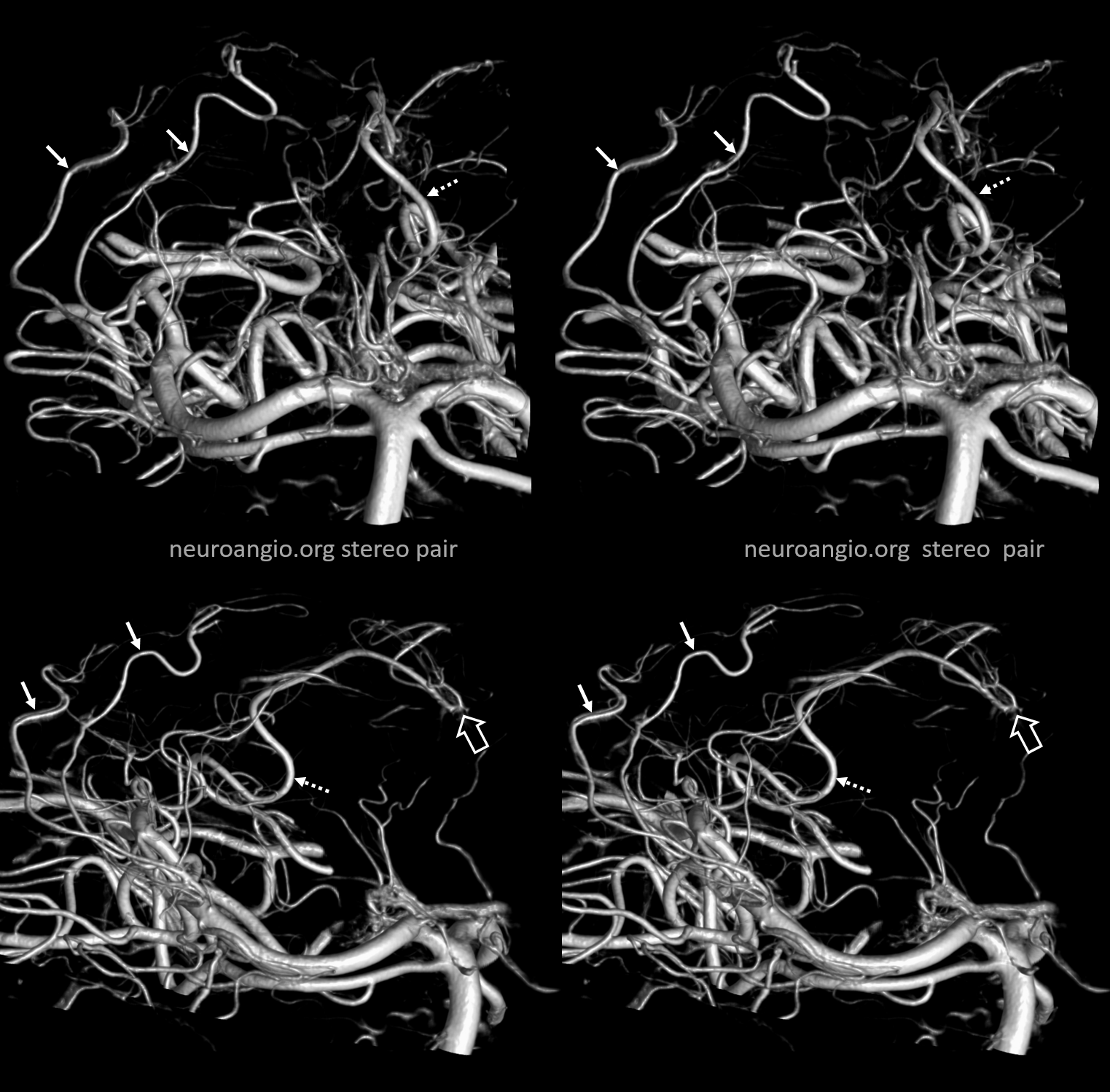
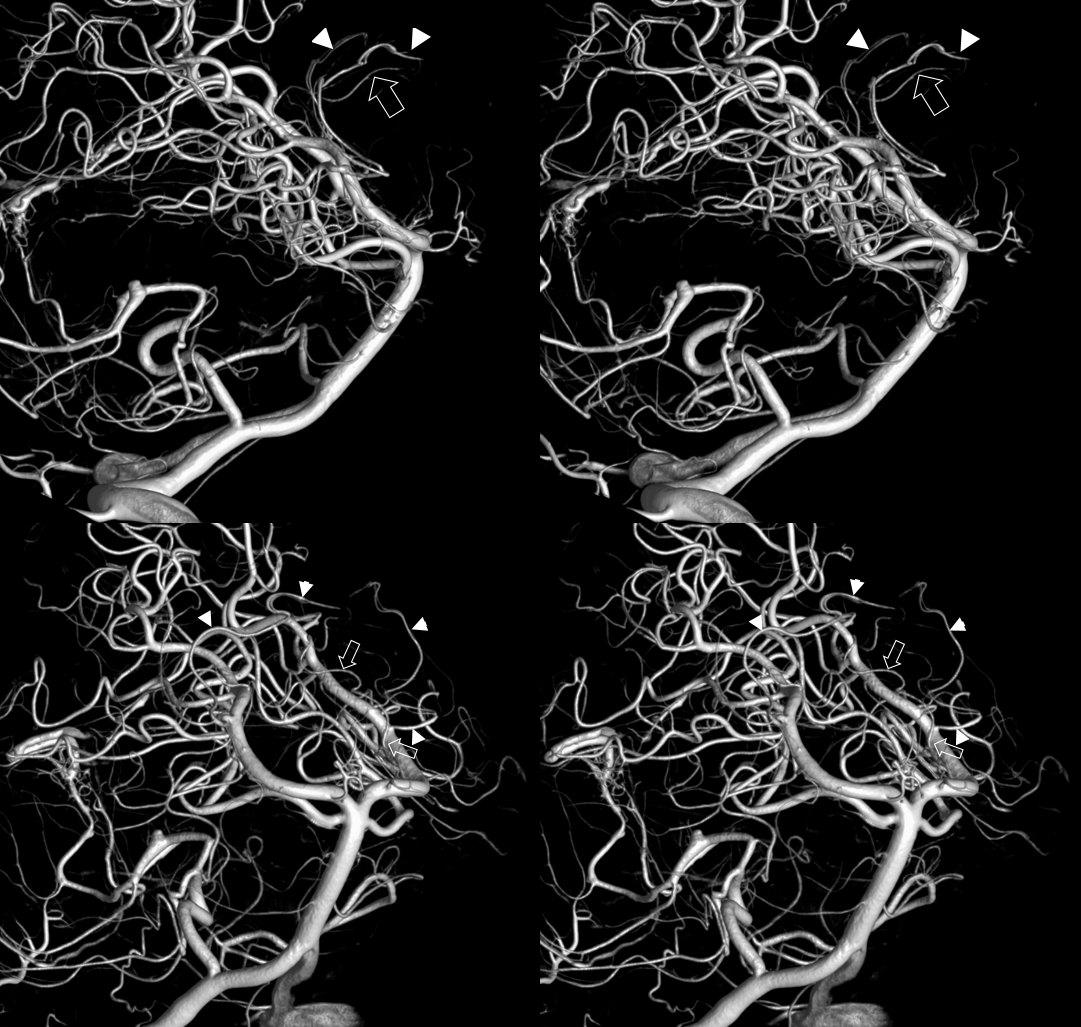
Duplicated Posterior Lateral Choroidal Arteries
This actually happens — rarely but it does. No such thing for anterior choroidal so far. Below are duplicated posterior choroidals — one supplying more of the posterior horn, the other the atrium. Interestingly, there is also a very large posterior medial choroidal, which extends into the body of the lateral ventricle across the foramen of Monro (open arrows). Much better landmark than the stupid venous angle
Angio — can’t see anything
DYNA — without arrows, stereo
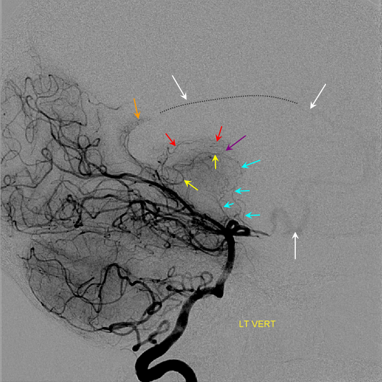
DYNA, with arrows — medial is dashed, laterals are solid
Variant supply of third ventricle choroid plexus and sequential identification
Having a series of images through which an arterial course and corresponding parenchymal and venous phases can be traced is very useful when questions of identity arise. Here, variation in foramen of Monro supply is present with distal origin of a branch coursing through velum interpositum (yellow) to reach foramen of Monro, with a hypoplastic medial choroidal artery. The lateral choroidal system (red) is well developed. Parenchymal and venous phases help identify the choroidal blush and venous counterparts of the arterial vessels, increasing one’s confidence in correctly identifying the anatomy.
Red=posterior lateral choroidal; yellow=fornix/choroidal branch in velum interpositum; purple=choroidal blush of the lateral (higher) and third ventricle (lower and anterior to the lateral ventricular blush); light and dark blue=internal cerebral vein
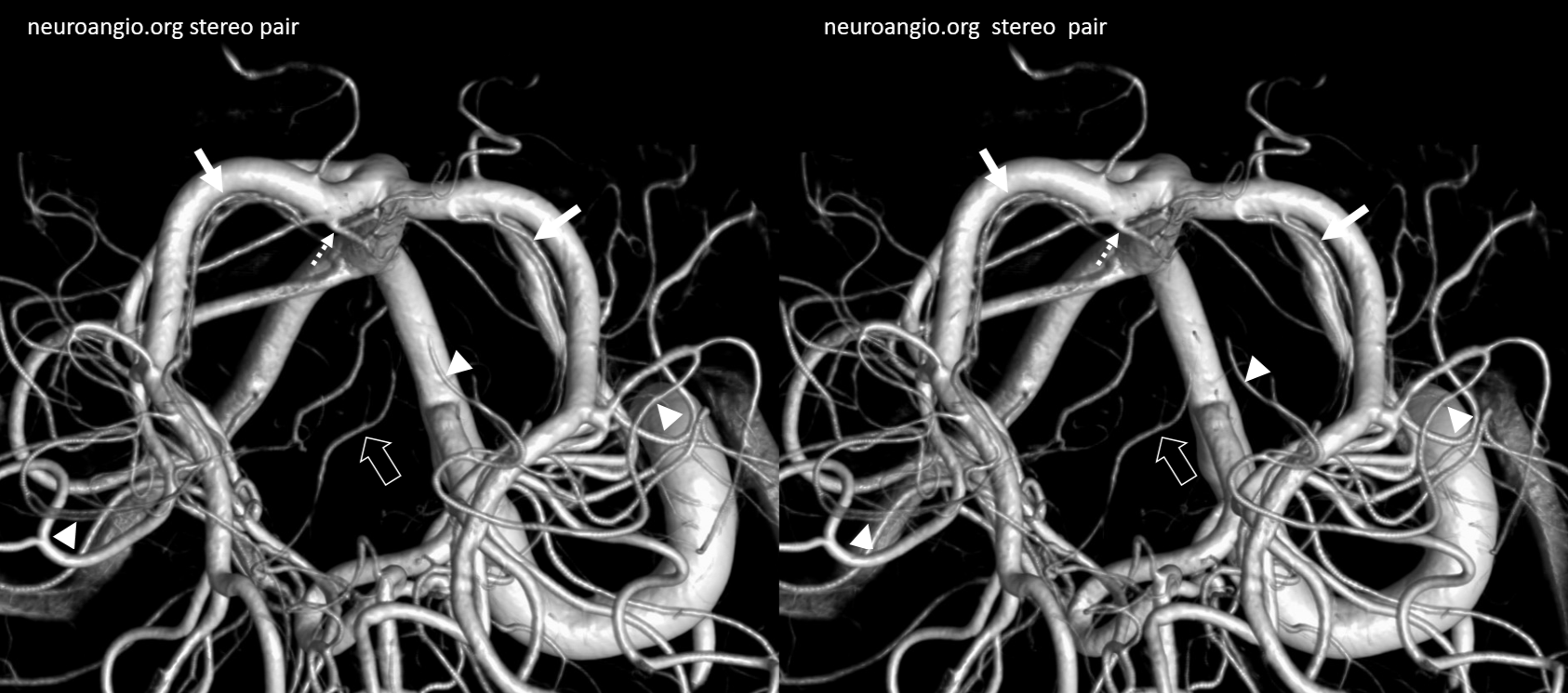
Anterior Choroidal – Posterior Lateral Choroidal anastomosis — the anterior choroidal beyond the plexal point supplies the plexus of the temporal horn, where it is in balance with the posterolateral choroidal going to the atrium region. This is elegantly shown in the following case of left choroidal plexus AVM, supplied by both vessels with beautiful illustration of draining veins.
Red=anterior choroidal; yellow = posterolateral choroidal; pink=choroidal vein; light blue=internal cerebral vein; brown=basal vein to sylvian veins; dark blue = atrial vein; white = superior petrosal sinus; green = mid basilar agenesis
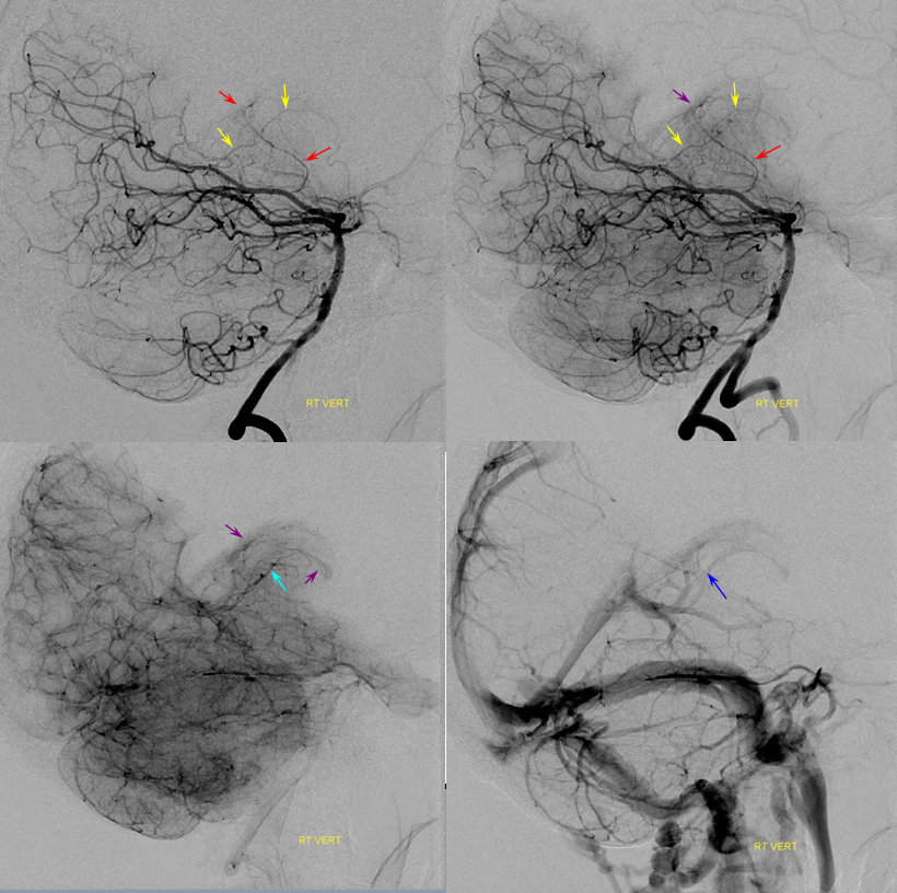
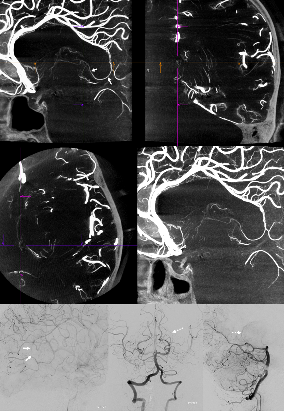
Posterior Lateral and Medial Choroidal Arteries Cone Beam Imaging
It happens… Check out this one as seen on DYNA CT — notice lack of posterior medial choroidal visualization off the vert injection — where a well-developed left posterior lateral choroidal (dashed arrows) is present. The point where posterior medial choroidal goes into the ventricle is just distal to the crosshairs — same epsilon sign
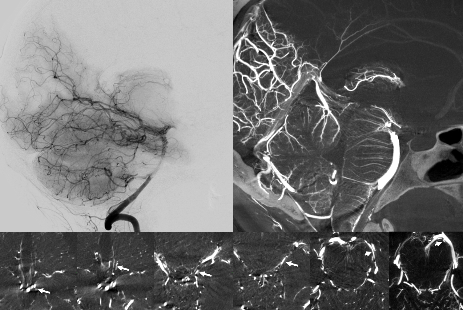
Dural Branches — Davidoff-Schechter
The artery of Davidoff and Schechter — defined as dural branch of the PCA — usually arises from the posterior medial choroidal, somewhere near the midline. It is rarely seen on 2D angio and more commonly on HR-CBCT. The dura of the falcotentorial junction is a kind of watershed — far away from the anterior, middle, and posterior meningeal arteries. This is probably why it has relatively frequent piodural supply. Here is an example — bottom row are axial MIPs tracing the artery to posterior medial choroidal.
Another Example — also from posterior medial choroidal — and of course also much better seen on HR-CBCT
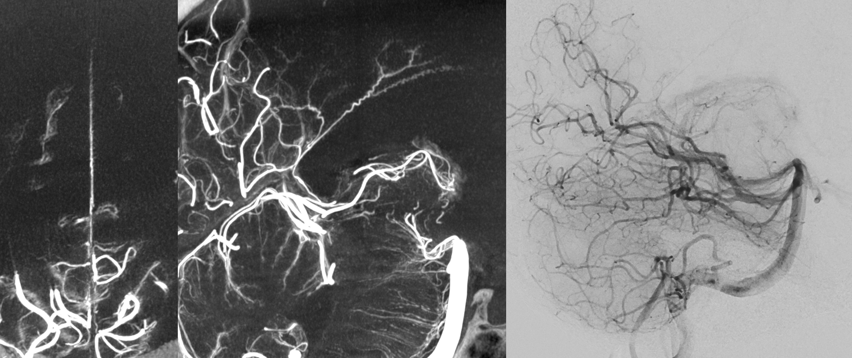
Stereo pair below — solid arrows — Davidoff-Schechter. Dashed arrows — posterior medial choroidal. The loop area of Davidoff-Schechter is where it picks up the dura
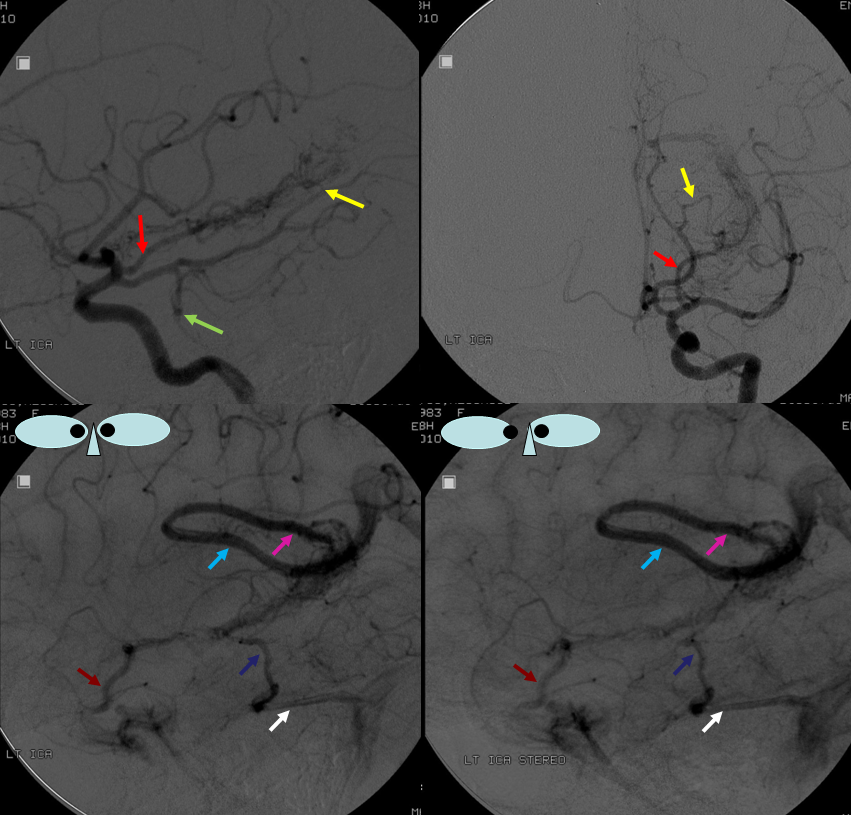
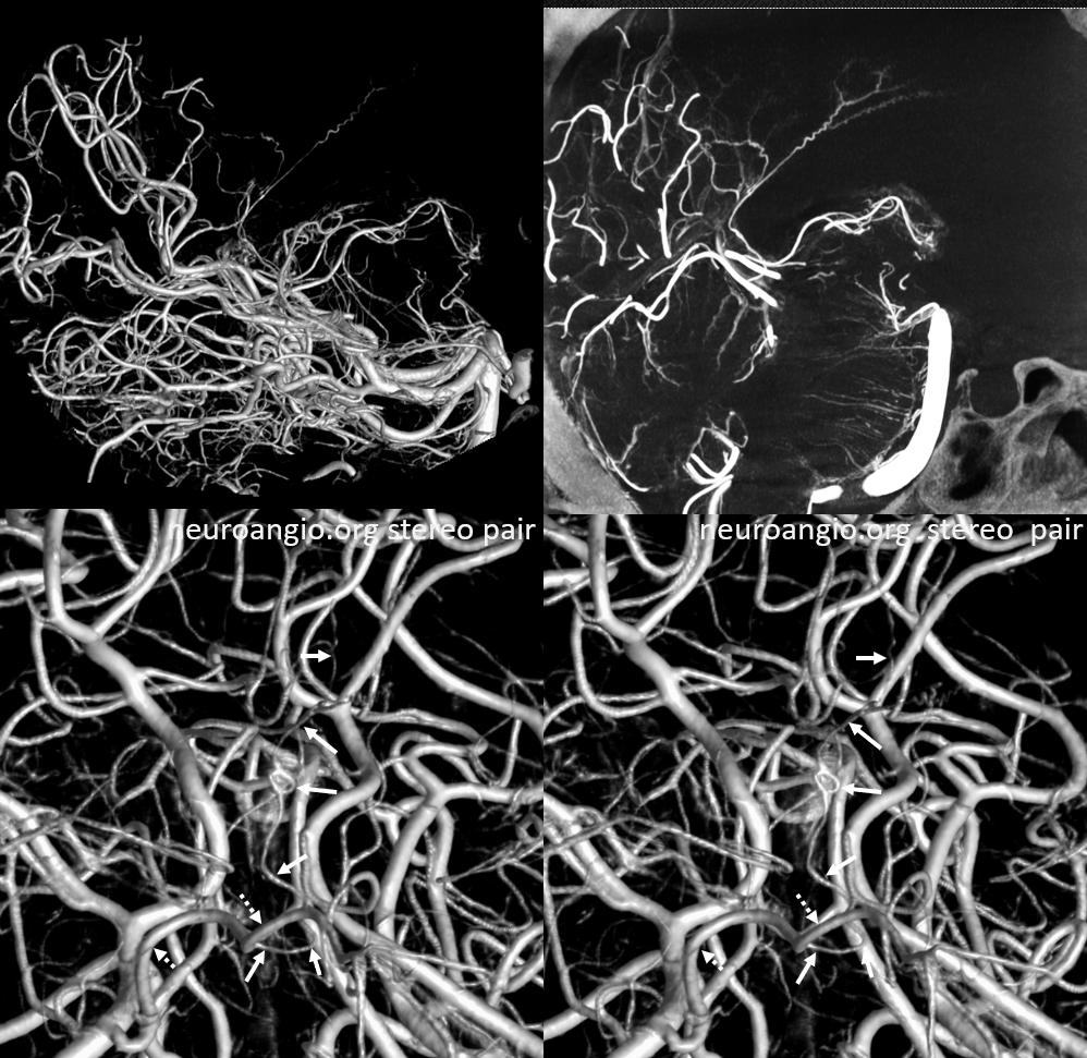
Collateral Supply of the Anterior Choroidal
Infrequently, the intraventricular anastomoses between posterior lateral and anterior choroidal arteries are sufficient enough to allow anterior choroidal resupply in case of latter’s occlusion. This is usually not the case though. Parenchymal branches of the anterior choroidal have very poor collaterals — one similarity with the lenticulostriates.
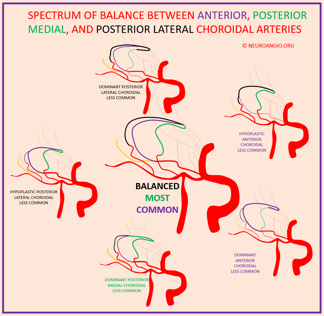
Below is an idea of the choroidal spectrum. A lot more of this discussion is found in the anterior choroidal page.
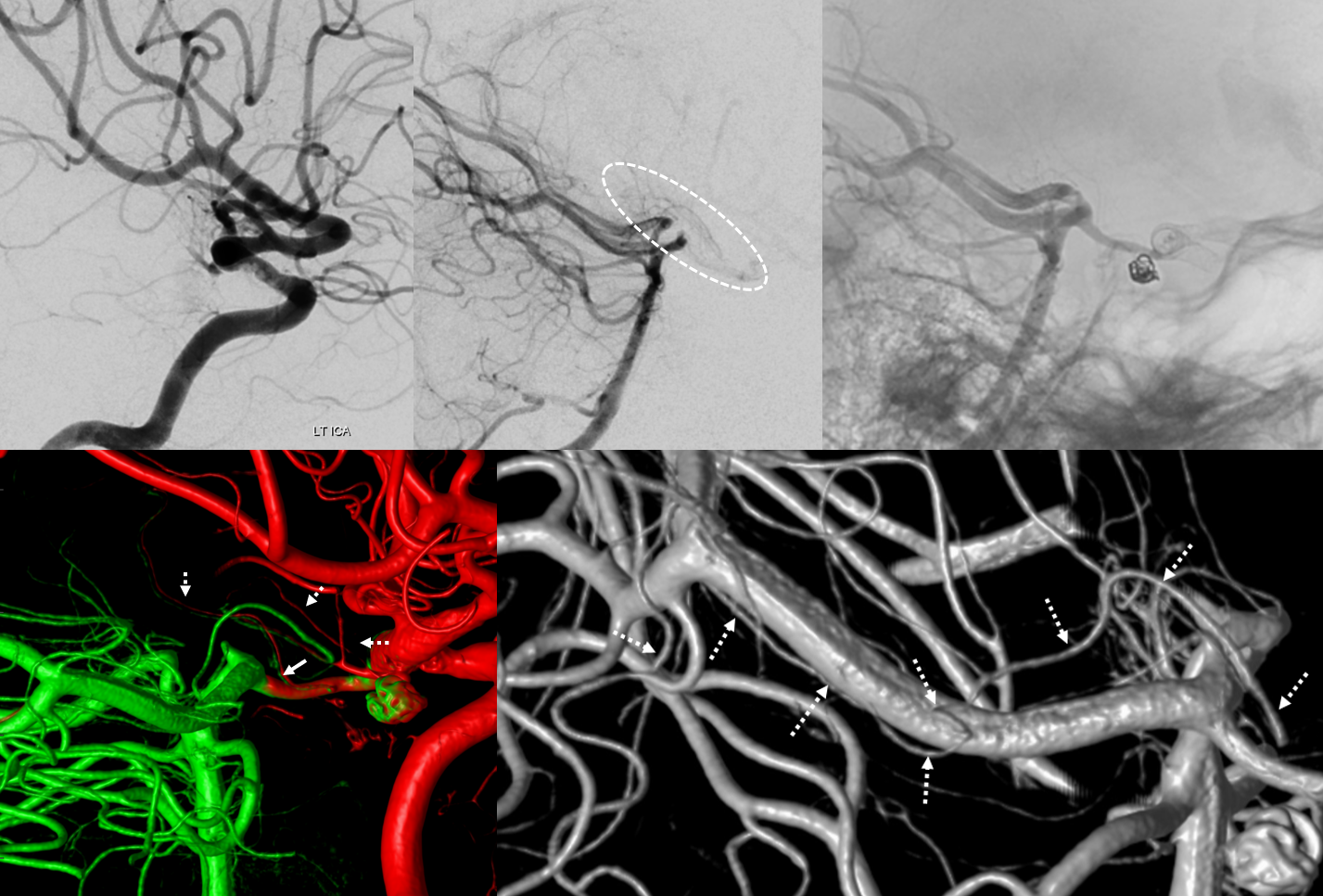
This is an example of posterior lateral choroidal reconstitution of the anterior choroidal artery in setting of treatment. There is thrombus in the ICA. The proximal choroidal is open (left top image). Center top image shows reconstitution of some branches in the PCOM-Choroidal area. Top right image shows retrograde filling of the PCOM. Bottom left image — fusion of HR-CBCTs — the fusion is imperfect, allowing one to see side by side the green of the vertebrobasilar injection and red of the carotid. The anterior choroidal region (dashed arrows) and PCOM thalamoperforator (solid arrow) are seen. Bottom right image traces the course (dashed arrows) of the posterior lateral choroidal reconstituting the anterior choroidal