
Posterior Meningeal Artery Region
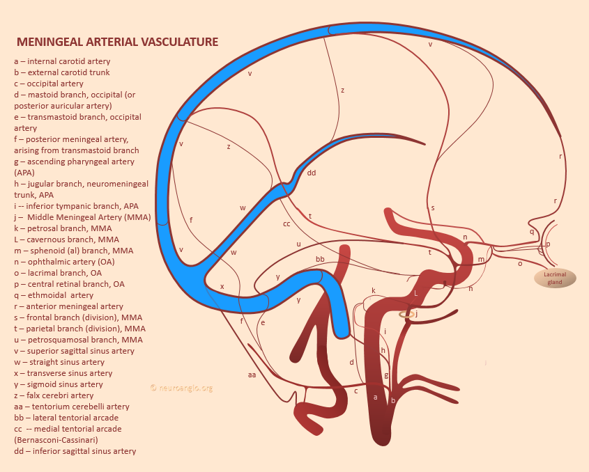
There is tremendous variability in here. Meningeal branches to this area come from occipital (either transosseous or occipital origin neuromeningeal trunk), ascending pharyngeal (neuromeningeal trunk), vertebral (either as tentorium cerebelli branch or a non-midline, non-tentorial posterior meningeal branch), PICA (also either tentorium cerebelli or more lateral). Finally, there may be no discrete posterior meningeal itself — the MMA (petrosquamosal, posterior / parietal) can do the trick.
Of particular interest is the area of falcotentorial junction — this is where some particularly interesting dural fistulas happen.
A key concept in understanding posterior meningeal vasculature is that meningeal arterial channels often run parallel to/ within walls of the major dural sinuses. We emphasize this repeatedly. Particularly these are easier to see when these vessels are enlarged in dural fistulas — one example here. There are arteries in walls of SSS, sigmoid, transverse, straight sinus — etc. See intrinsic dural vasculature page. The posterior meningeal artery often does that — for example — originating from neuromeningeal trunk, entering skull via jugular foramen, then running along sigmoid/transverse sinus toward midline.
Below are multiple examples
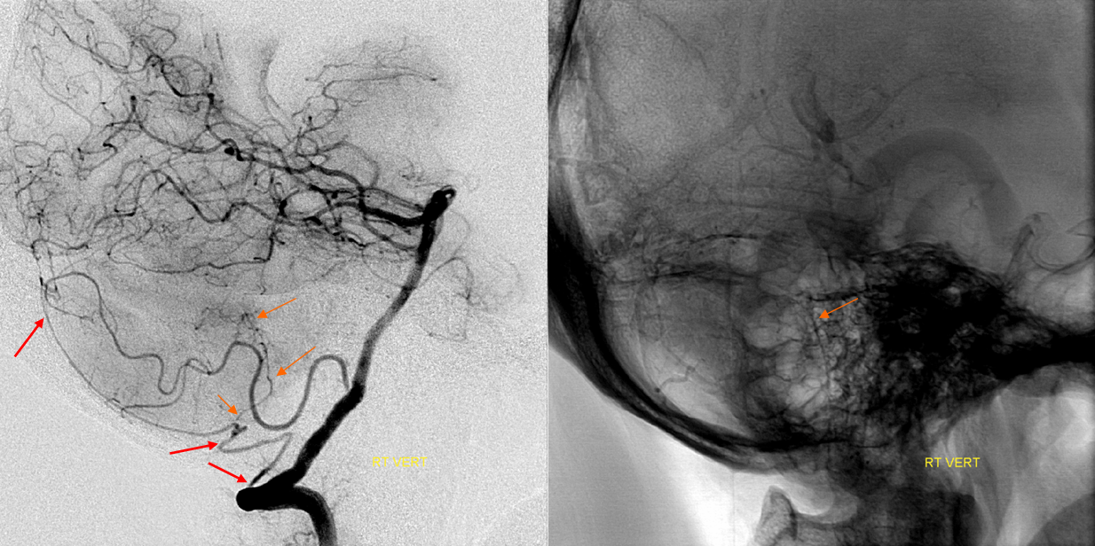
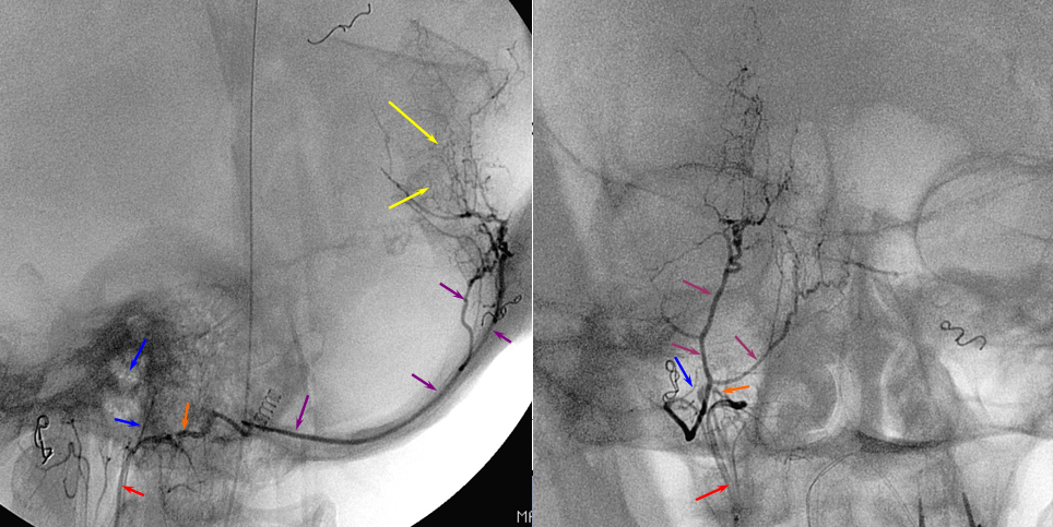
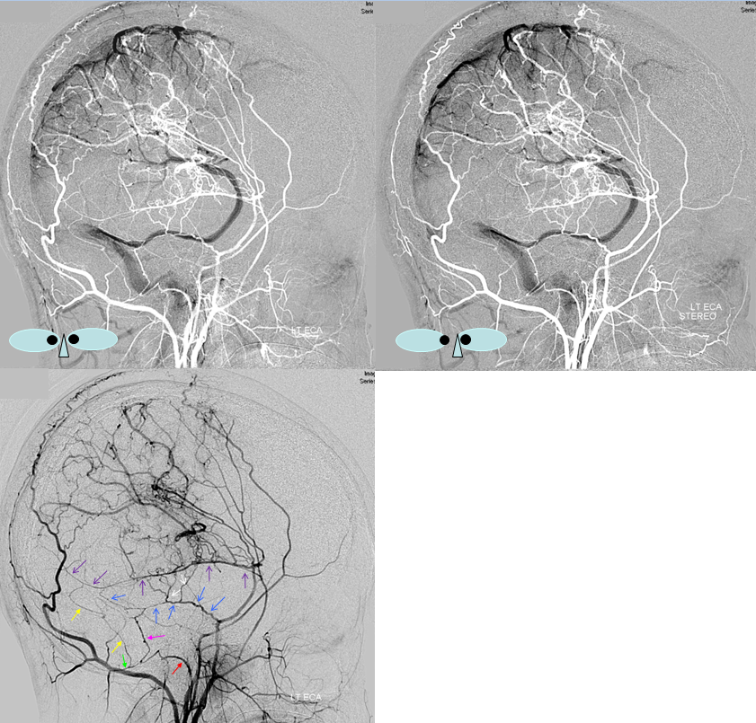
Lateral views — posterior meningeal (red) from vert — notice small branch in wall of the sigmoid sinus (orange)
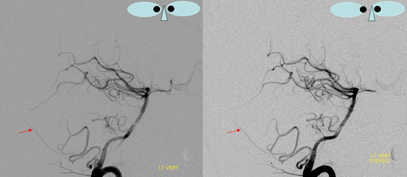
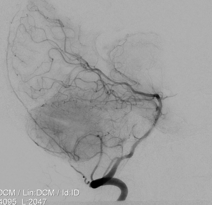
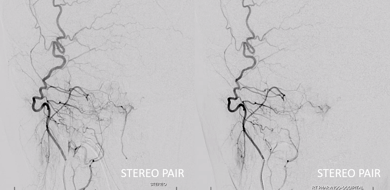
PICA origin posterior meningeal (red). Stereo views.
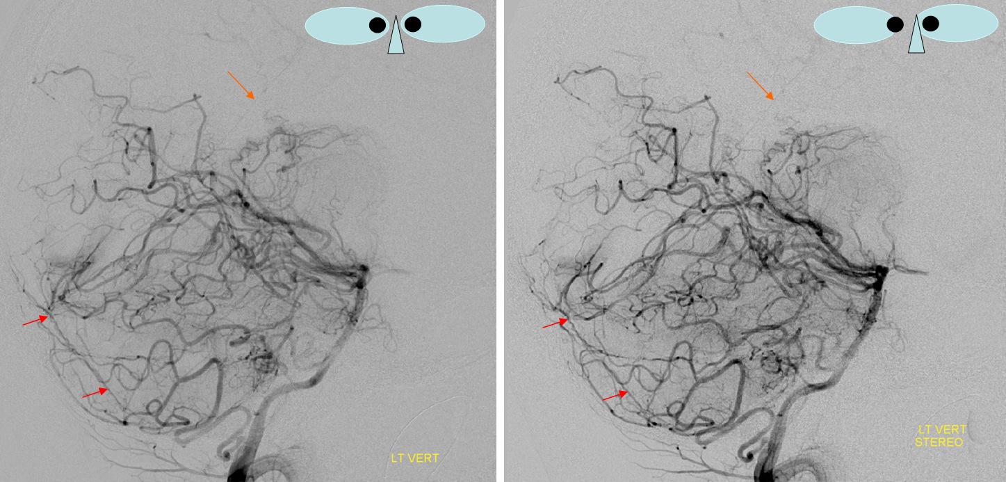
Below — later phase, same patient. Vascular resistance in meningeal branches is higher than brain – you have to wait a bit to see more — here the posterior meningeal extends to torcular. Also present is a small but relatively famous artery of Davidoff and Schechter (orange). It refers to a number of PCA to dual connections supplying the falcotentorial region. Its not really a distinct artery — more like a propensity for pial to dural connections there — patients with falcotentorial fistulas have hundreds of Davidoff and Schechter mini-mes…
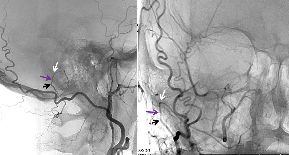
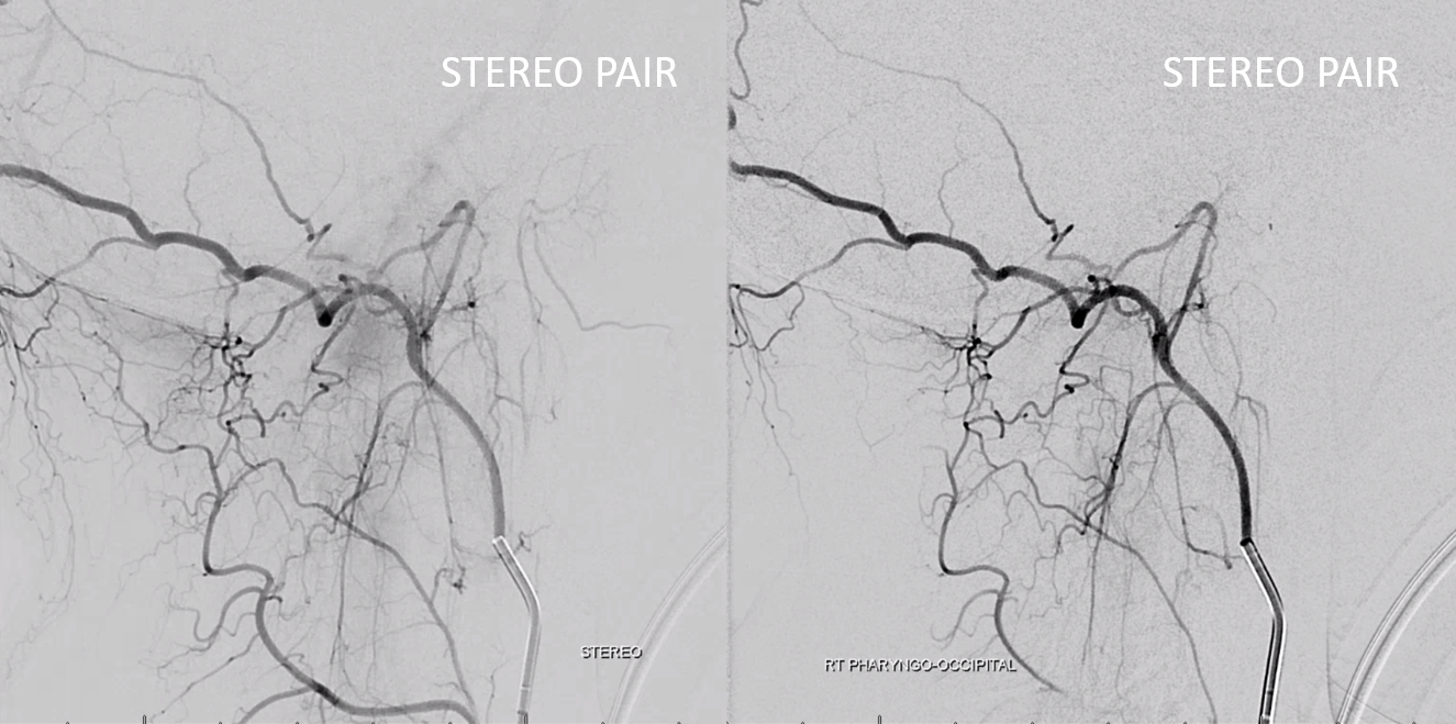
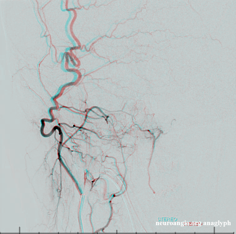
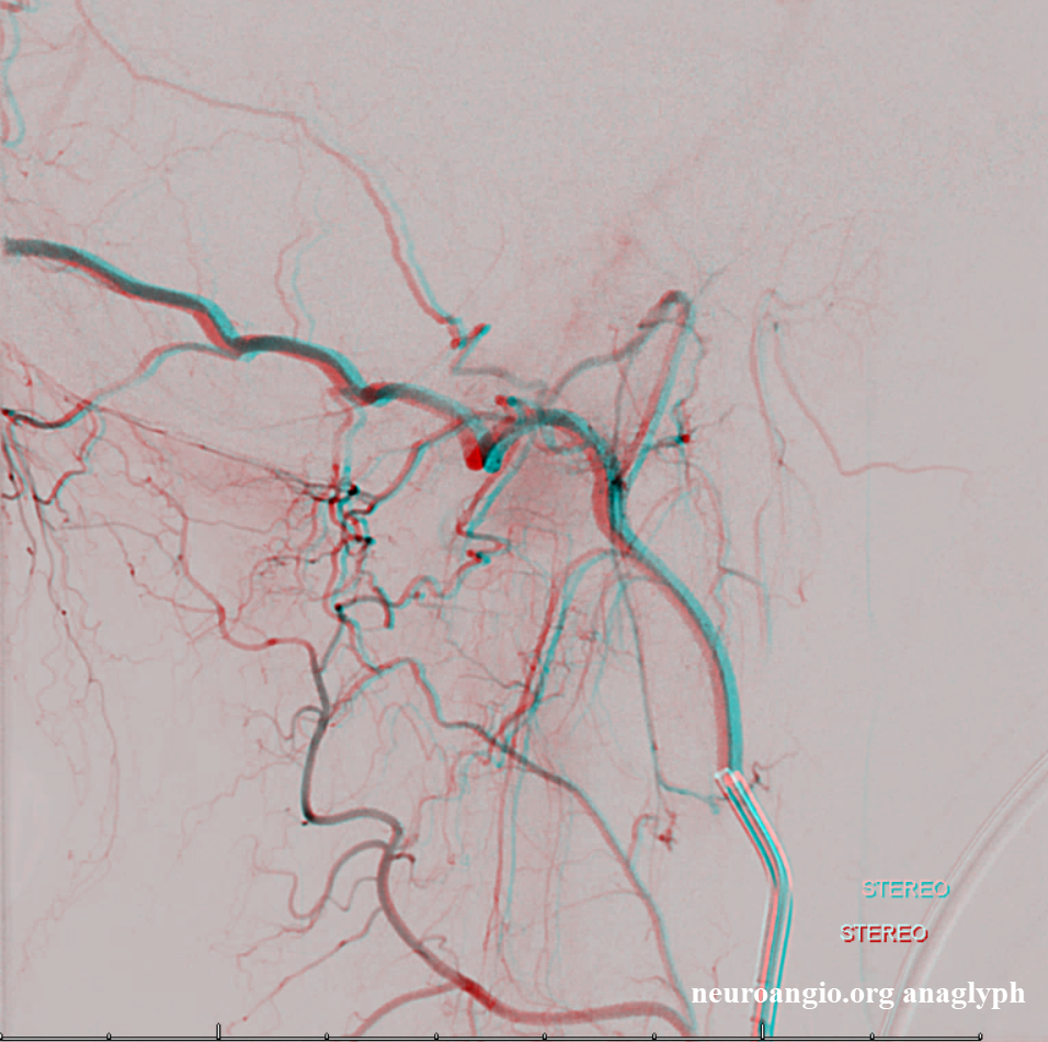
Posterior meningeal artery origin from jugular division of the ascending pharyngeal artery
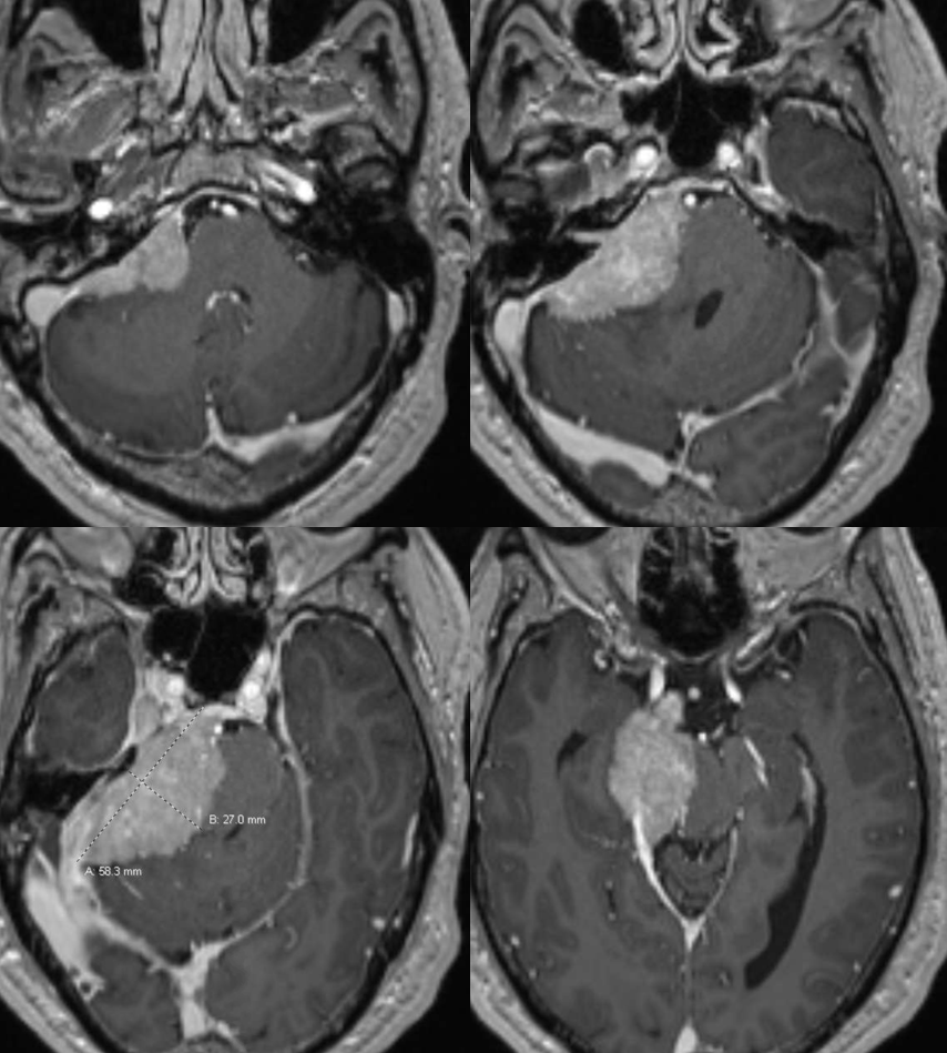
Very common. In a patient with en-plaque meningioma of the parieto-occipital region, an enlarged posterior meningeal artery coming off the jugular division of the neuromeningeal trunk of the ascending pharyngeal artery participates in supply of the tumor. The tumor was embolized with particles and pedicle closed with pushable coils (contralateral parietal division MMA branch coil is visible as well) Inferior tympanic branch in a patient with parietooccipital meningioma. The inferior tympanic branch (blue) projects over the auditory canal, and follows the expected course of the aberrant carotid artery. Very nice.
Neuromeningeal trunk=red; Jugular division=orange; posterior meningeal artery=purple; inferior tympanic artery=blue; meningioma blush = yellow
Falx Cerebelli
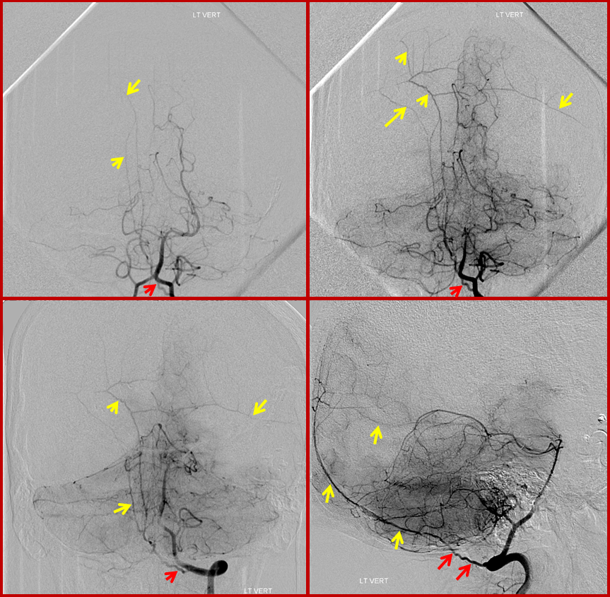
Prominent falx cerebelli artery arising from the C1 segment and extending along the dura of the superior sagittal sinus (no arrows). It really needs a frontal view to be sure though…
Posterior Meningeal Artery Network (yellow) arising from C1 branch of the vertebral artery (red). Why is this network so extensive — clue is lower right image…
Occipital Artery Contributions
This one almost invariably participates in supply of posterior fossa dural structures via transosseous channels originating from its mastoid branch. When the transosseous branch is large enough (i.e. gives rise to supply of posterior meningeal region), its corresponding hole in the skull is recognized as the “mastoid foramen”. The territory supplied is usually posterior to petrous pyramid base, in region of sigmoid sinus. Consequently, dural fistulas of the sigmoid sinus usually demonstrate a component of occipital supply. Usually, middle meningeal and ascending pharyngeal arteries represent more fruitful venues for transarterial embolization.
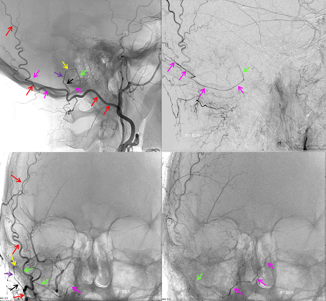
Mastoid branch supply of the posterior meningeal artery. Non-pathologic role of occipital artery (via its mastoid branch) in supply of dural structures is demonstrated in this patient, where the OA (red) gives gives rise to posterior meningeal artery (green and pink). The mastoid branch consists of a short extracranial segment (black), followed by transosseous segment (purple). The intracranial portion consists of a short anteroinferior dural segment (yellow), and a long medial segment (green) towards the lateral aspect of the foramen magnum, and then heading superiorly (pink) as the posterior meningeal artery.
Occipital origin neuromeningeal trunk
See occipital and ascending pharyngeal artery pages for more info. Either or both hypoglossal and jugular divisions can come from occipital. Either one can contribute to the posterior meningeal network — usually its the jugular one. Hypoglossal often supplies clival dura via clival branches.
See below
Separate origins of pharyngeal trunk (ball arrow) and hypoglossal division (white arrow) from the occipital artery. The hypoglossal contributes to supply of posterior meningeal territory (a.k.a. ascending pharyngeal origin of posterior meningeal artery)
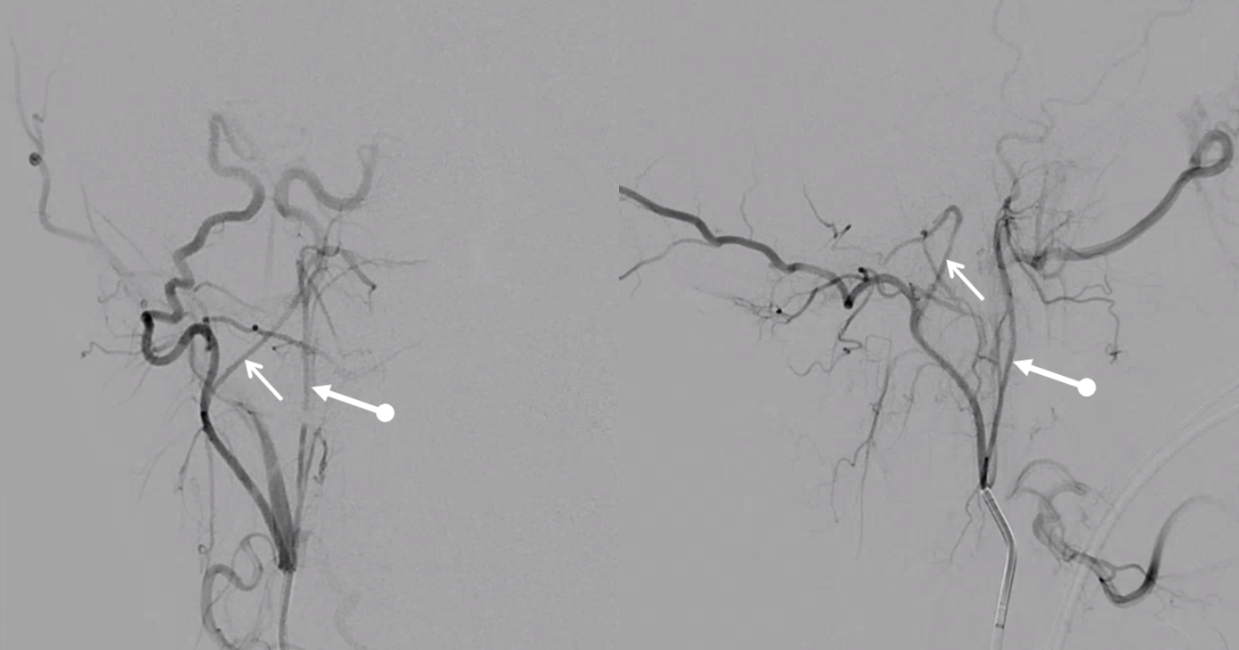
A diagnostic catheter position distal to the pharyngeal trunk isolates the hypoglossal division (white arrows), which opacifies the odontoid arcade (dashed arrows) via several anastomoses (white arrowheads) inside and outside the skull
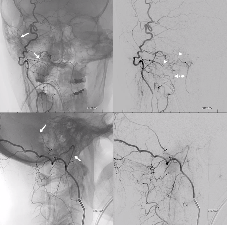
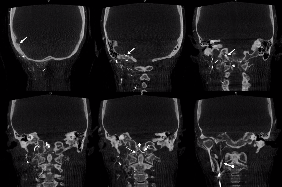
DYNA CT — same arrows as above. Notice the posterior meningeal artery in the groove of the sigmoid sinus (aka artery of sigmoid sinus — see meningeal arteries and middle meningeal artery pages for more info)
Cross-eye stereos
Anaglyph stereos
Another example — another nasty meningioma
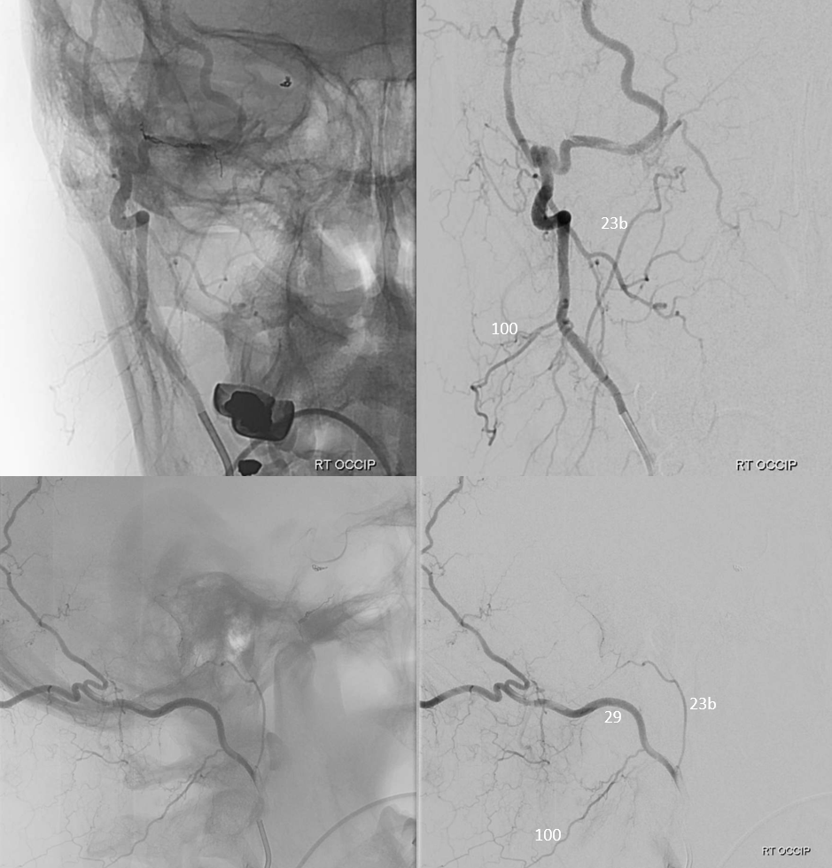
Jugular division (23b) origin from occipital (29) artery
Axial DYNA MIPs
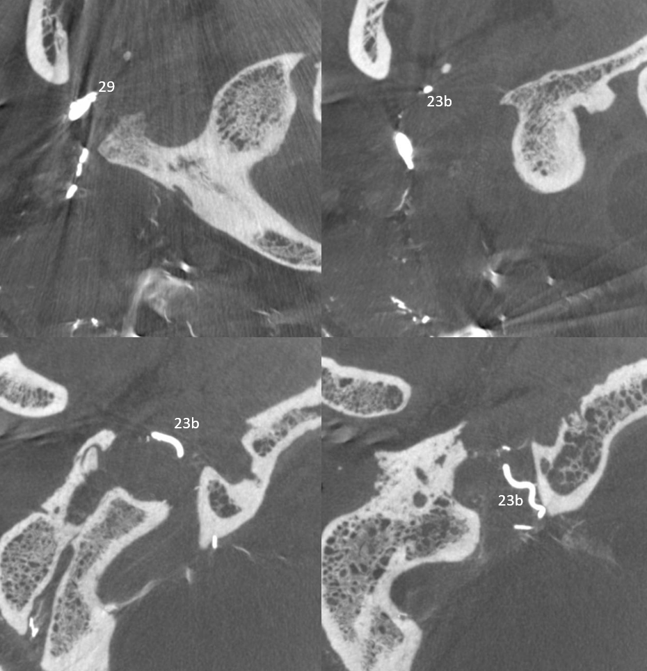
Another example:
Movie of DYNA CT — both jugular and hypoglossal divisions are off occipital — supplying posterior fossa meningeal tissues. Nice odontoid arch connection with hypoglossal. You can scroll through individual movie frames.
Ascending Pharyngeal origin
Again same idea. AP origin of neuromeningeal trunk. See movie below. You can stop and scroll through it. The posterior meningeal reaches all the way to wall of straight sinus. Connections are everywhere…
Below is a nice example of arteries in walls of sigmoid sinus — double mask technique
•Red – ascending pharyngeal artery, neuromeningeal trunk
•Pink – ascending pharyngeal artery, jugular branch
•Blue – middle meningeal artery, basal tentorial branch
•Purple – middle meningeal artery, petrosquamosal branch
•White – anastomotic connections between basal tentorial and petrosquamosal branches
•Green –mastoid branch, occipital artery
•Yellow –cerebellar fossa branch, usually from occipital artery.
Back to Meningeal Vessels page