With Eytan Raz MD PhD
The role of advanced imaging (3D imaging) continues to evolve — from early applications in identifying the right projections for aneurysm coiling to the wealth of information leading to safer and more effective treatments for essentially everything in neurointerventional arena. One of important educational frontiers in neurovascular space is to facilitate utilization of these readily available capabilities. This work is being spearheaded at our institution by Dr. Eytan Raz. The following represent examples of DYNA CT (This is Siemens Q). These are continuously improving and will be obsolete soon enough. So we will keep updating them.
We emphasize these contributions in multiple publications
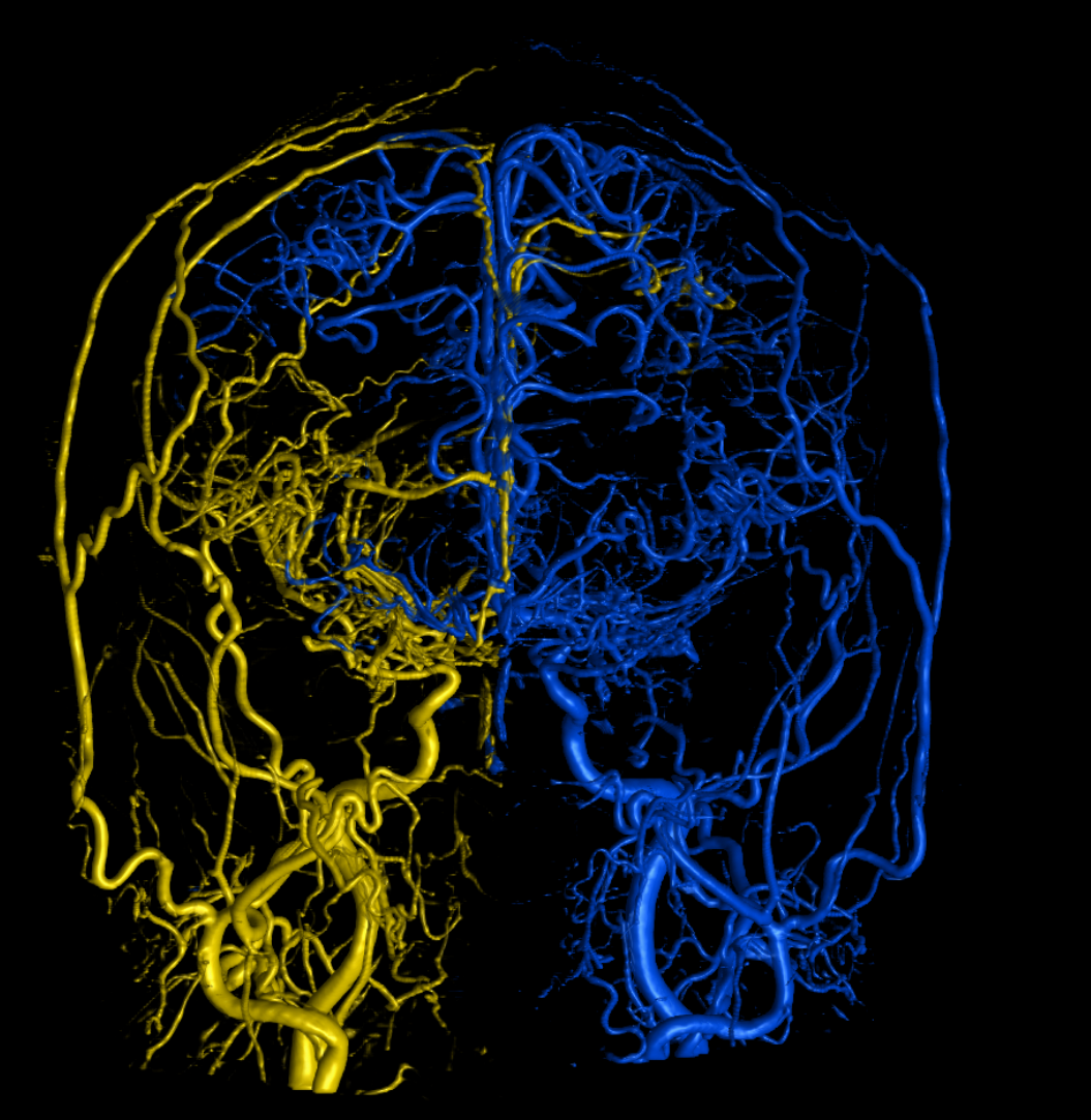
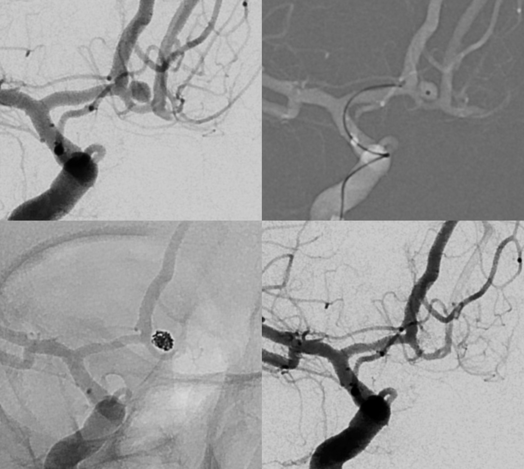
Below are some examples. These, and many more, in context of interventions / treatments, are found on the Case Library page
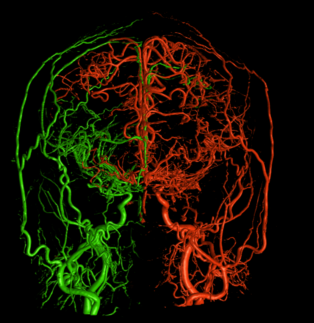
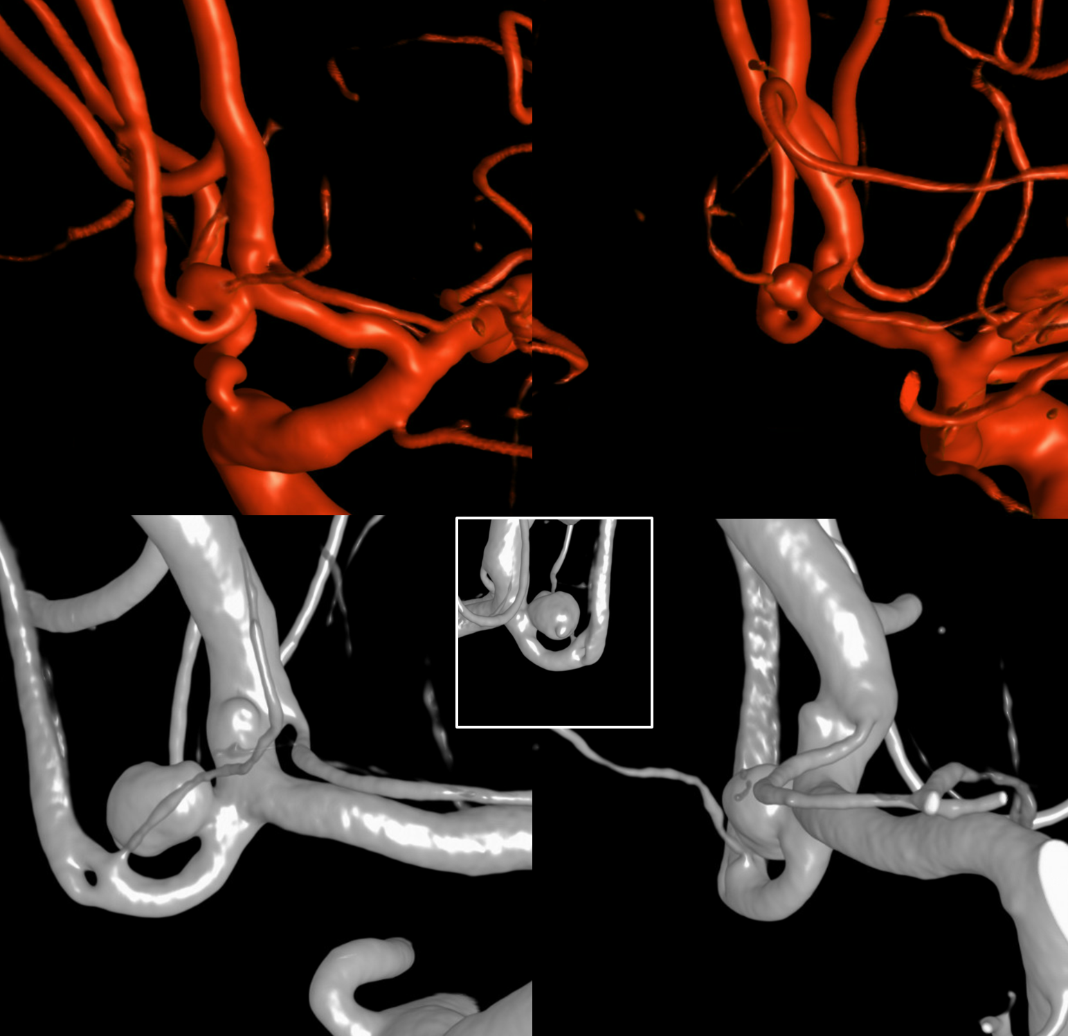
Moya Moya Fusion
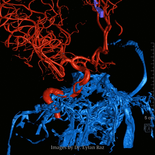
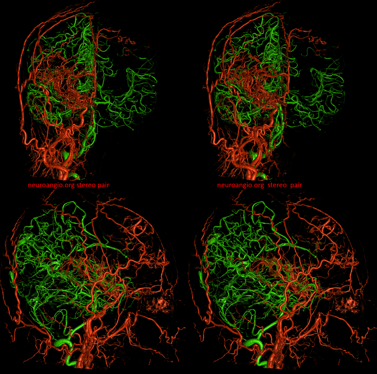
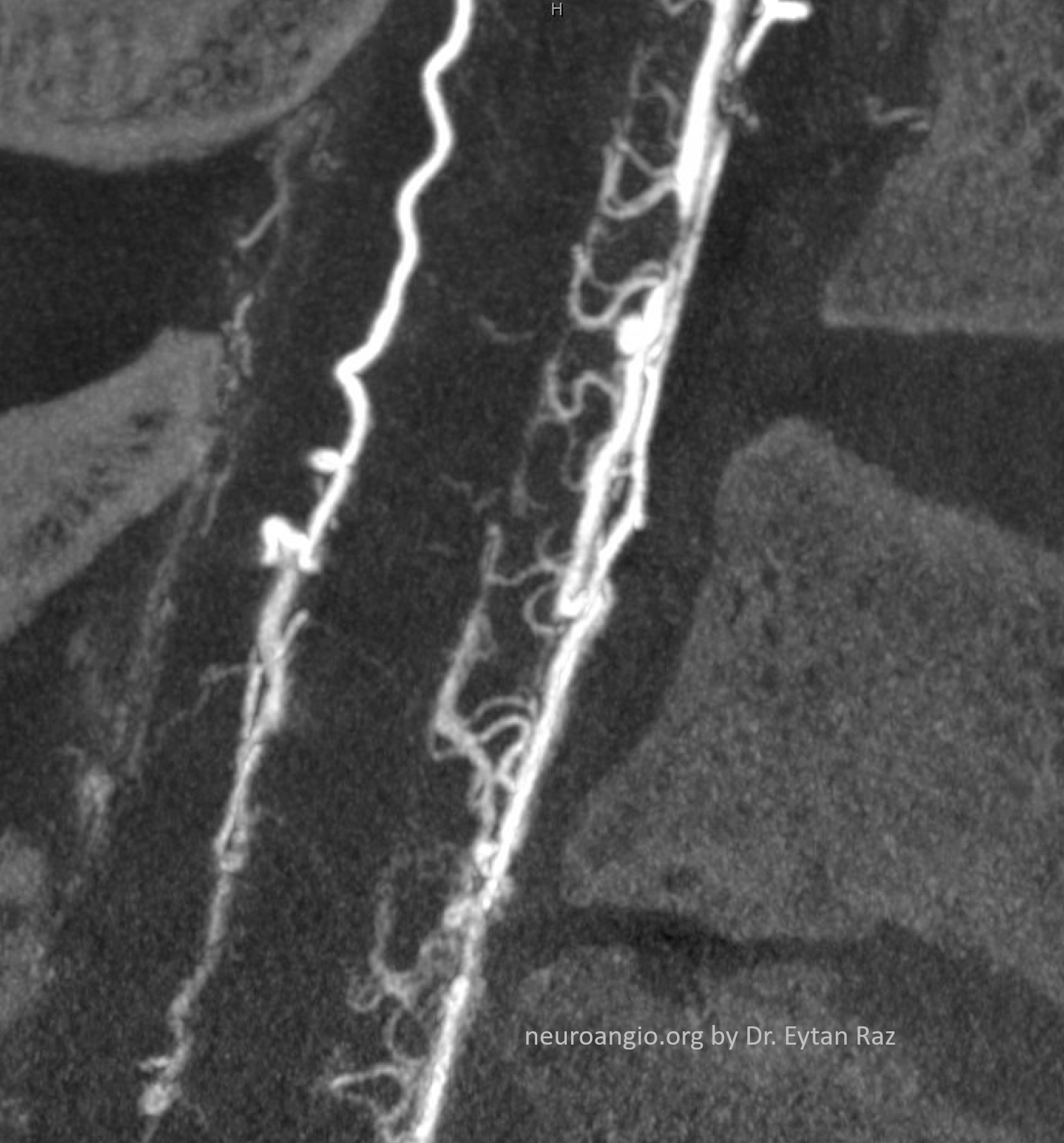
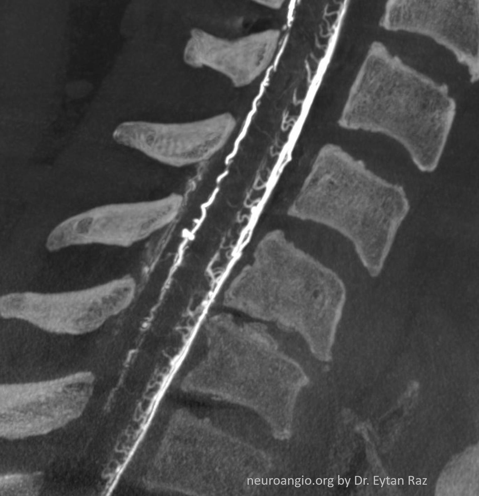
Fusion same case vert and right ICA
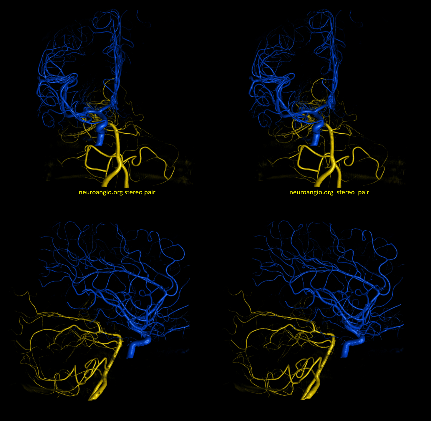
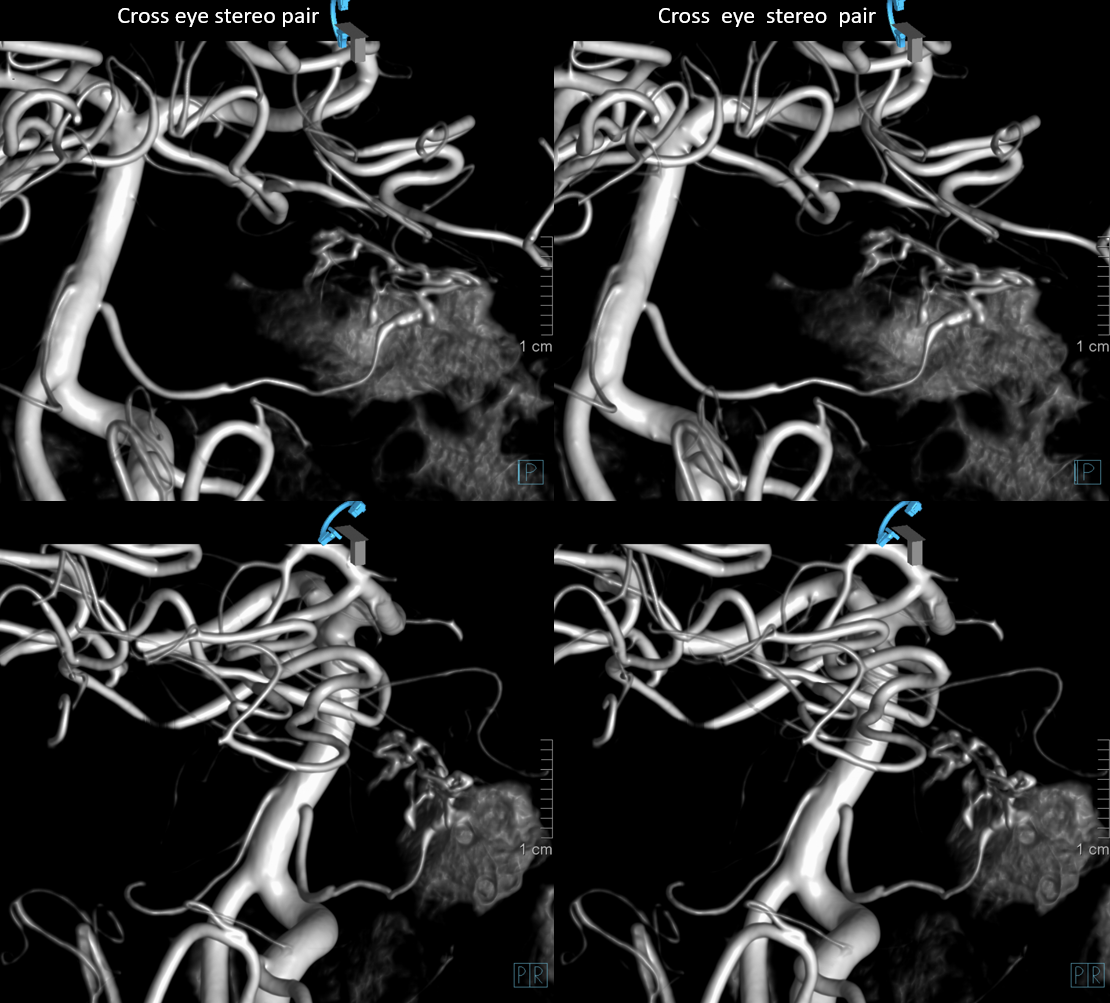
Stereo ICA vert fusion
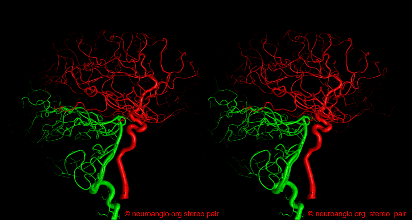
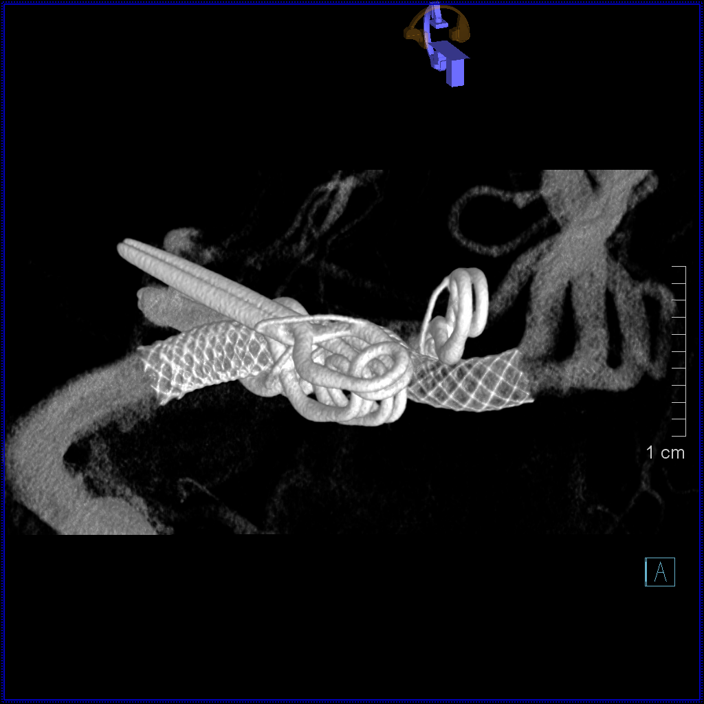
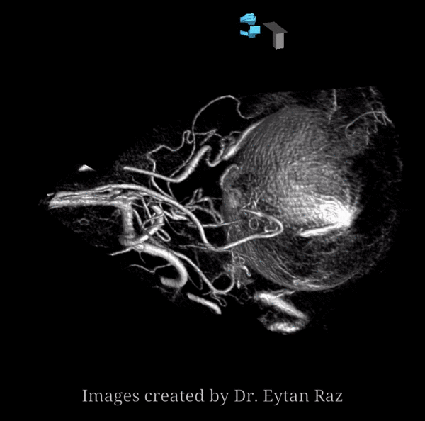
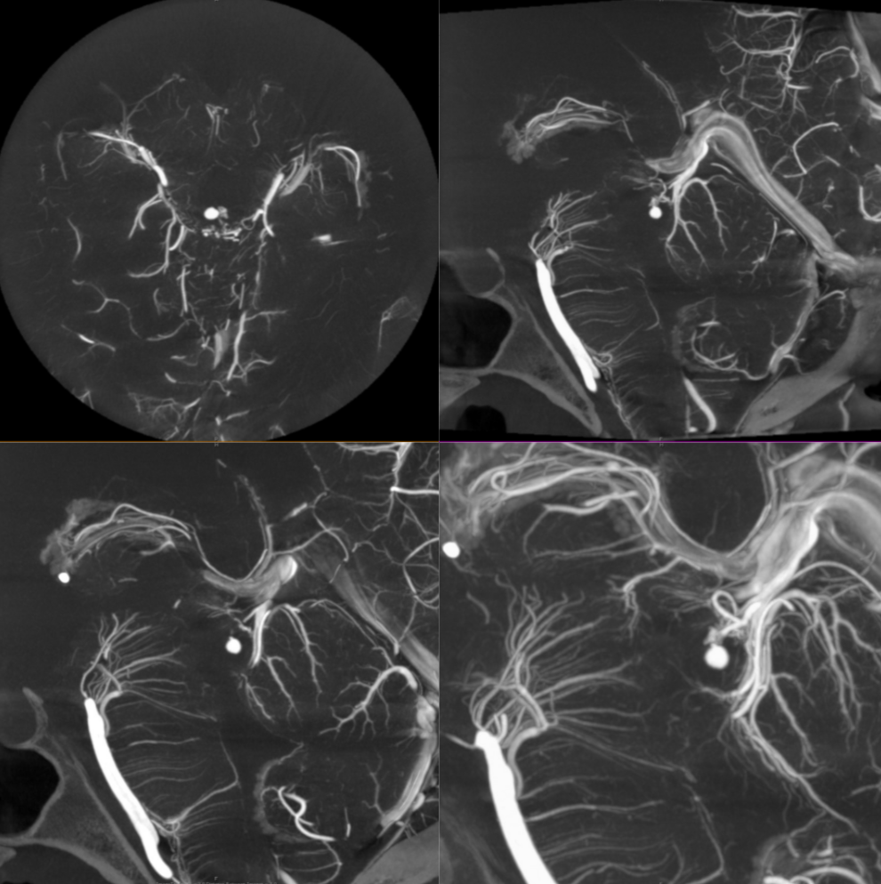
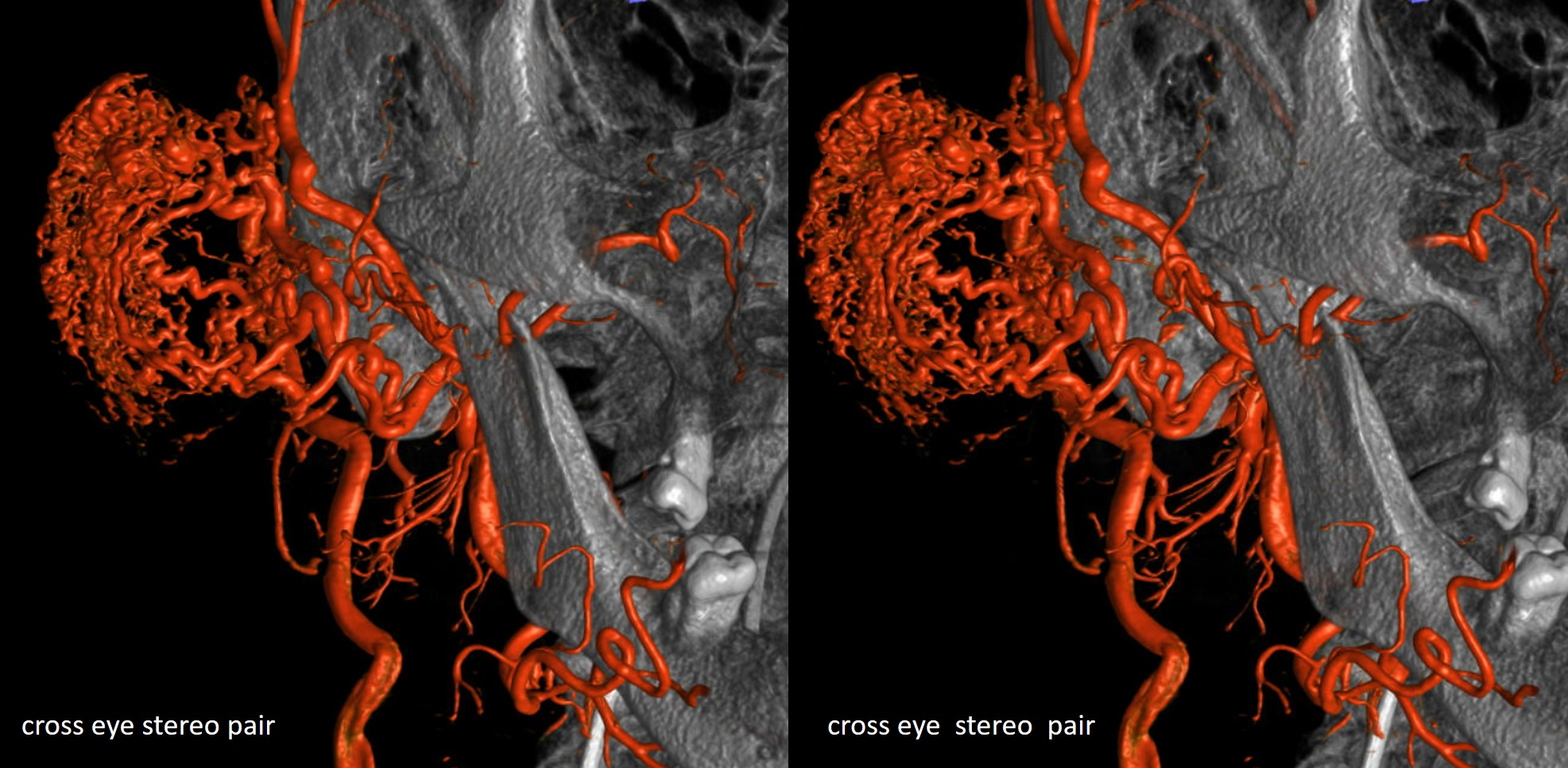
Giant Cavernous Aneurysm
Video

Technical points
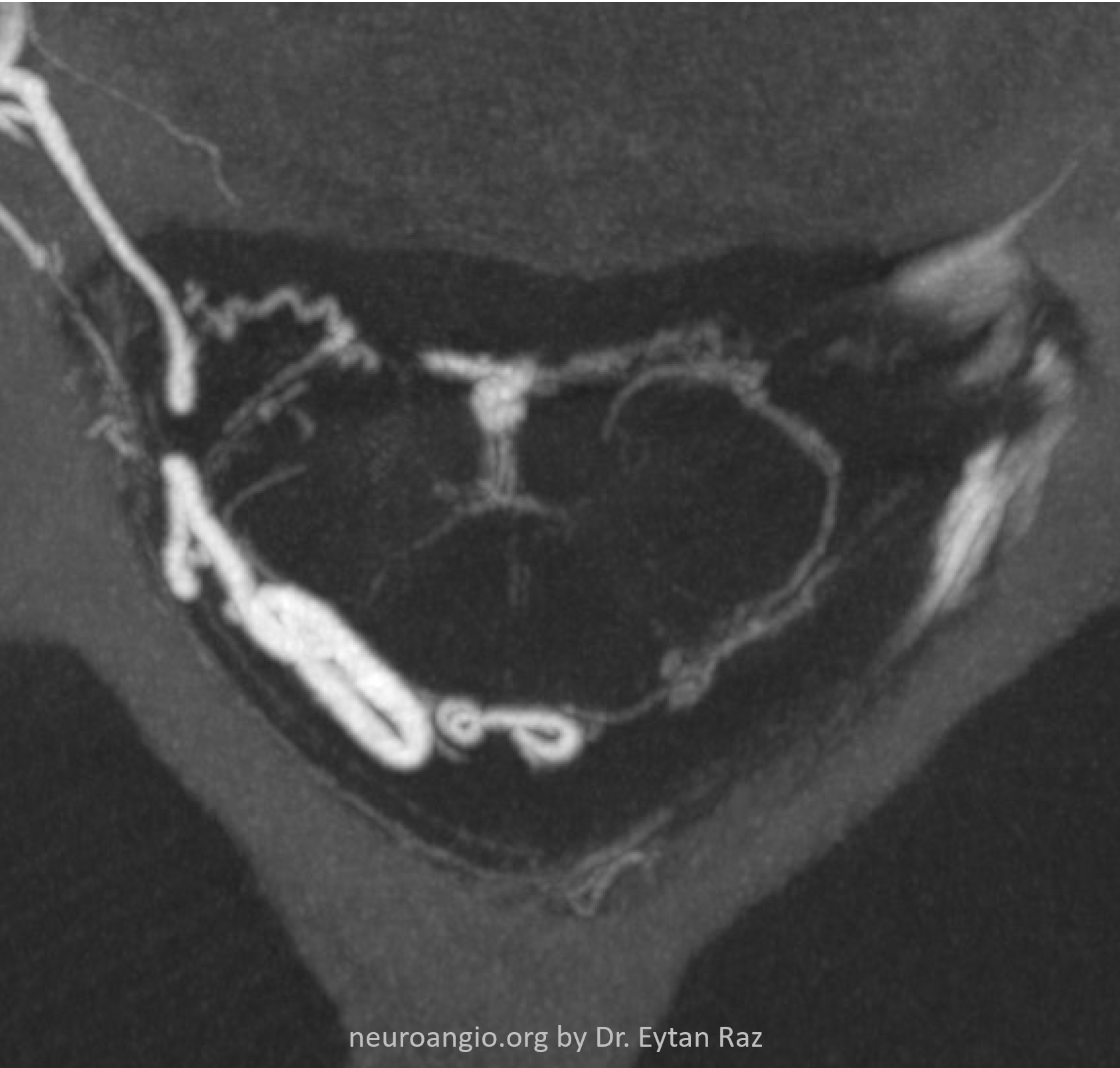
These ones are important — otherwise images are not good. Machine must be calibrated and camera cant be wobbly. When it is, you can imagine that a point in space will no longer be represented by a point if the camera wobbles. Below is an example — you can visually see that the head “moves” at both ends of rotation. it is of course the camera and detector moving, not vise versa.
The resulting reconstruction is very bad. Since the machine assumes that everything is stationary, and only its camera is moving in a pre-detemined way, if the camera were to move in a way not anticipated by design, a distortion in reconstruction will happen (arrows). Below is what this looks like on VR and MIP images. Note that the unsubtracted (MIP bottom row) images are less affected than those with contrast (top row)

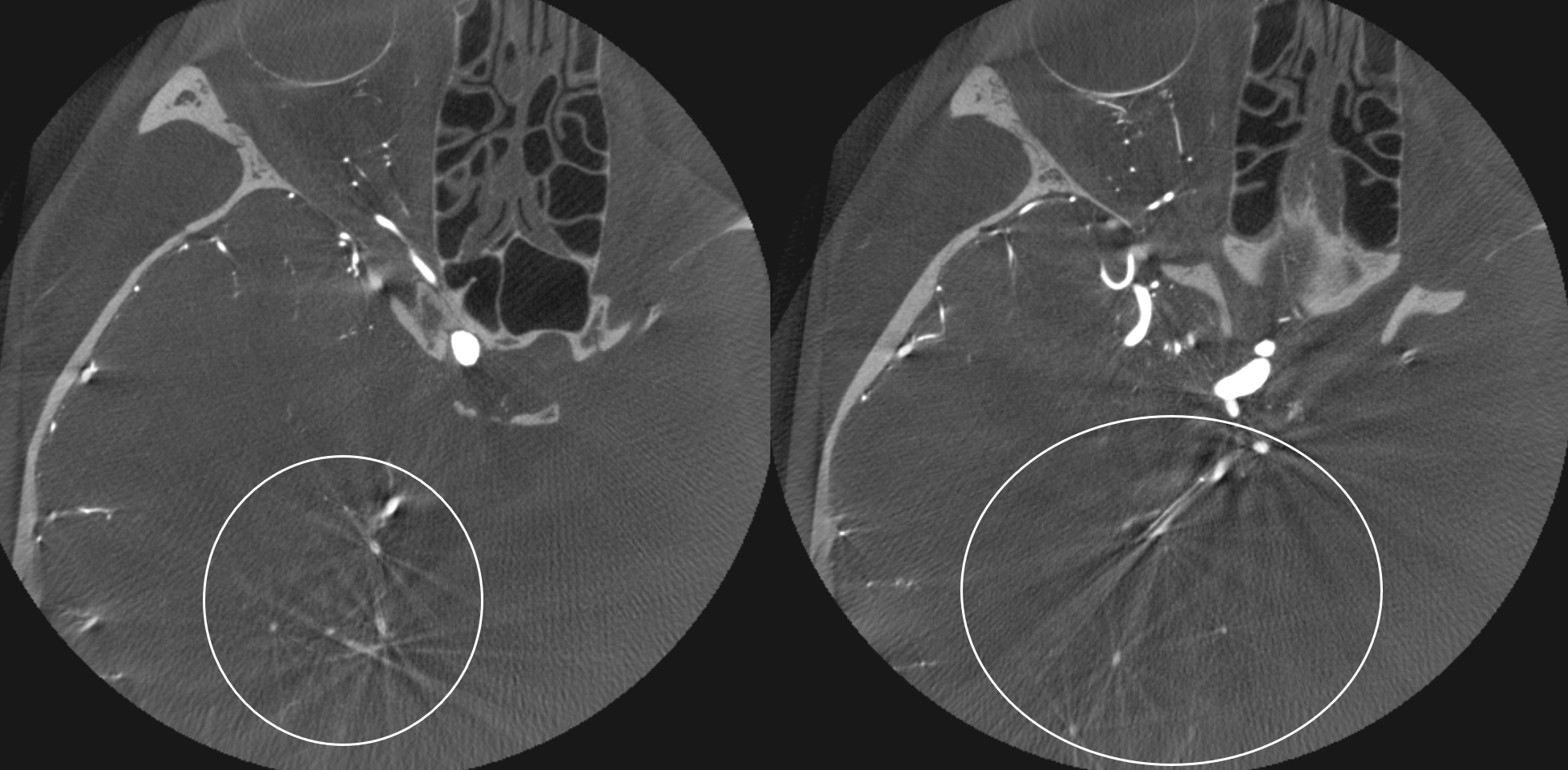
Intermittent Vessel Filling
Another issue with cone beam CT is that the reconstruction algorithm can be very sensitive to data being present at all points during acquisition. For example, if a vessel is opacified only intermittently, it will be “present” during some points in rotation and “absent” in others. The reconstruction algorithm is not good at this point with this kind of confusion, and produces star or cross type artifacts. See below for the intermittently filling PCOM