
Numbers 8 above. And 67 below
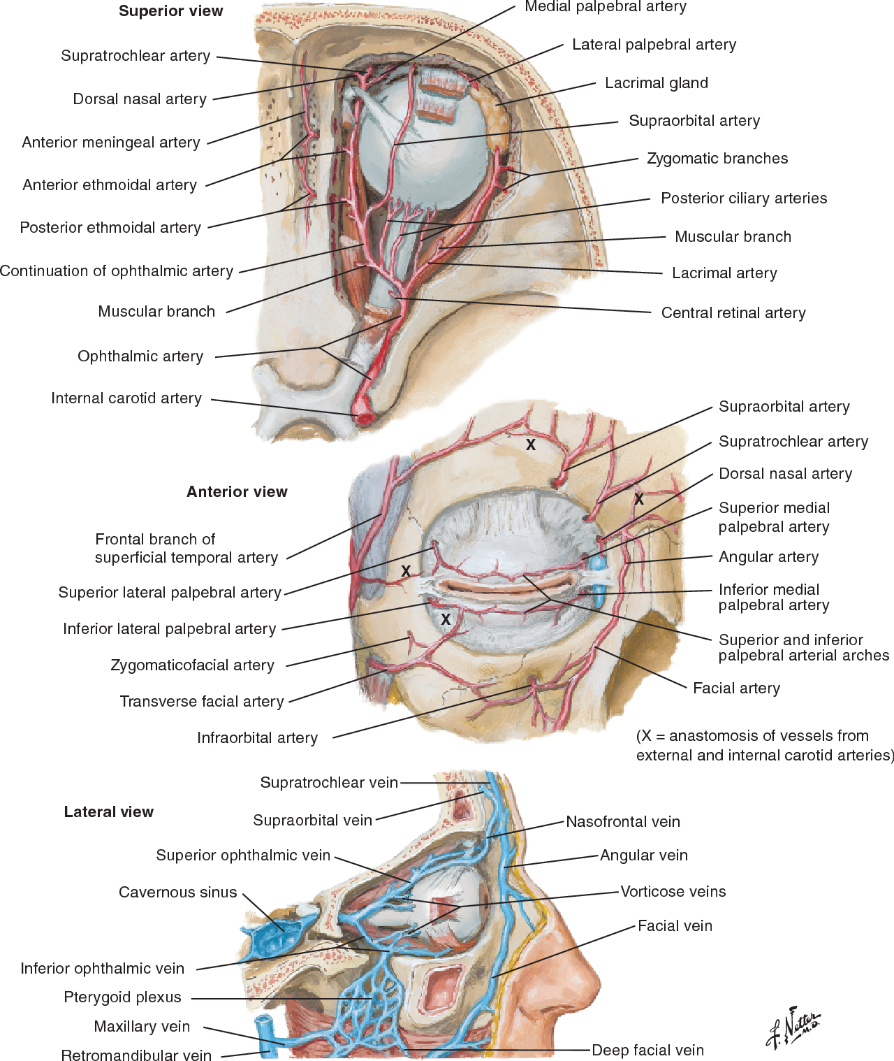
The great Netter depiction of the ethmoidals
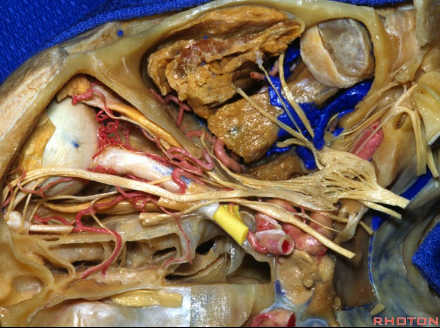
Beautiful dissection from Rhoton
Number: Usually anterior and posterior ethmoids. Variable dominance, occasionally 1 or 3 vessels.
Origin: Distal Ophthalmic Artery
Territories: Nasopharynx (septum, upper parts — ethmoid sinuses), anterior parasagittal dura (via anterior meningeal arteries).
Anastomoses: contralateral ethmoids, septal, sphenopalatine, anterior meningeal, anterior cerebral (via frontal base autosynangiosis).
Importance: Participate in supply of all regional vascular lesions (ethmoidal dural fistulas, frontal base meningiomas, nasal tumors. Can be source of epistaxis, including recurrence after IMAX embolization.
Angio / In Vivo Imaging
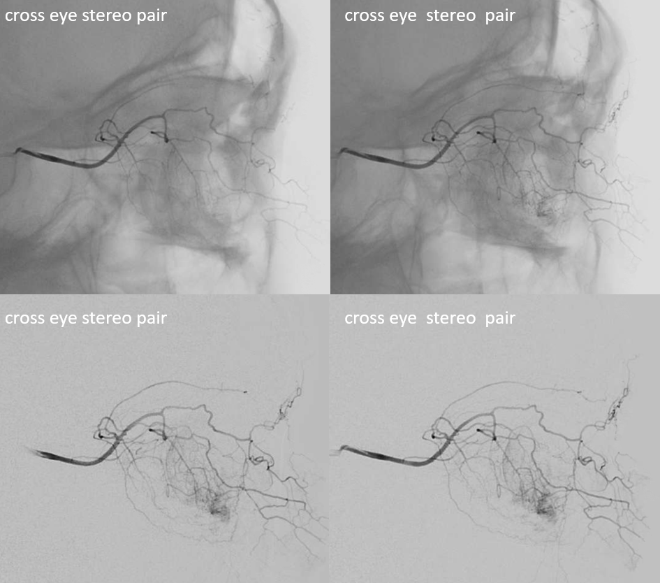
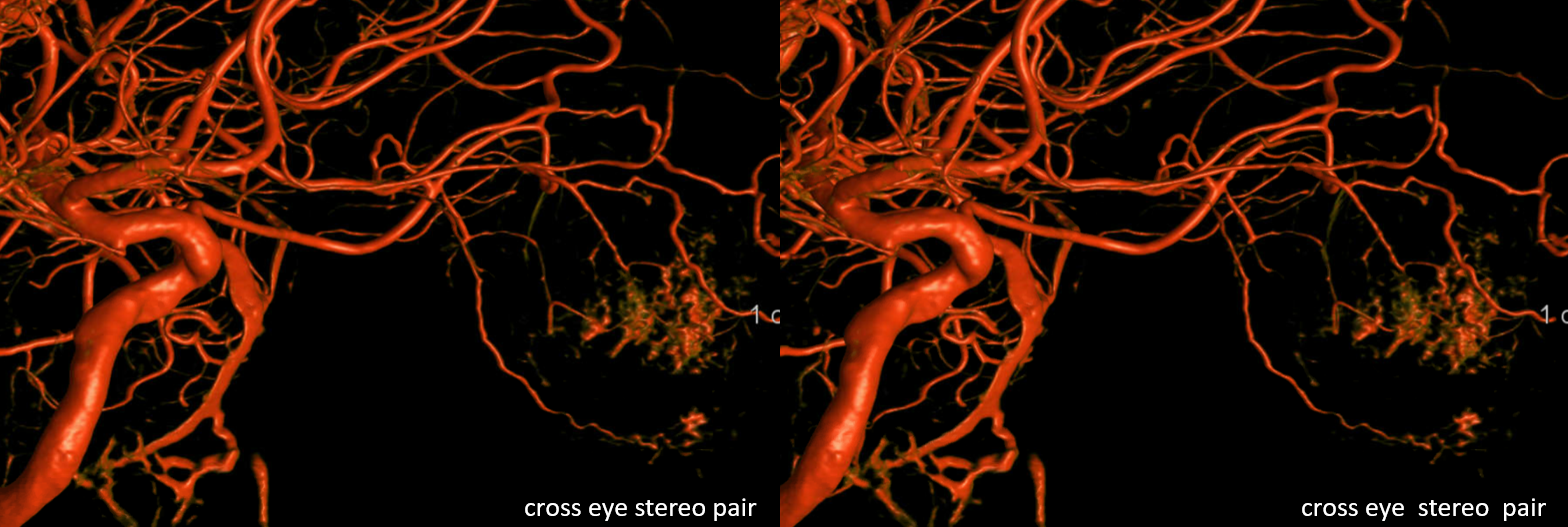
Opthalmic artery injection sereos — anterior ethmoidal artery supply of the septum and upper nasopharynx
Movie
Another Case
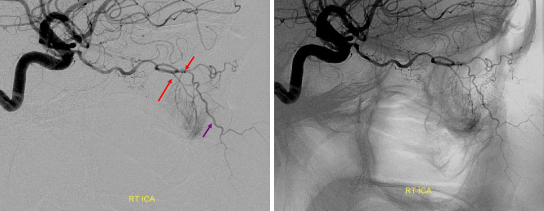
Septal system (purple) here is mainly supplied by the ophthalmic via anterior ethmoidal (red) artery. When particularly big, ethmoidal supply can contribute to failure of epistaxis embo. In this case, the ethmoidals must be surgically closed
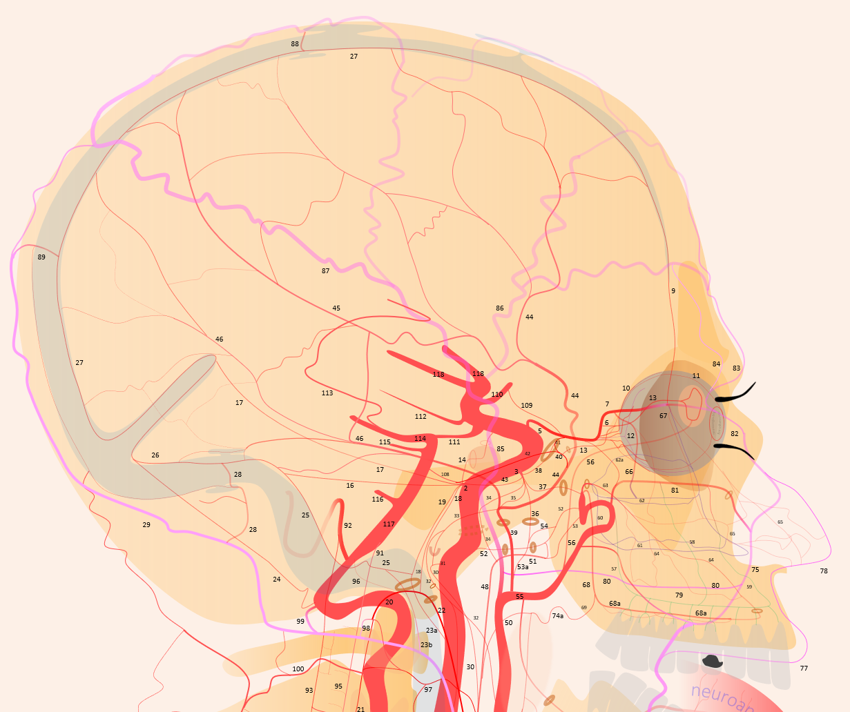
Another example, large ophthalmic because it supplies lots of nasal septum (pink), cutaneous nasal branch (white), and anterior forehead tissues (purple, supraorbital/supratrochlear 83/84)
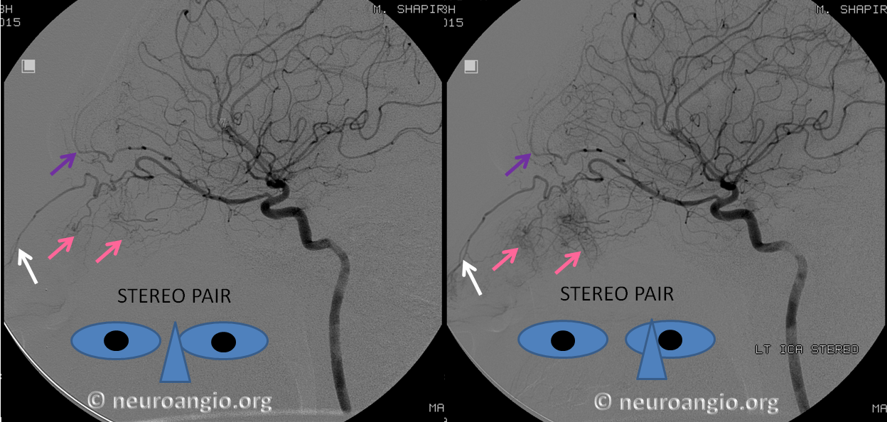
Another example of supply to the upper nasal region (superior turbinate, upper septum) — the best way is on frontal views, ethmoid branch reaching to midline, with mucosal blush
Notice branches to the upper and lower eyelids on lateral views! — not labeled, try to stereo
Posterior Ethmoidal Artery Opacified via ECA injection (meningophthalmic variant). See full case on Ophthalmic Artery page. Anterior is much smaller here
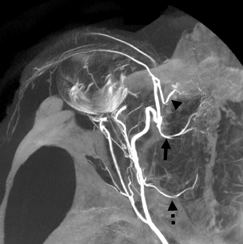
Triplicated Ethmoidals
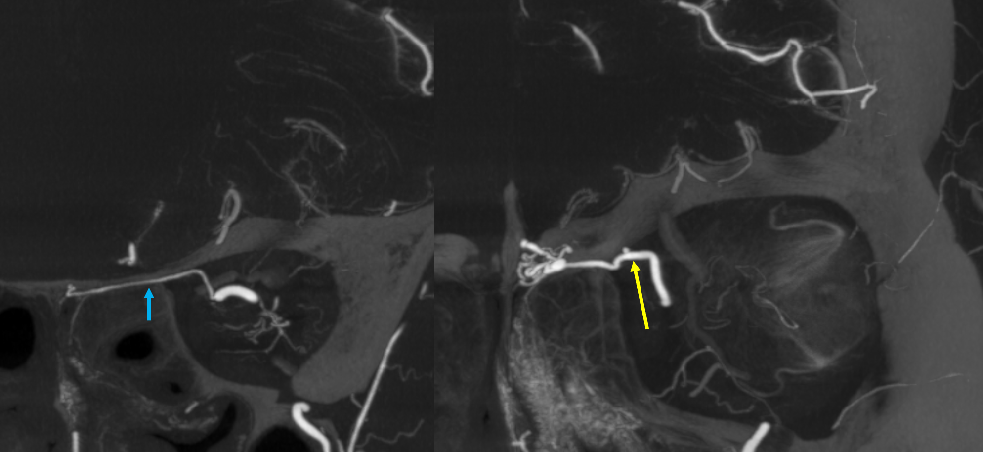
Of course, vessels don’t read books or websites. There are 3 ethmoidal sets of arteries — posterior (dashed arrow), anterior (arrow), and whatever else we want to call them — lets say anterior-most (arrowhead)
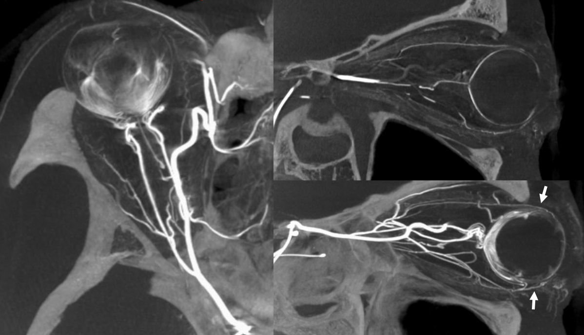
Movie of the same subject below — this is a case of penetrating sharp object orbital trauma — notice major swelling of the anterior orbit / eyelids, and an irregular “guitar pick” shape of the globe due to increased intraorbital pressure, which is maintained in perfusion range by prior canthotomy. The extra-ocular muscle vessels are particularly well-seen, and their anterior ciliary tributaries to the anterior chamber, thanks to post-traumatic hyperemia.
Same subject DYNA MIPs. Anterior ciliary arteries are labeled
Another example — large anterior (arrow) and two posterior (oval) ethmoidals — common trunk, two posterior ethmoidal foramina
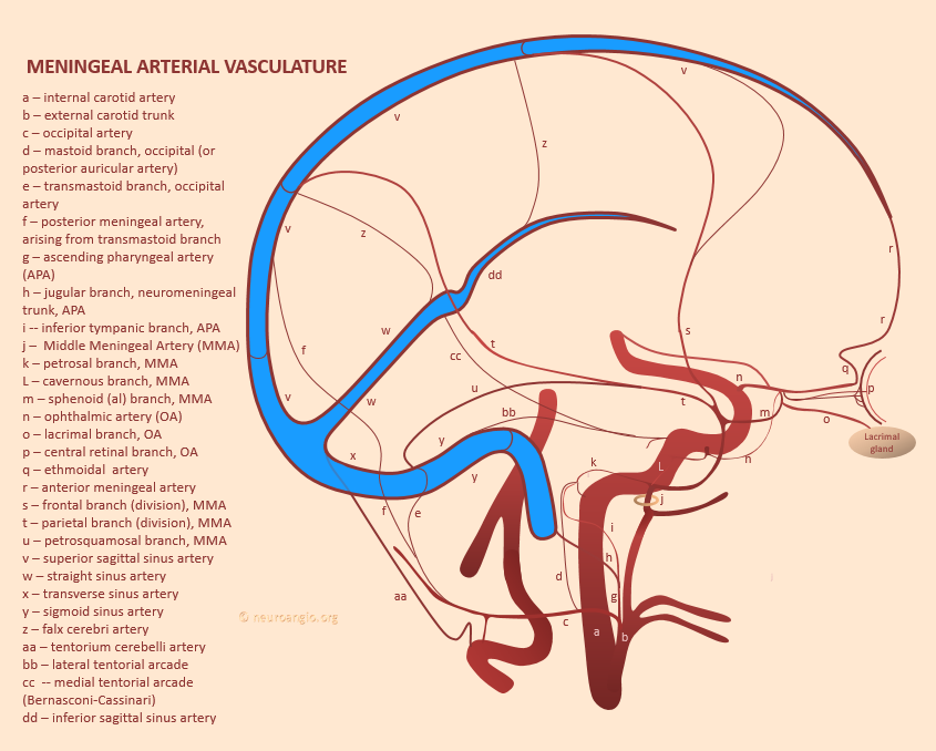
Supply of Anterior Meningeal Artery
See Meningeal Arteries page for that. Its important. This meningeal network is key. A case of Anterior Meningeal Route Embolization is a great example of this kind of connectivity. It is also a potential danger when liquid embolics are used for MMA embolization, as the liquid can travel a great distance along the anterior meningeal artery so we are very careful to watch that area.
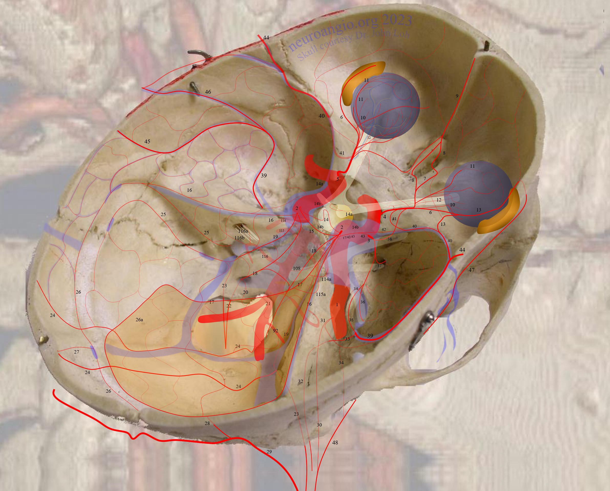
The diagram below gives the idea
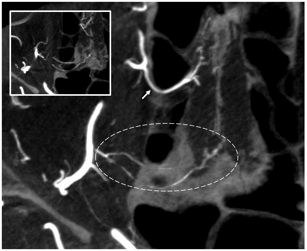
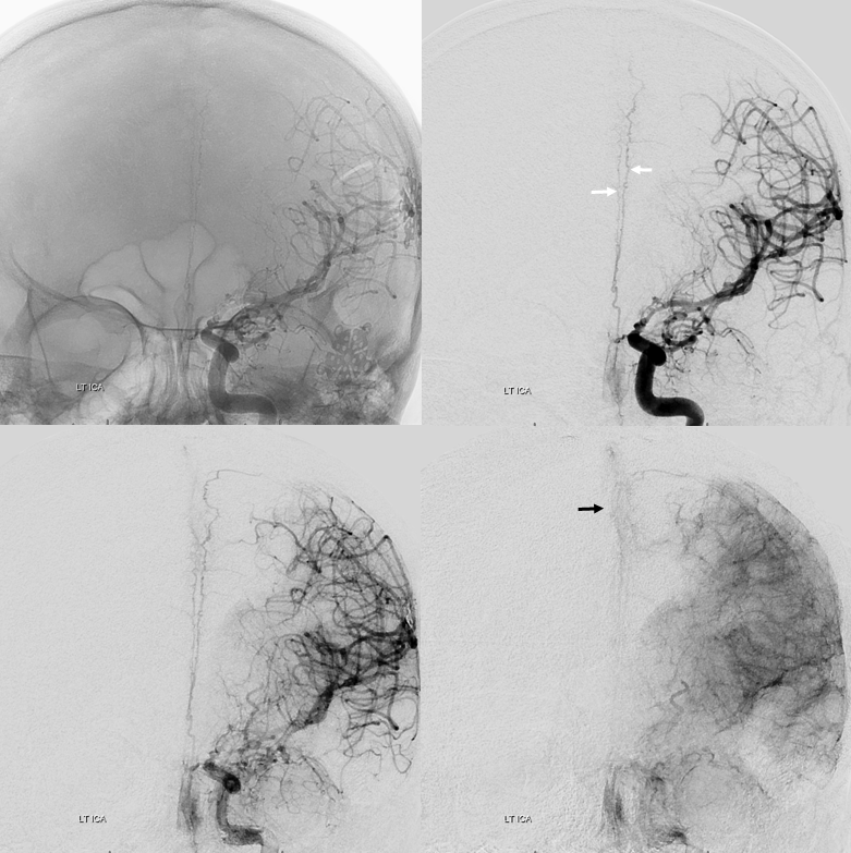
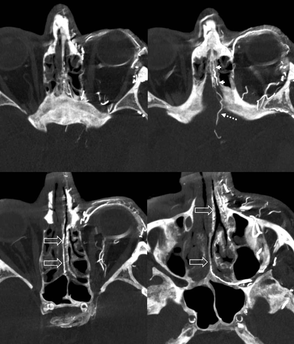
A well-seen anterior meningeal network shown below — even bifurcating (white arrows) with both arteries in wall of sagittal sinus. There is a meningeal blush (black arrow). The size of anterior meningeal artery no doubt is enlarged to compensate for loss of the MMA post left pterional craniotomy and clipping of a ruptured PCOM. Angio is done for spasm Rx.
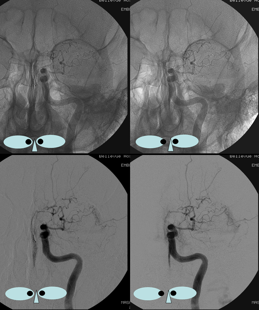
Ethmoidal Autosynangiosis in Moya Moya
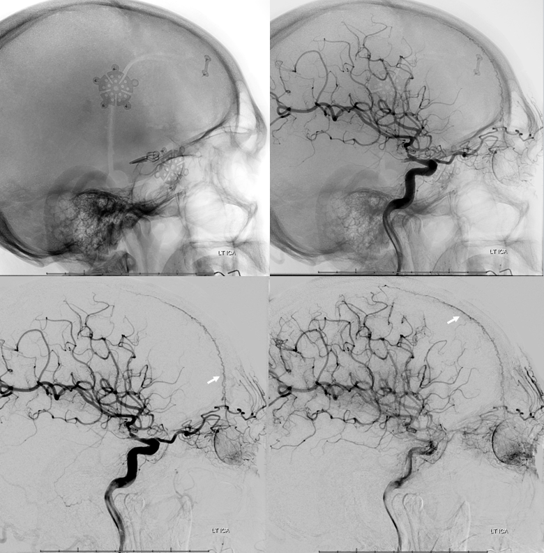
Ethmoid arteries participate in dural supply — as evidenced in their supply of cribriform plate meningiomas for example, or dural fistulas. Another example is an autosynangiosis, typically with the anterior frontal / gyrus rectus branches of the anterior cerebral artery in cases of Moya-Moya. In example below, courtesy Hima Pendharkar, the supply mostly comes from anterior ethmoidal group — notice however that there are two branches present, both anterior — again, they don’t read books.
The other side. Anterior ethmoid group is marked by arrows. The posterior group also participates, and is unmarked.
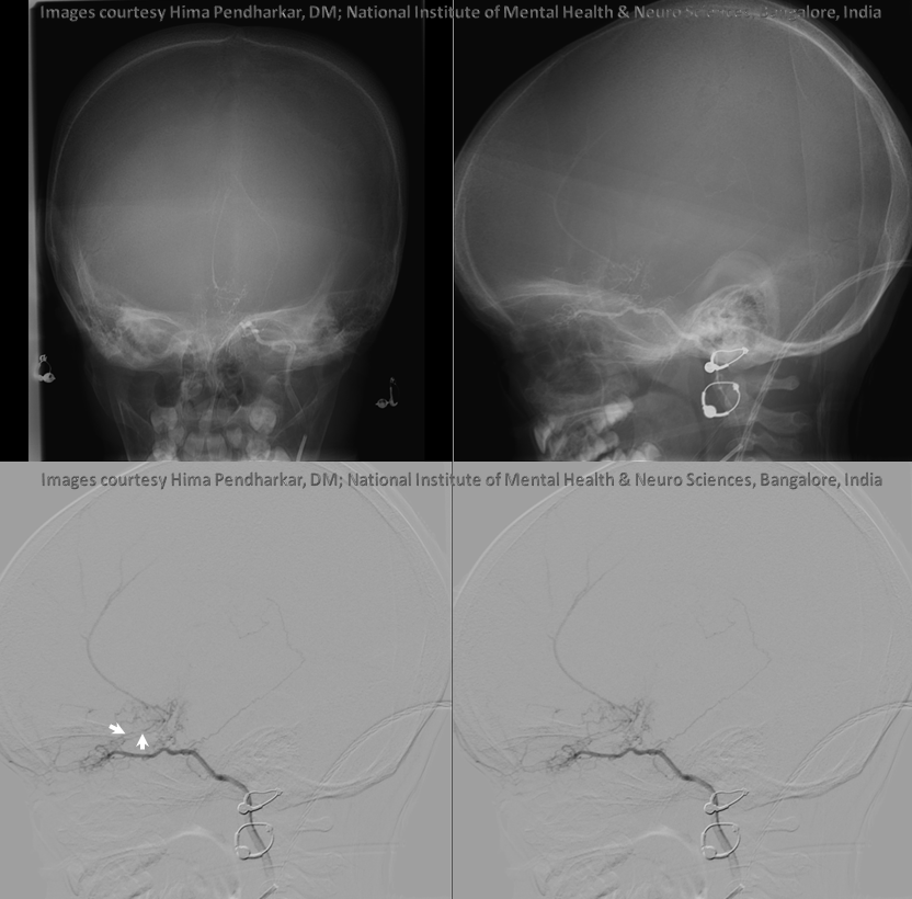
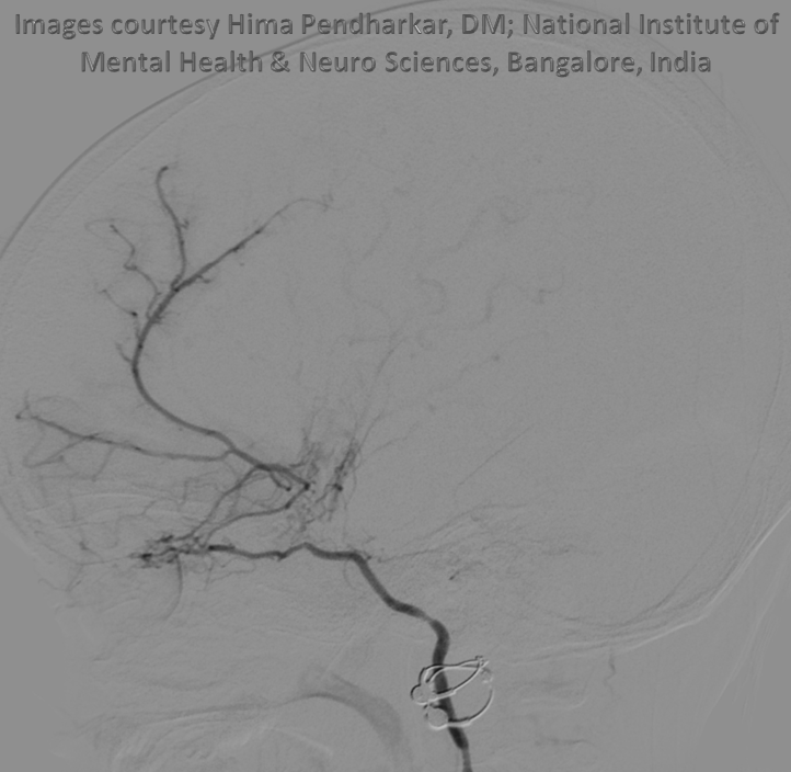
Another case of Moya Moya
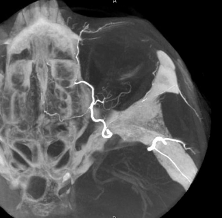
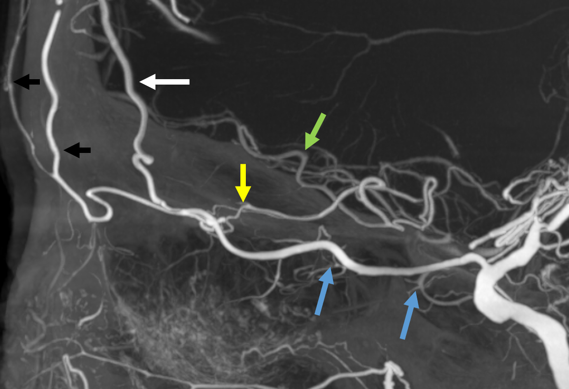
Some amazing DYNA imaging by Dr. Eytan Raz — anterior ethmoidal = yellow; recipient frontopolar/ rectus branches of ACA = green; posterior ethmoid branches supplying the nasal septum = blue; anterior meningeal artery = white’ supraorbital/supratrochlear branches = black
Movie
Stereo angios of yet another example
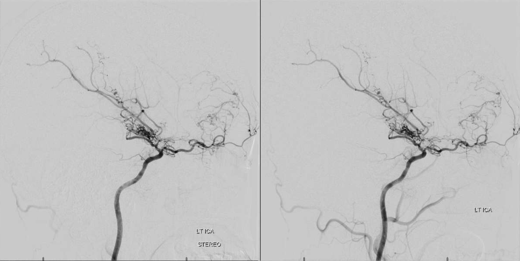
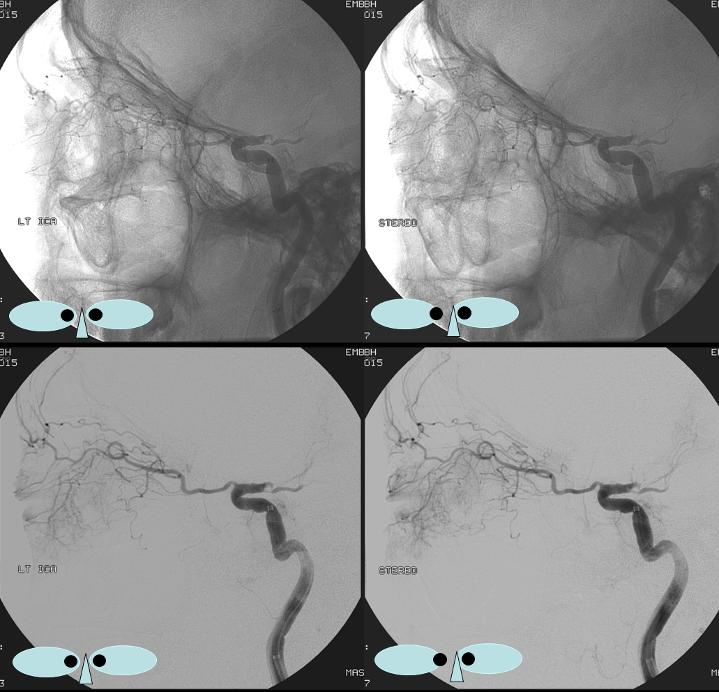
Another case — Ethmoidal to Gyrus Rectus ACA dural anastomoses in setting of ICA occlusion. Posterior ethmoidal = arrow. Gyrus rectus branch = dashed arrow. Septal branches = arrowheads.
ICA reconstitution via superficial temporal – Ophthalmic Artery. Small contribution from IMAX via septal to ethmoid anastomoses is present also.
The most efficient by far ECA to ICA cervical carotid reconstitution is via the Ophthalmic Artery. Tons of possibilities — via MHT, anterior deep temporal, facial, ethmoid/anterior falcine, etc. Here, in case of proximal right ICA dissection, there is a really nicely seen STA to supraorbital anastomosis, and an anterior deep temporal to lacrimal branch also.
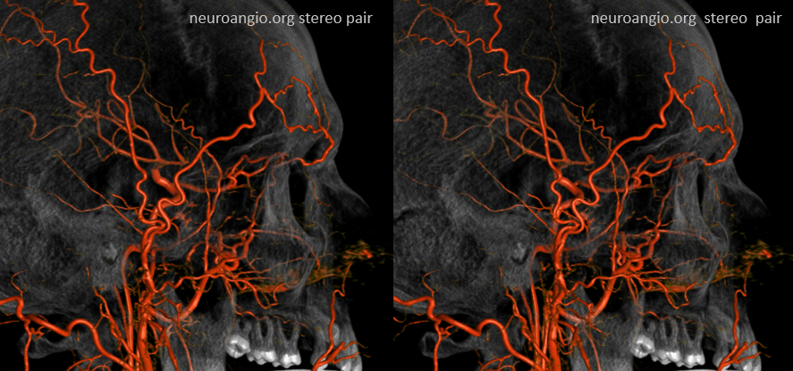
Can you see a myriad other EC-ophthalmic anastomoses on this MIP movie?
Ethmoidal Dural Fistulas
- Ethmoidal Dural Fistula middle meningeal – anterior meningeal approach embolization
- Ethmoidal Dural Fistula Trans-ophthalmic nBCA Embolization and Amazing Globe Images
- Ethmoidal Dural Fistula Trans-Ophthalmic nBCA Embolization Case 2
- Ethmoidal Dural Fistula Transvenous Embolization