
Otospongiosis (also known as Otosclerosis) is a rare condition where the normally super-dense bone around the cochlear (it is normally the densest bone in the body) becomes “spongiotic” or soft. The condition used to be called “otosclerosis” because in some stages of the disease the bone appeared “sclerotic” on x-ray imaging. It is however a misleading term because sclerotic implies more dense, whereas in fact the bone becomes less dense. So, the modern term is otospongiosis.
The cause is unknown. Presentation is usually with hearing loss. Often, hearing loss is “conductive” — meaning the nerves of the inner ear are intact, and the issue is with transmission of sound. The vast majority of patients with otospongiosis do not have pulsatile tinnitus. They usually have nonpulsatile tinnitus, like so many others with hearing loss. However, occasionally presentation is with pulsatile tinnitus. Why is unclear to me. Two possibilities are:
- Conductive nature of hearing loss leads to decreased ear sensitivity to external sounds and increased sensitivity to normal internal sounds, which include flow in the nearby carotid artery and jugular bulb. Beethoven, for example, who most likely had otospongiosis, would put a stick between the cover of a piano and his forehead to get the vibration of sound to transmit to his skull, and could hear it somewhat this way.
- Otospongiotic bone is actually somewhat hypervascular and perhaps pulsatile tinnitus is due to actual sound of increased regional blood flow. Certainly this mechanism is responsible for PT in patients with glomus tympanicum and glomus jugulare tumors. However, otospongiosis is not a tumor and is nowhere near as vascualr as glomus. The bottom line is that we don’t know why a minority of people with otospongiosis have pulsatile tinnitus anymore than we know why some people with other forms of hearing loss have pulsatile tinnitus. I think reason (1) is still possible.
Treatment is surgical. Sometimes, ossicular chain reconstruction works. Sometimes, its bad enough to require cochlear implant. Most patients with tinnitus due to hearing loss (pulsatile or not) report improvement after cochlear implant. It is however a very major step and must be discussed with a cochlear implant specialist.
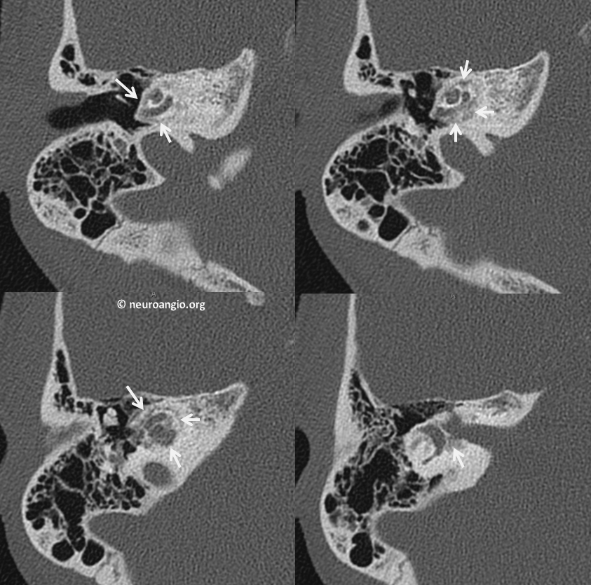
Here is a typical normal CT of middle ear cavity with bright dense bone around the cochlea (white arrows). The bone around cochlear is known as the capsule
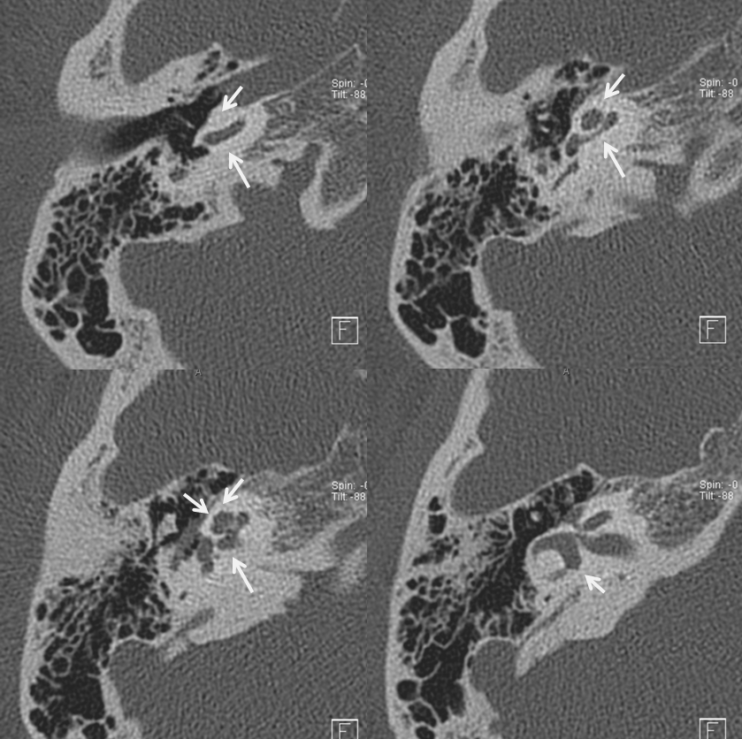
Below, in contrast, is a patient with advanced otospongiosis. There is a ton of lucent, spongiotic bone (white arrows) around the cochlear. Similar changes were seen on the other side. Typically, otospongiosis is bilateral
Back to Diagnosis and Treatment of Pulsatile Tinnitus