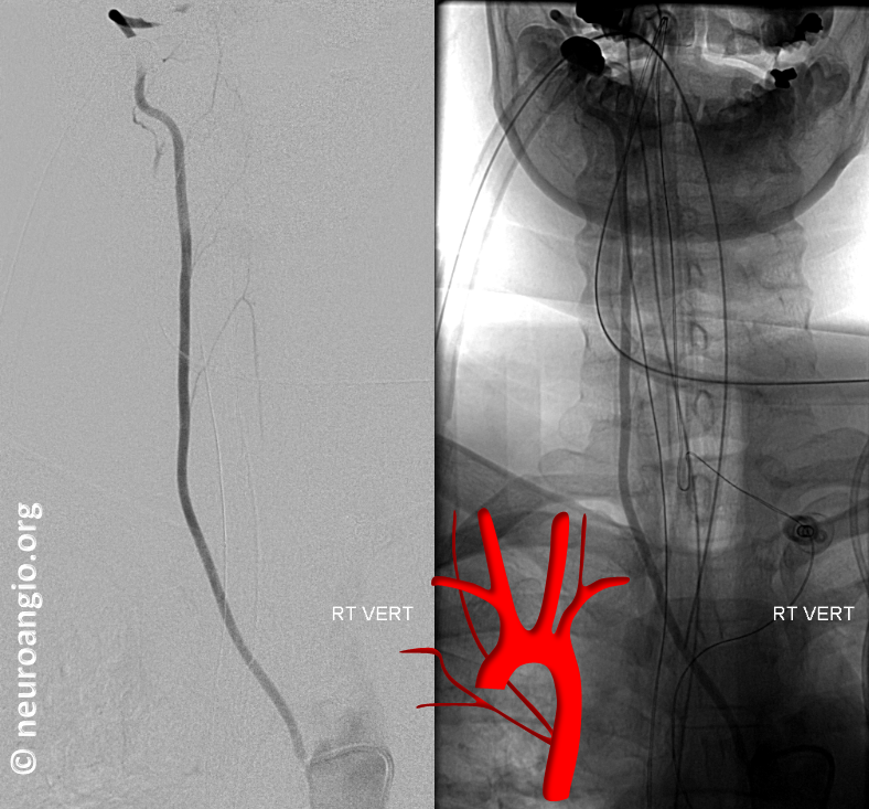
A rare but sometimes critical anatomical variant to know (See Aortic Arch — A Guide to the Perplexed for more).
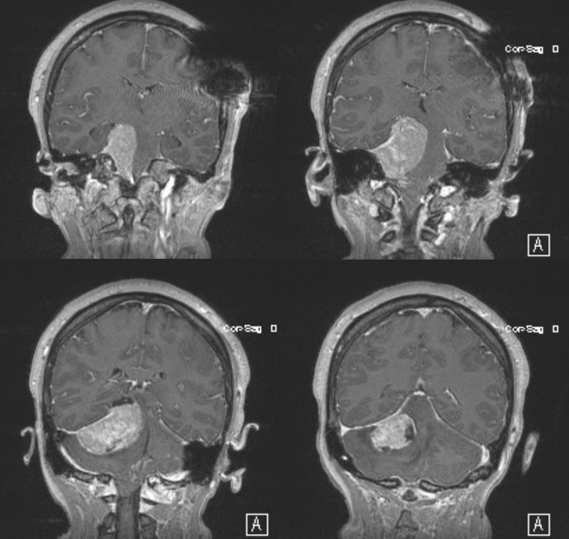
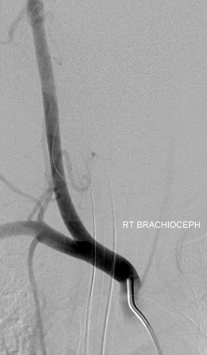
Sometimes you need to find that right or left vert and its not enough just to inject everything else. This patient presents for preoperative angiographic evaluation
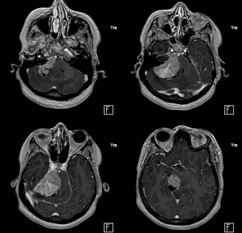
It is essential to find the right vertebral artery, for many reasons.
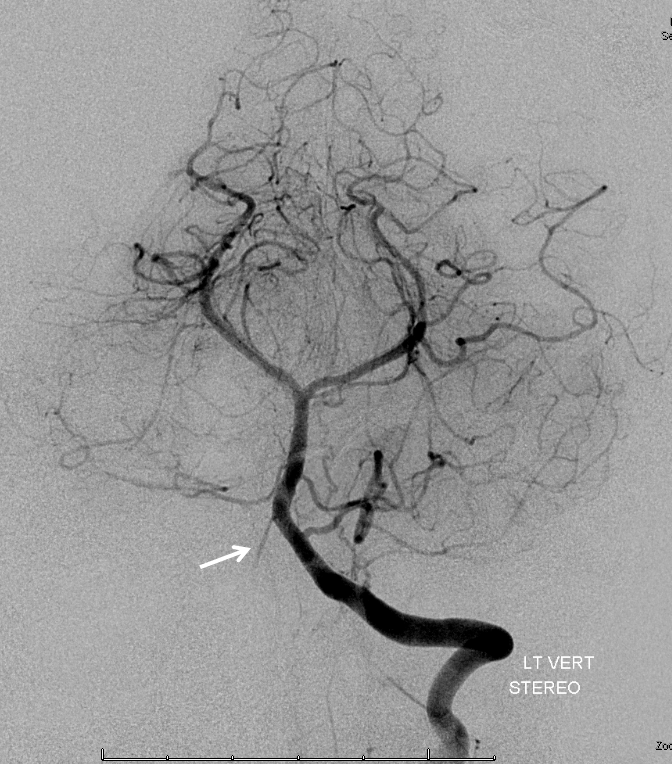
Here is the left, notice reflux into hypoplastic distal right vert (arrow)
The vert is not found in its usual location
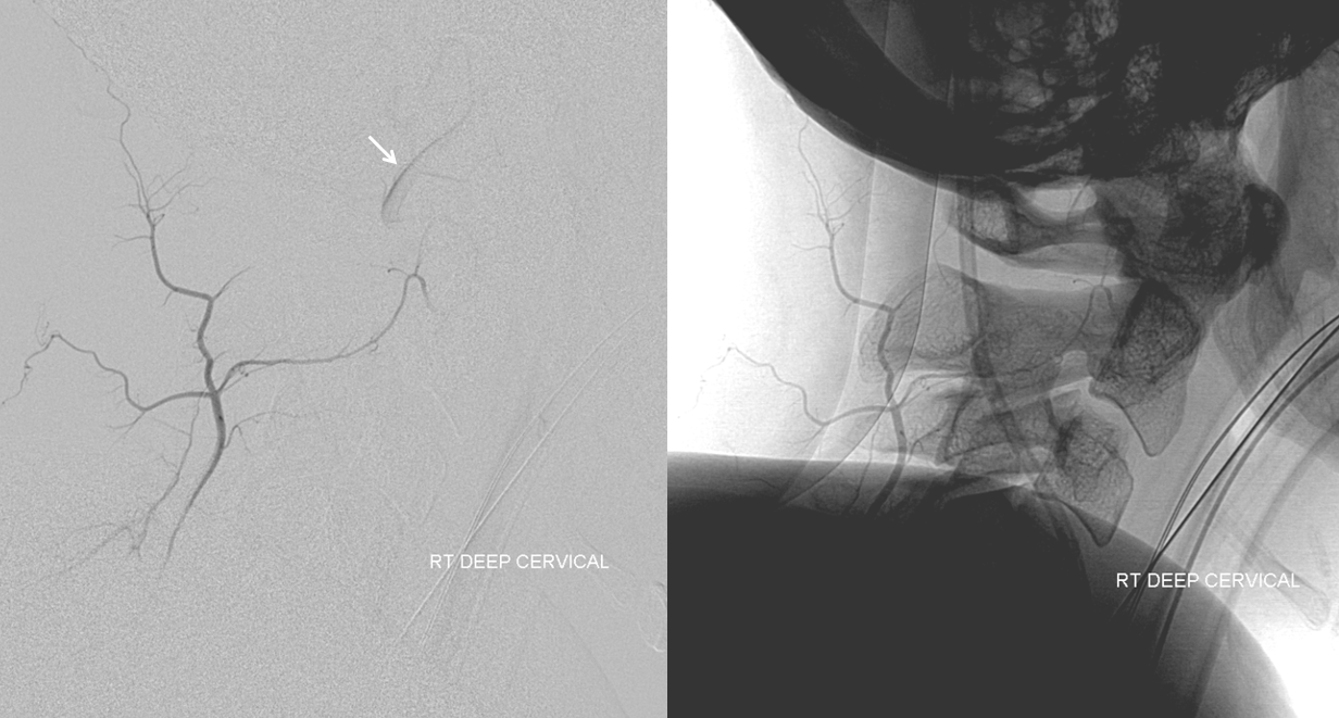
An injection of the right deep cervical artery shows transient opacification of the vert (arrow) via the C2 muscular anastomosis; we know the vert is there
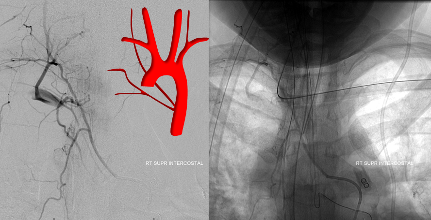
Where is it? The answer is aortic arch. When the right vert is apparently missing in action, the origin is usually in the upper thoracic descending aorta — at the supreme intercostal artery

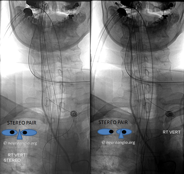
Stereo views are below
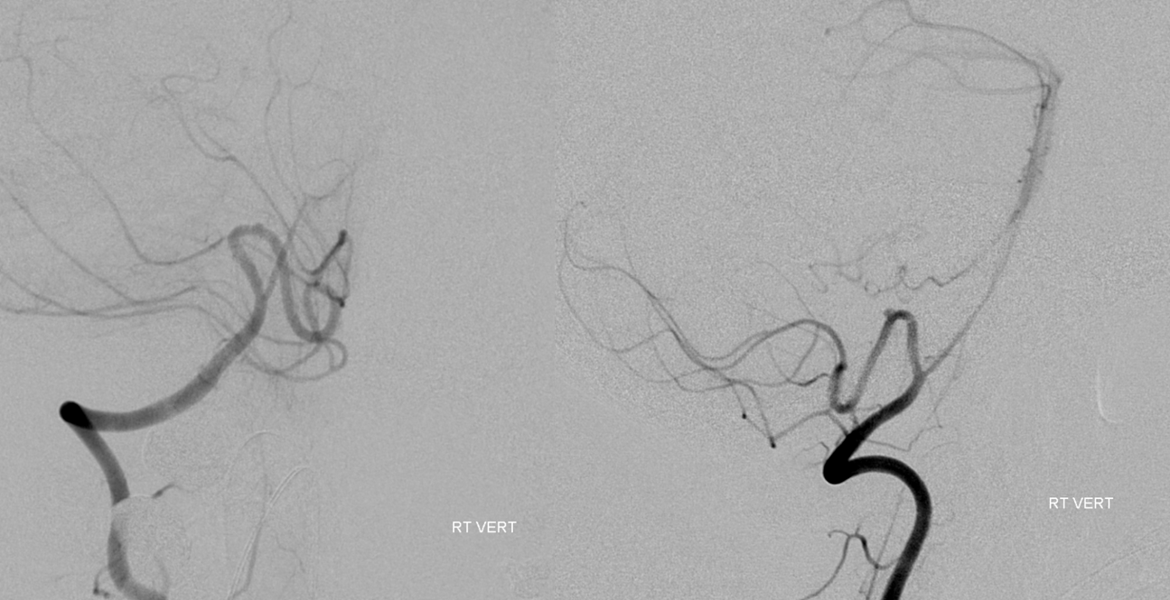
Distal vert images prove the usefulness of finding this vessel. Notice anterior spinal artery origin from the distal intradural vert (above the PICA), and a small aneurysm at the apex of the medullary PICA loop.
The vert appears to come off by itself. However it is not the case — the rest of the supreme intercostal trunk is just a small turn of catheter away. Notice how the supreme intercostal supplies the T1 intercostal artery and anasomoses with the proximal deep cervical artery, transiently opacifying the subclavian.
See Aortic Arch — A Guide To The Perplexed for more important arch variants. Spinal Arterial Anatomy. Vertebobasilar System. Vertebral Artery
