
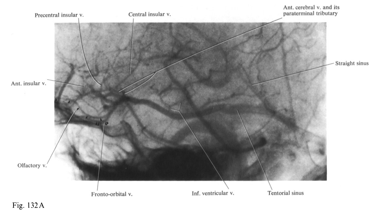
In collaboration with Dr. Kittipong Srivatanakul , Dr. Eytan Raz, and Dr. Matthew Young
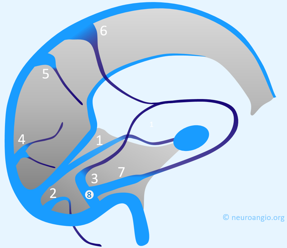
Dural venous channels — venous sinuses in the tentorium cerebelli that connect cerebellar and inferior cortical veins to the named venous sinuses (transverse, sigmoid) are well known. There is surgical literature on being careful not to cut them and risk venous infarction. However, there is nothing magical about tentorium cerebelli — these can be seen in all parts of dura, including falx cerebri, dural convexity etc. Many times the Labbe group of veins dos not drain directly into a venous sinus but instead into a dural venous channel in the supratentorial dura which then empties into the sinus.
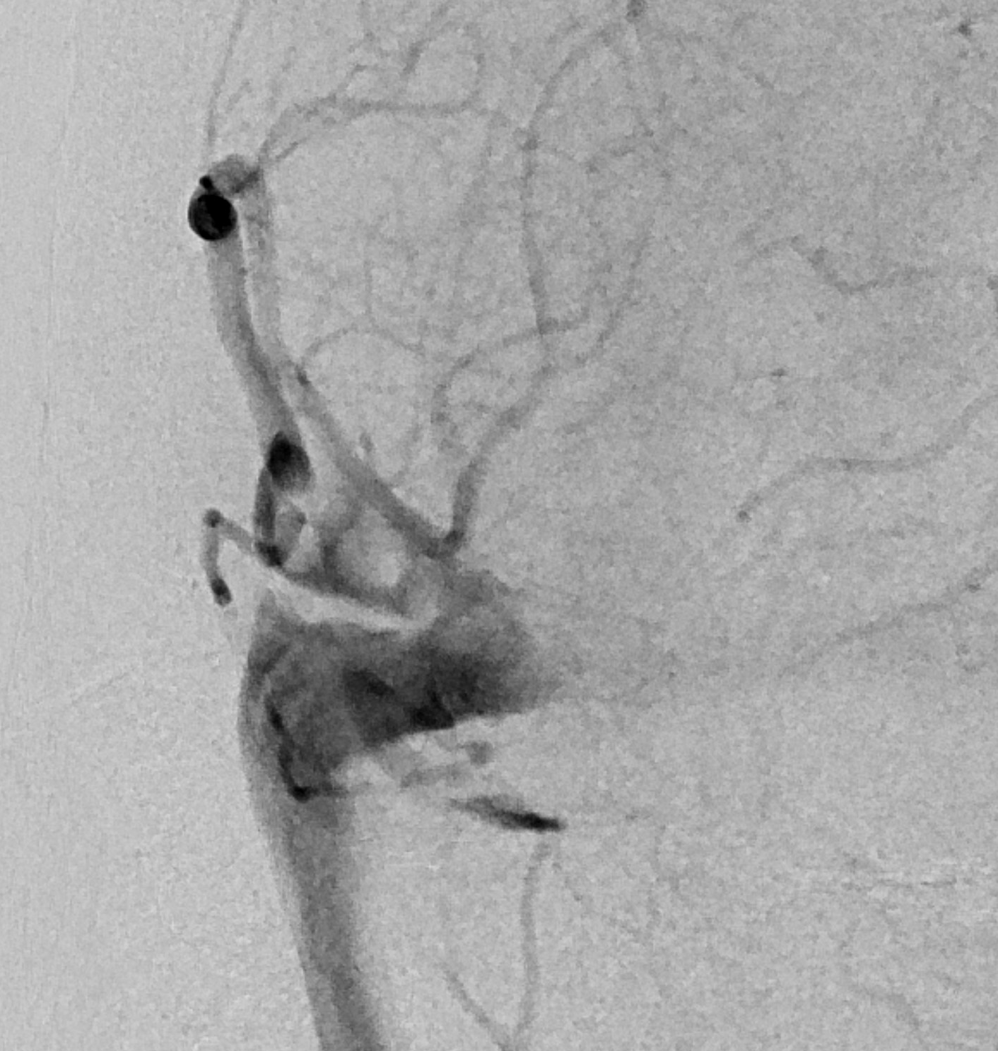
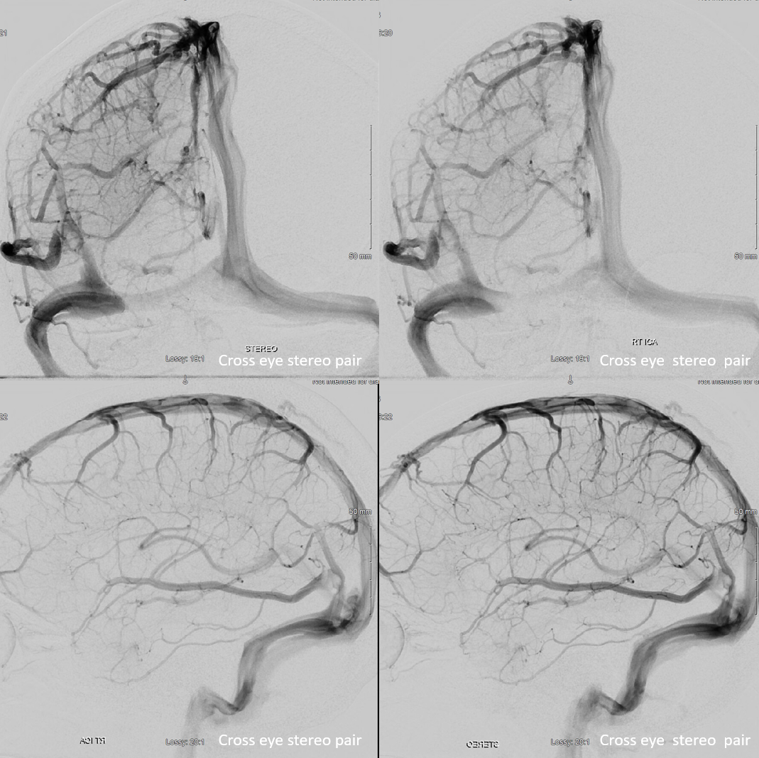
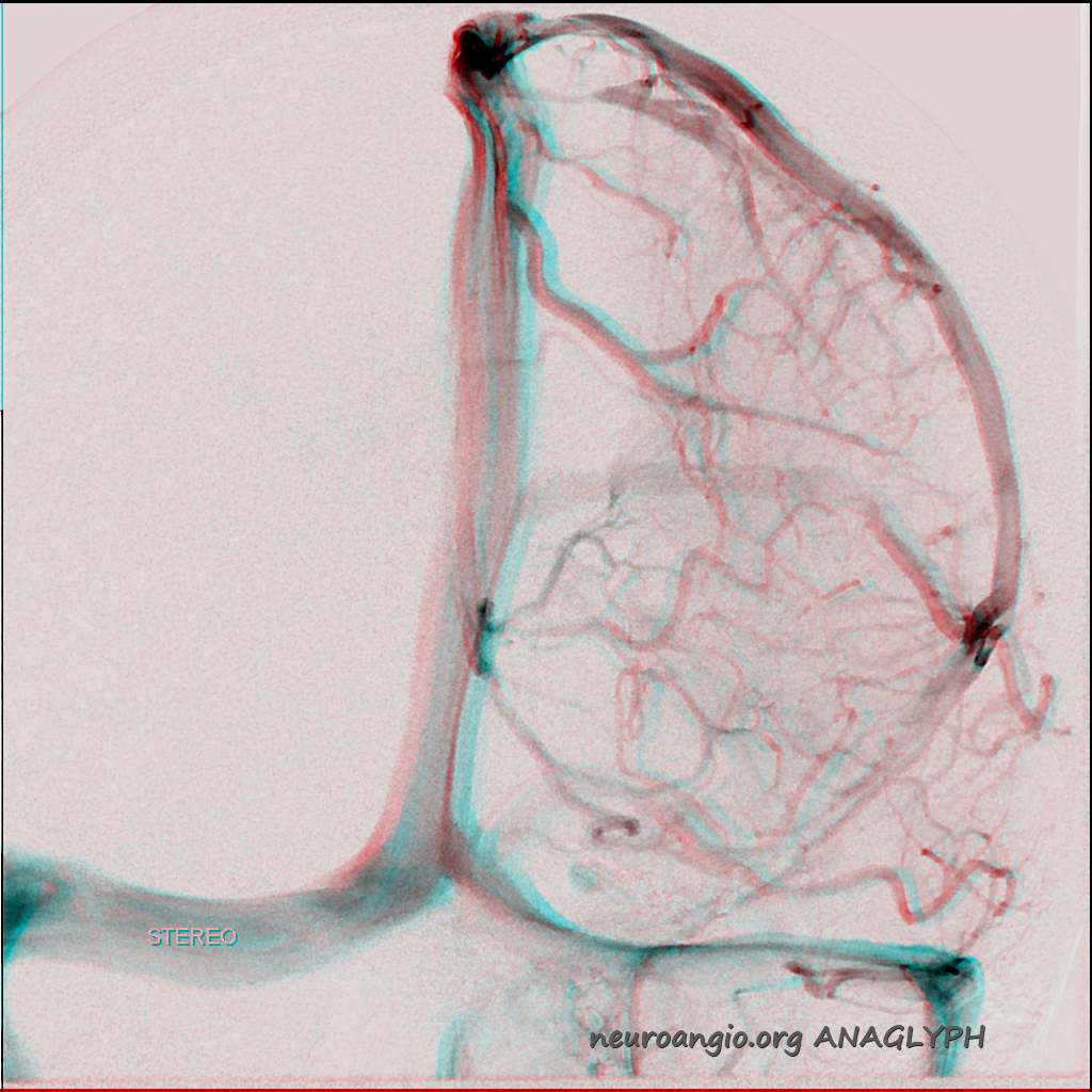
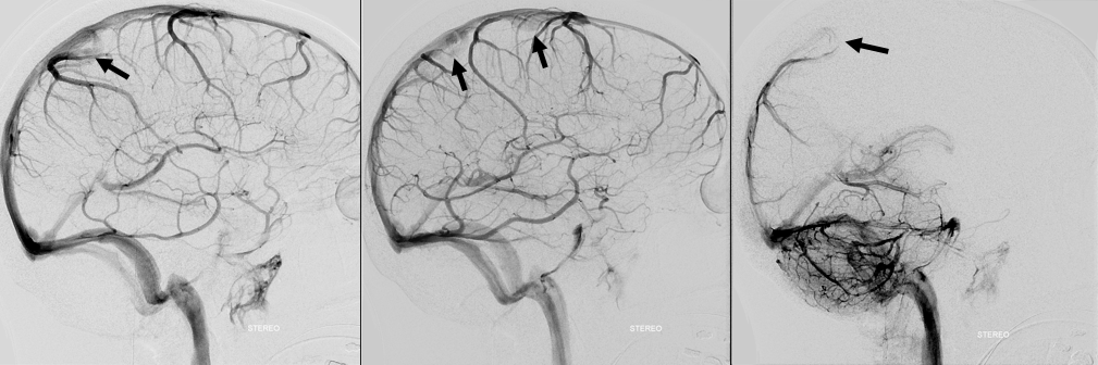
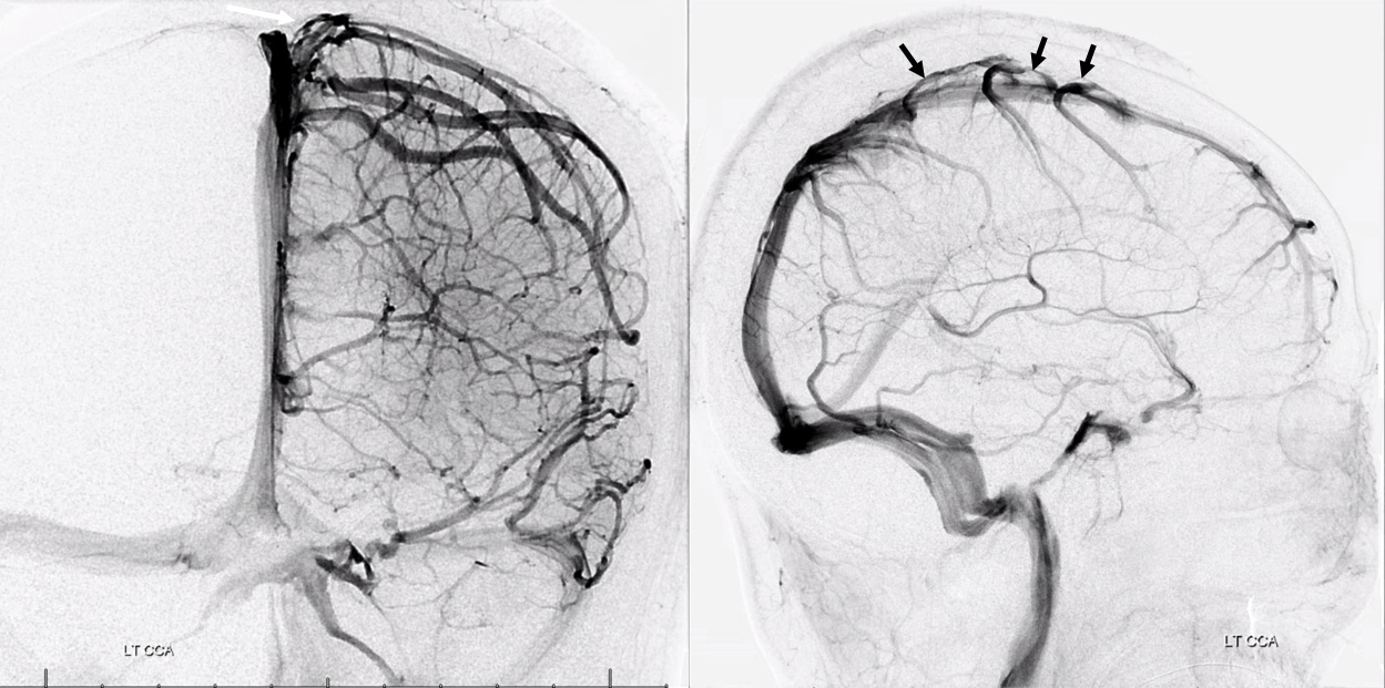
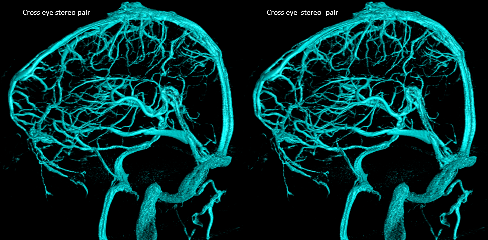
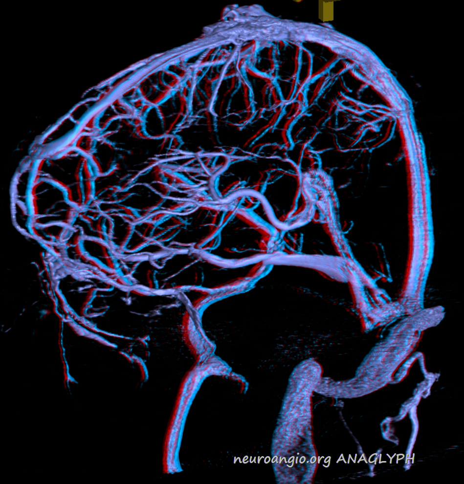
These dural channels are way more common than most people realize. They are easiest to see on angio because of a characteristic transition from a rounded cross-section of a cortical or bridging vein to a flattened, biconvex shape of a dural channel that runs within the dural covers.
These dural channels are important for two reasons. First, injuring them is like injuring the vein that drains into it — risking venous infarction.
Second, they seem to explain why some dural fistulas do not involve a sinus but instead drain seemingly directly into a cortical vein. Why? Because the fistula involves a dural venous channel. When this channel looses its connection to the sinus, the only way to drain is retrograde into cortical veins.
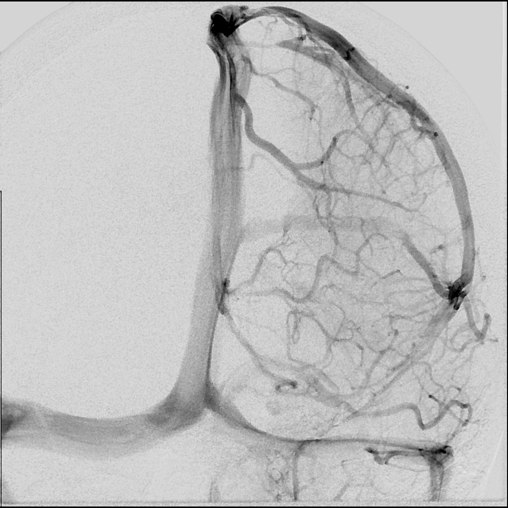
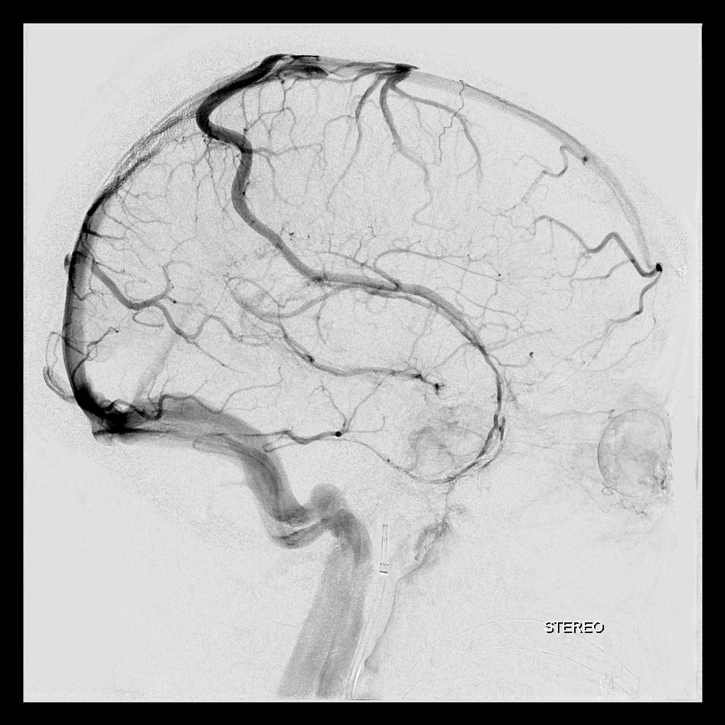
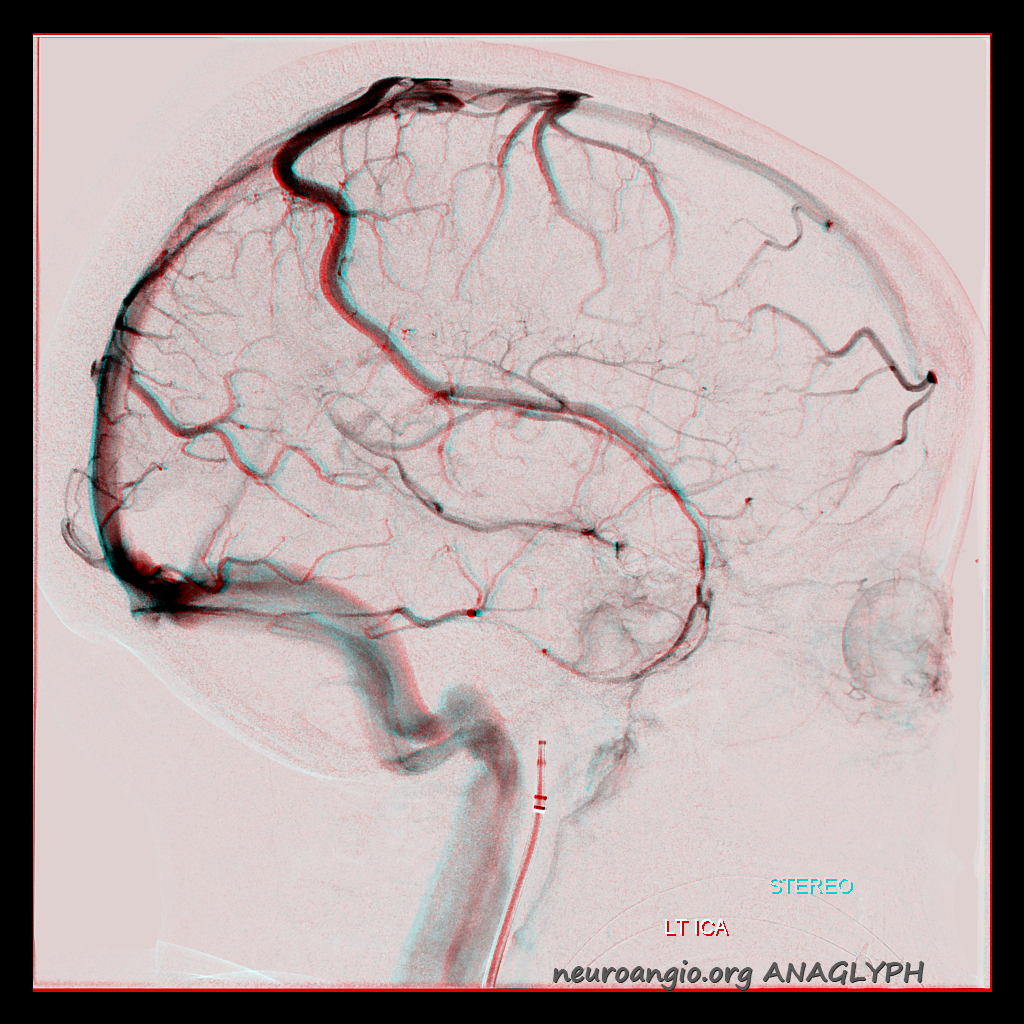
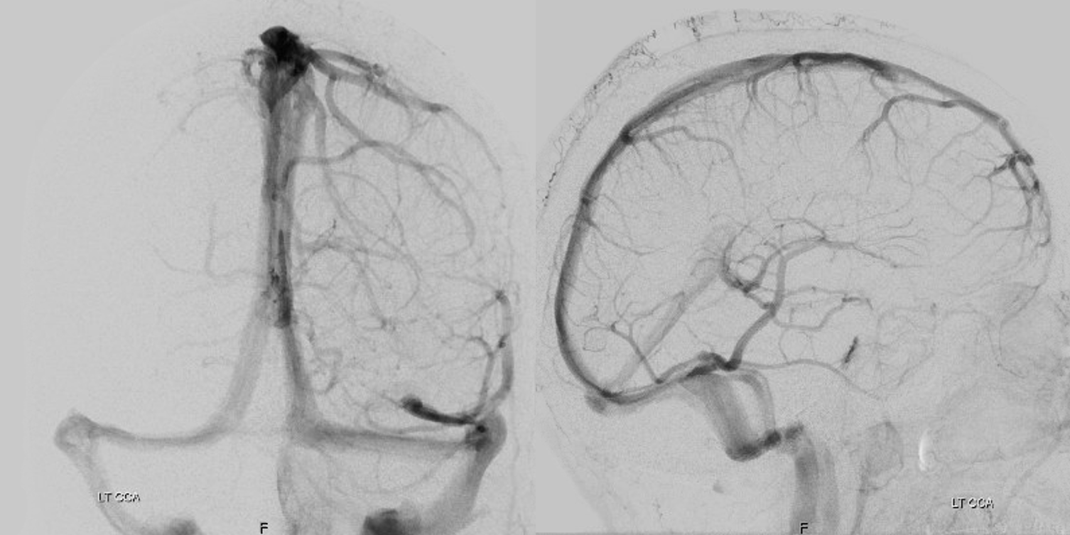
Examples of dural channels are below.
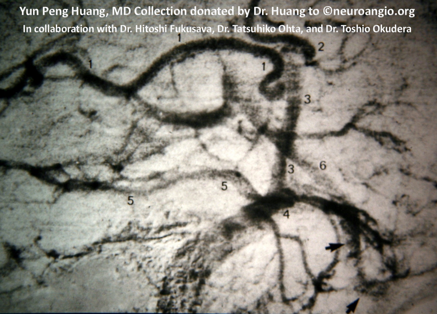
The earliest known example, according to Kittipong Srivatanakul, is by the great Yun Peng Huang and Toshio Okudera (numeral 3)
Another demonstration of a basal vein tentorium cerebelli dural venous channel from Salamon and Huangs Radiologic Anatomy of the Brain
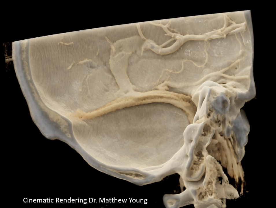
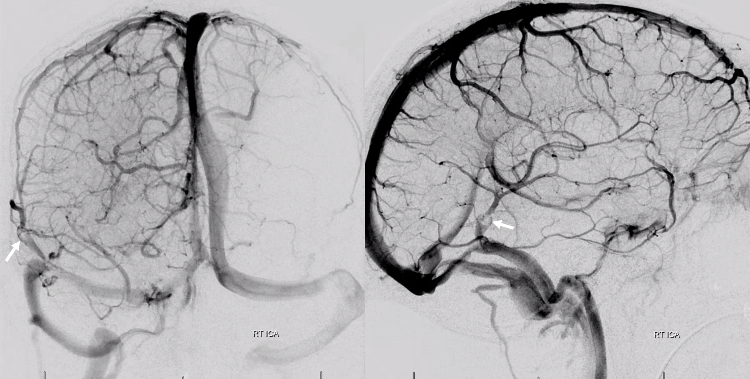
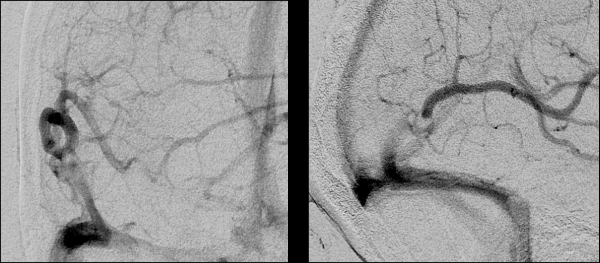
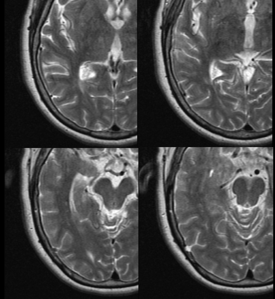
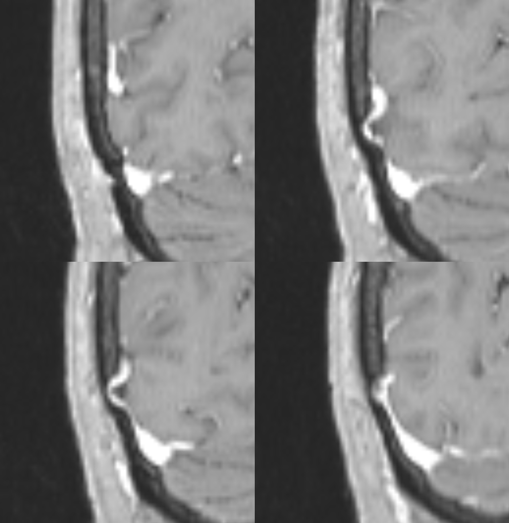
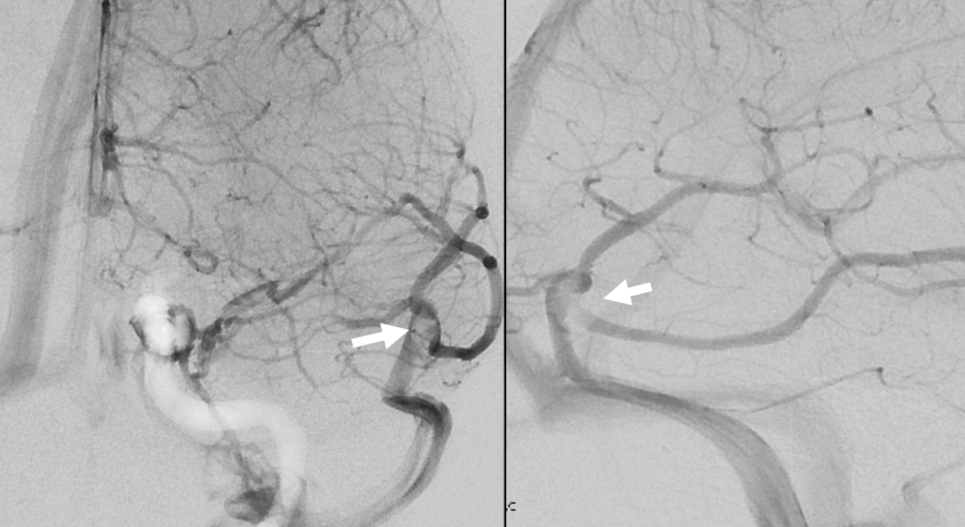
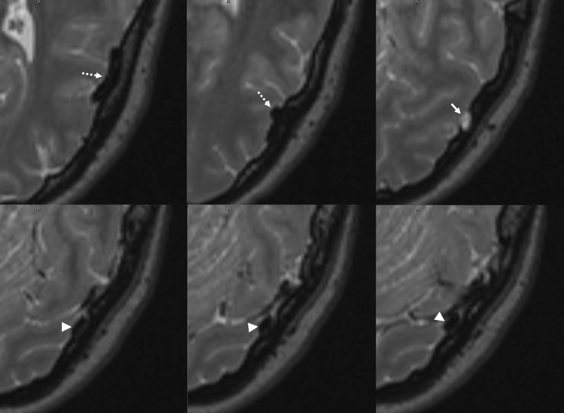
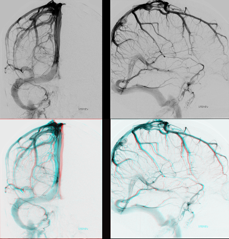
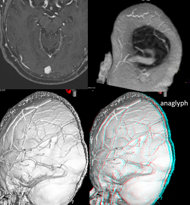
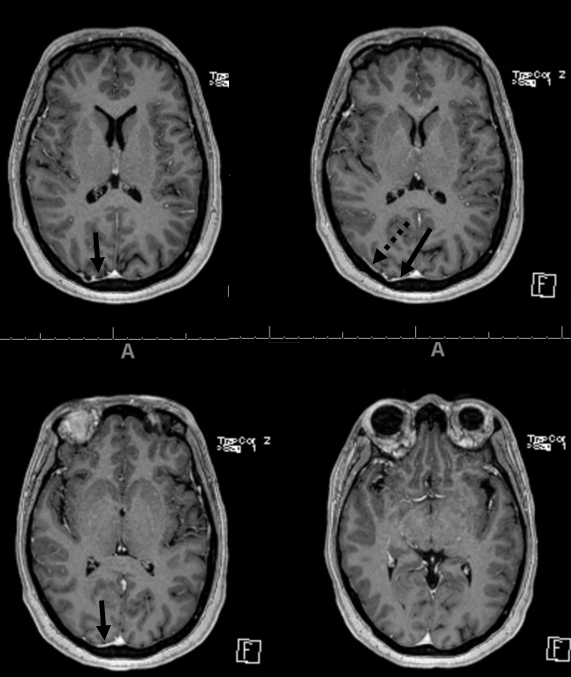
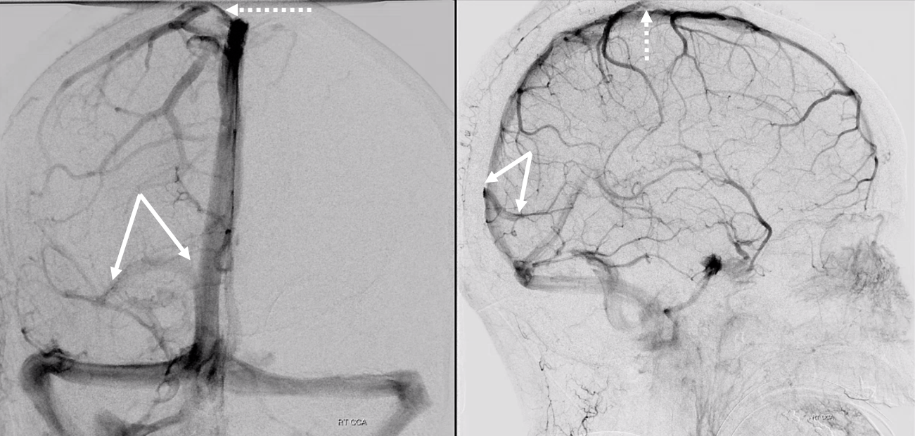
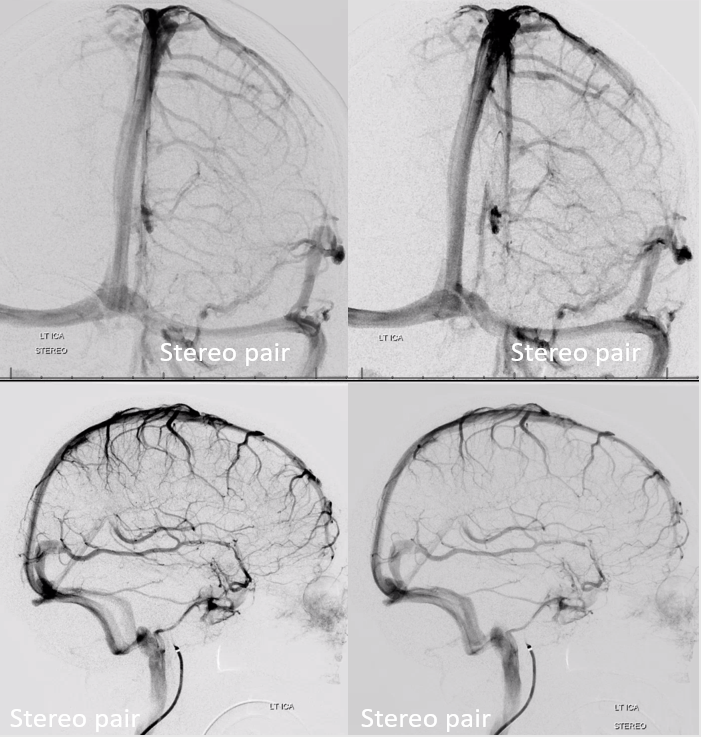
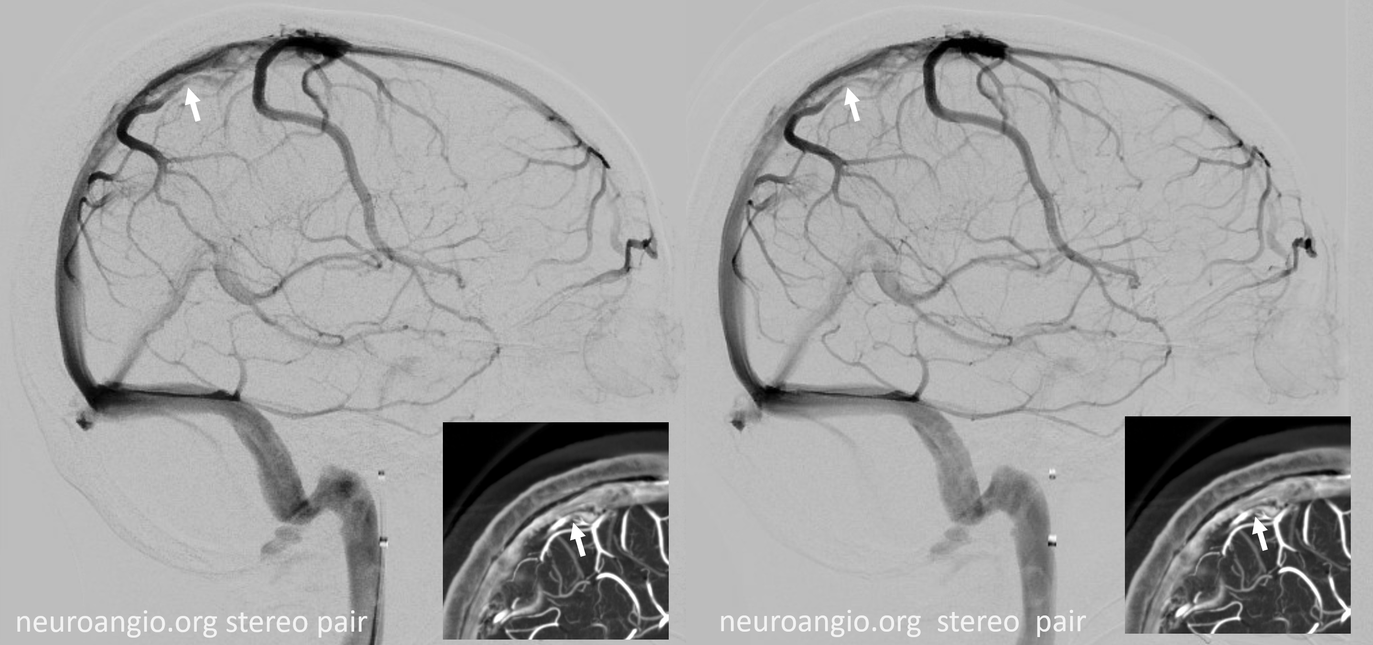
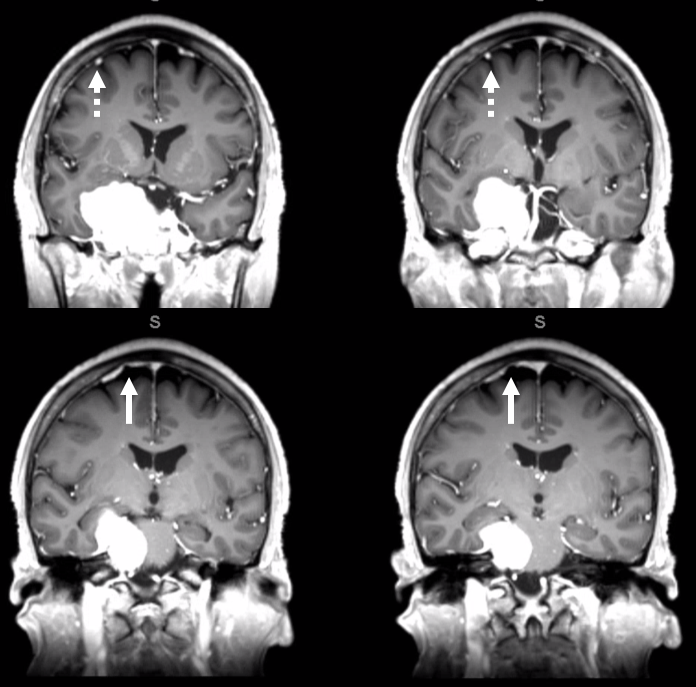
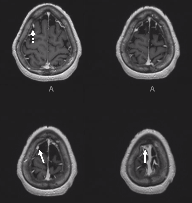
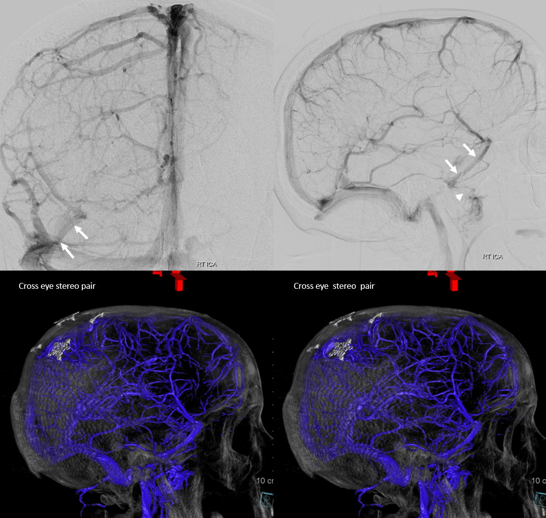
A typical case of Labbe (black arrows) draining into a dural venous channel (white arrows). Notice transition from rounded morphology of the Labbe to a biconvex flattened appearance of the dural venous channel (white arrows). Bottom row are volume rendered reconstructions of the same post-contrast T1 weighted MRI
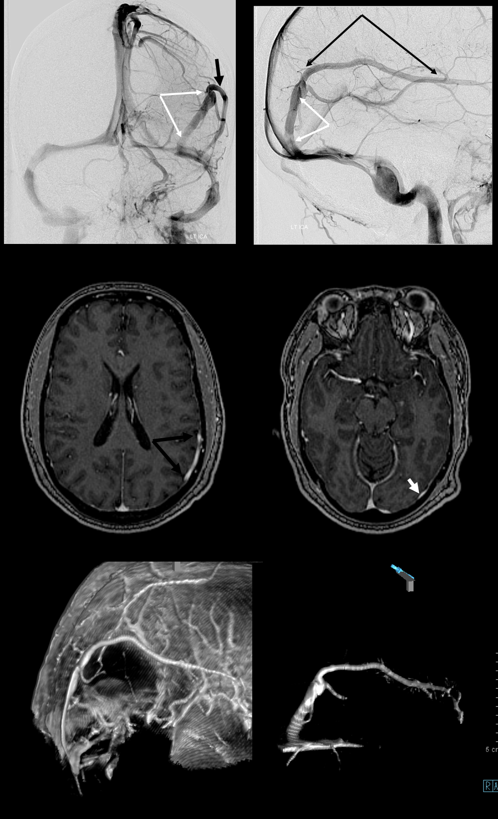
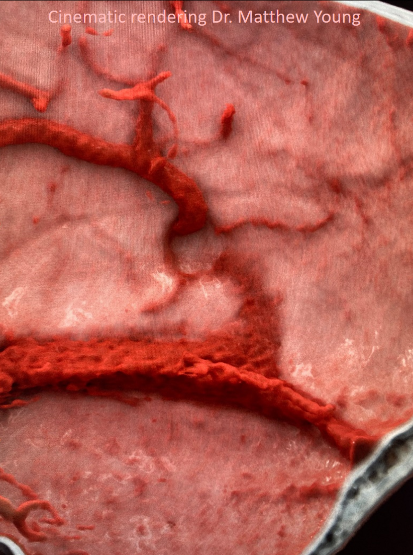
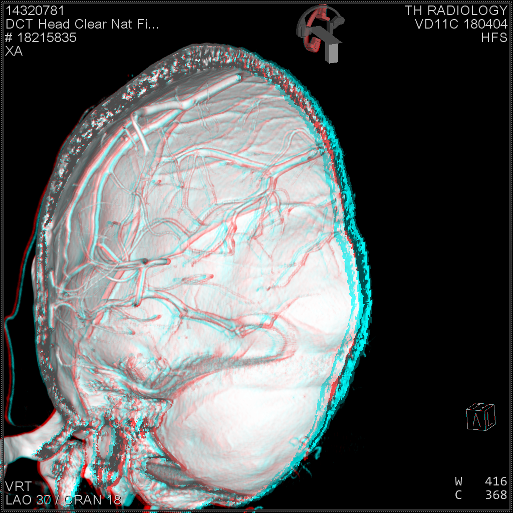
Cinematic rendering of this case, courtesy Dr. Matthew Young
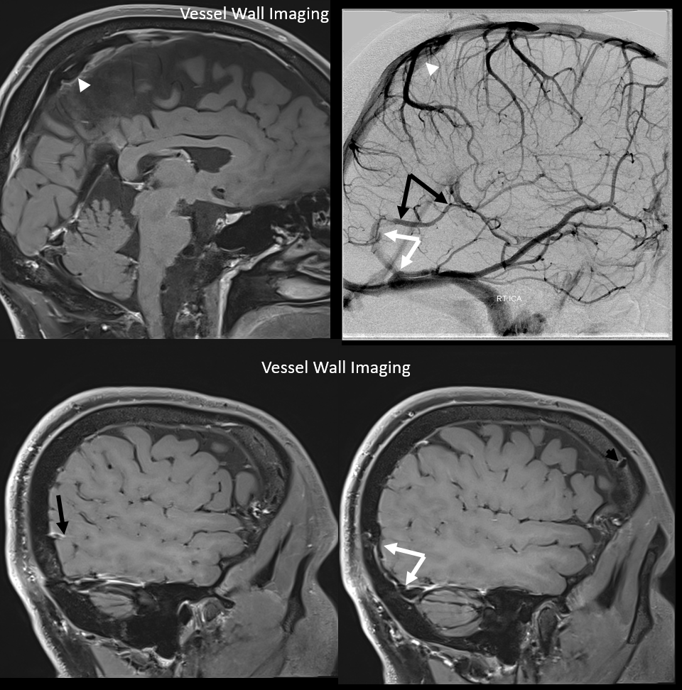
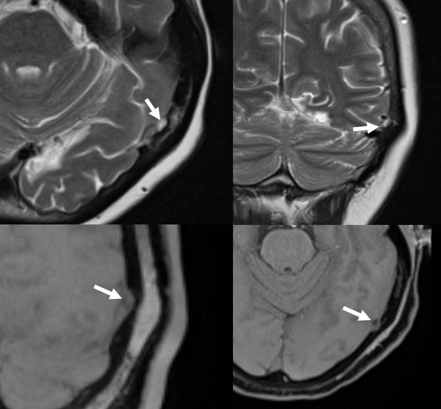
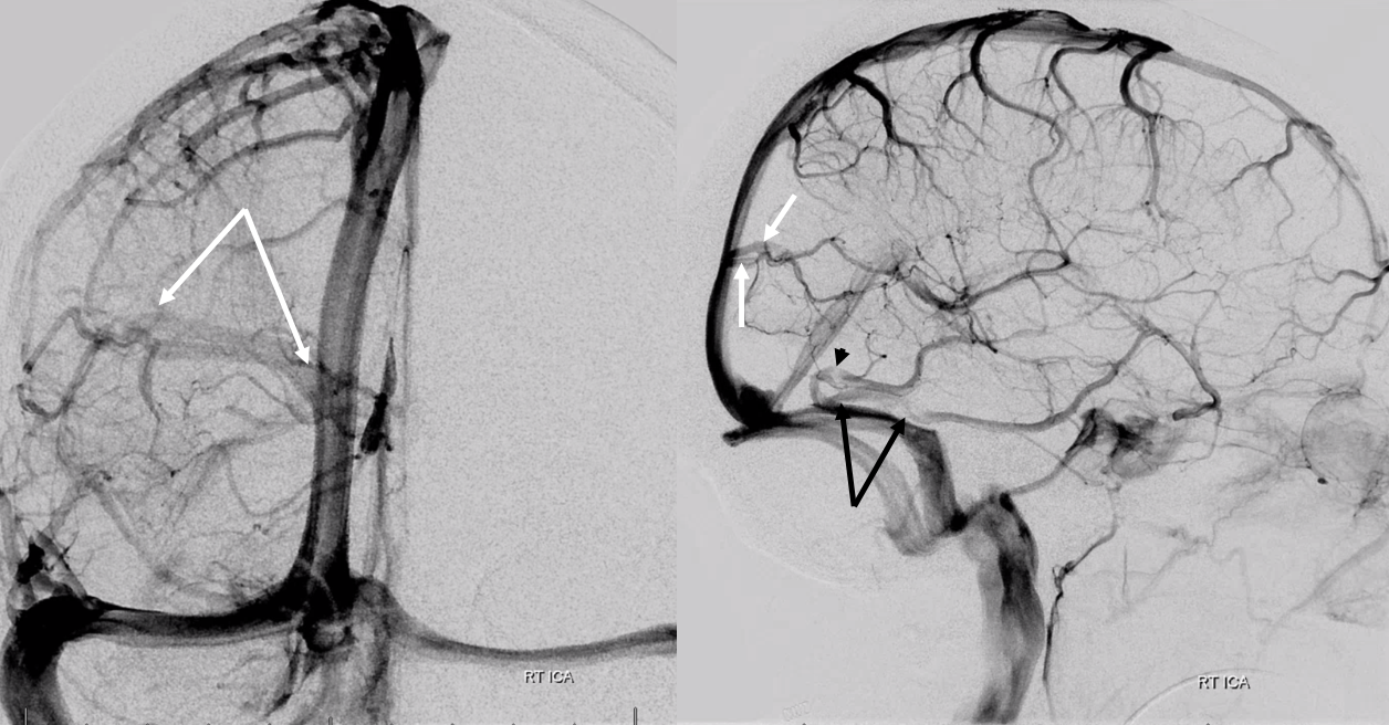
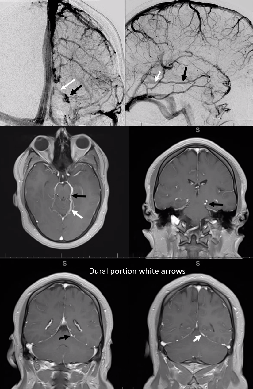
Vessel wall imaging is excellent at showing dural venous channels. There is one in falx cerebri draining the trolard (white arrowheads). The Labbe (black arrows) drains into another one (white arrows) that empties into the transverse (not sigmoid) sinus. Vessel wall is also excellent at depicting the emissary veins (black arrowhead)
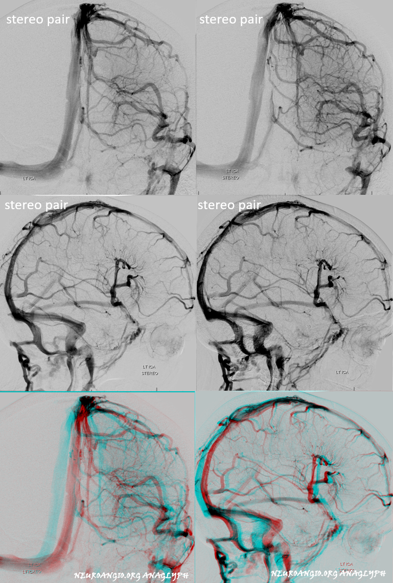
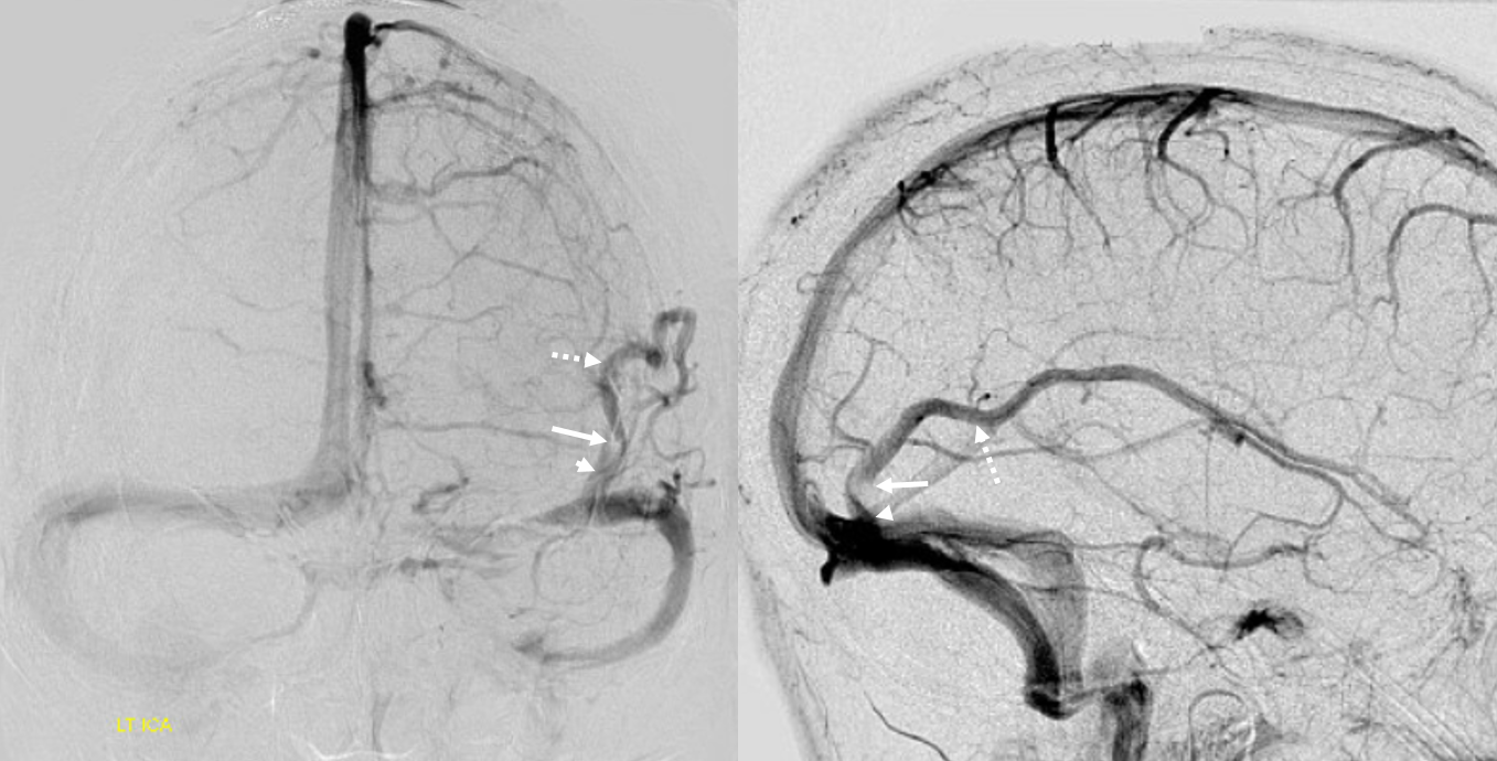
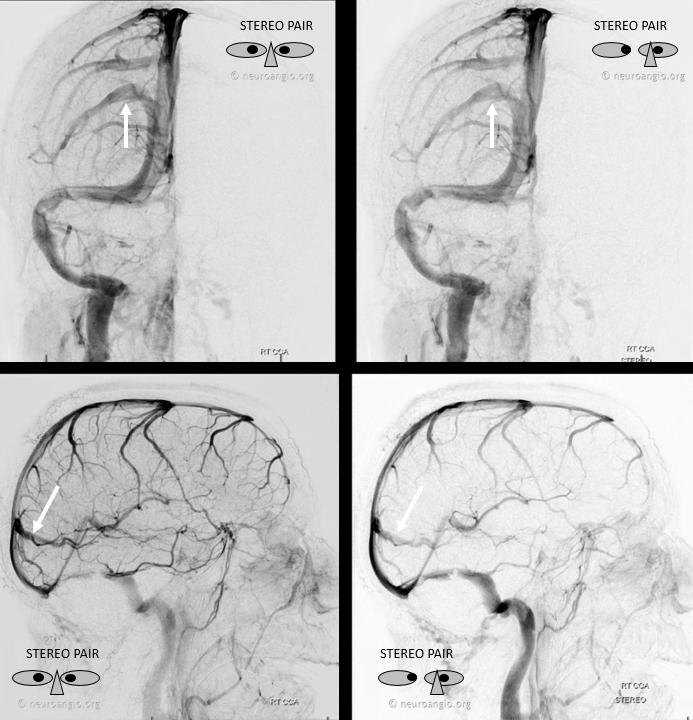
Here is one associated with a frontal opercular DVA. Bottom row are anaglyphs, top rows are cross-eye stereos. Another dural venous channel is drains a “normal” temporo-occipital vein (none are labeled, good luck!)
Arachnoid Granulation inside dural venous channel
Another proof that these are dural. Angio showing granulation (white arrow) inside channel
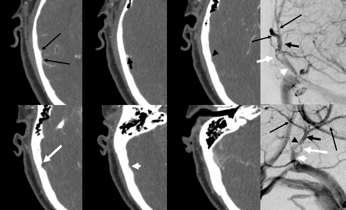
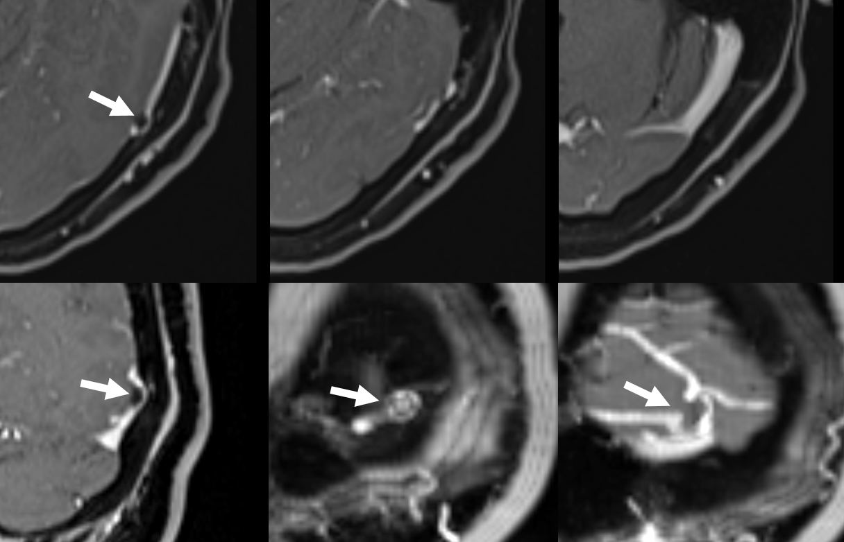
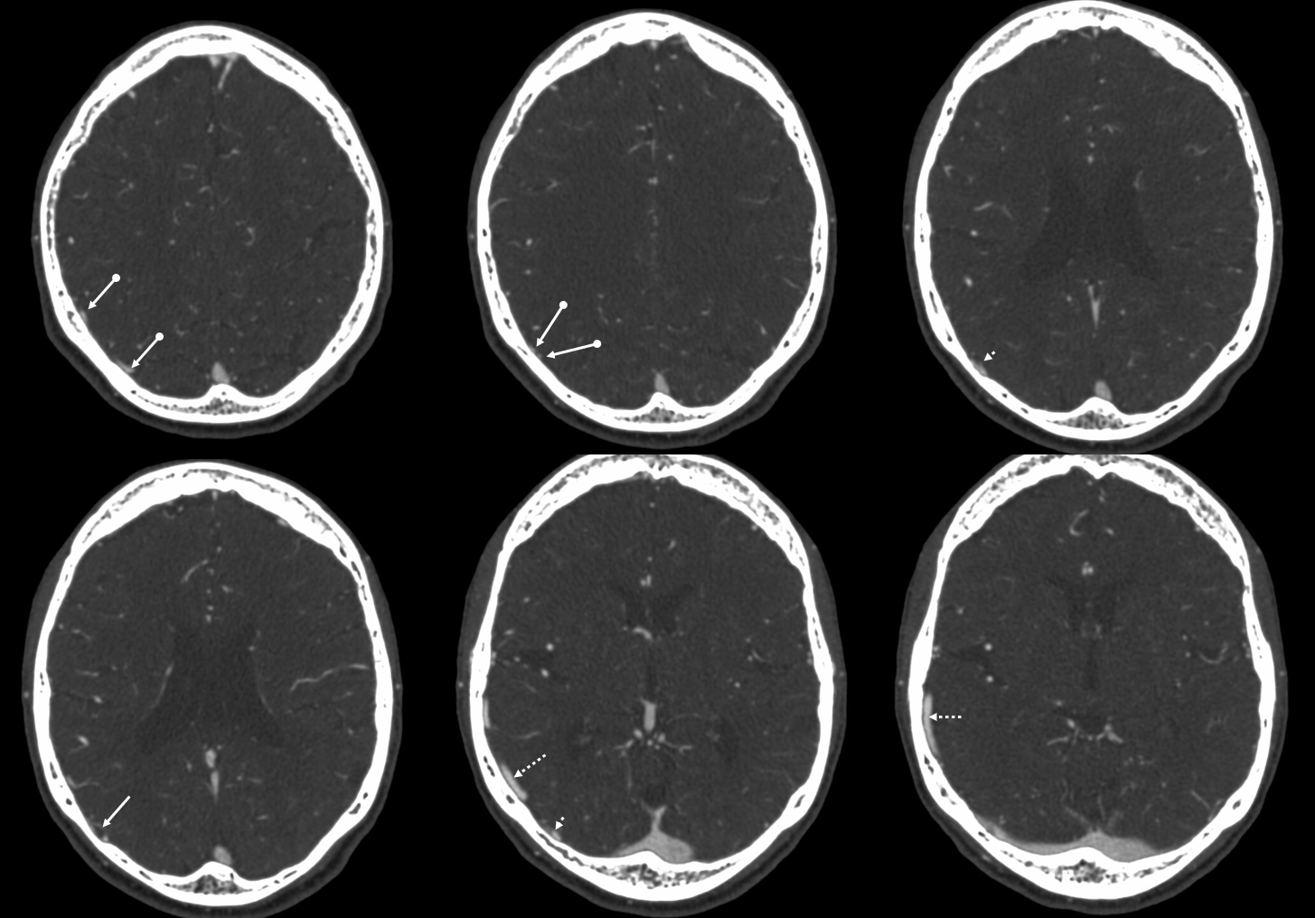
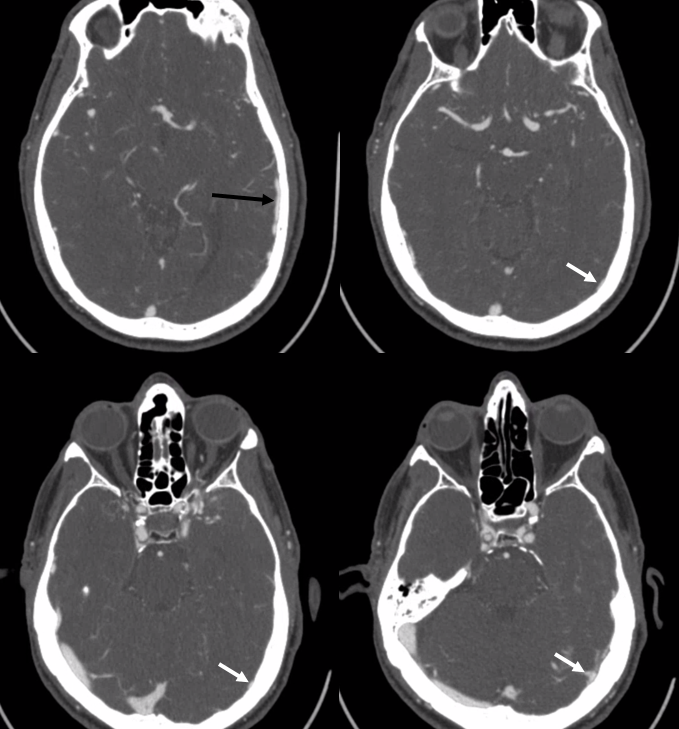
Could it be inflow from another vein? Below is venously contaminated CTA images next to angio. Same structures labeled with same arrows. Long skinny black arrows — tributary veins. Black arrow — common cortical venous segment. Black arrowhead — distal dural channel segment. White arrow — granulation (notice corresponding hypodensity on CT and scalloping of the skull inner table). White arrowhead — proximal dural channel segment just above the sigmoid sinus.
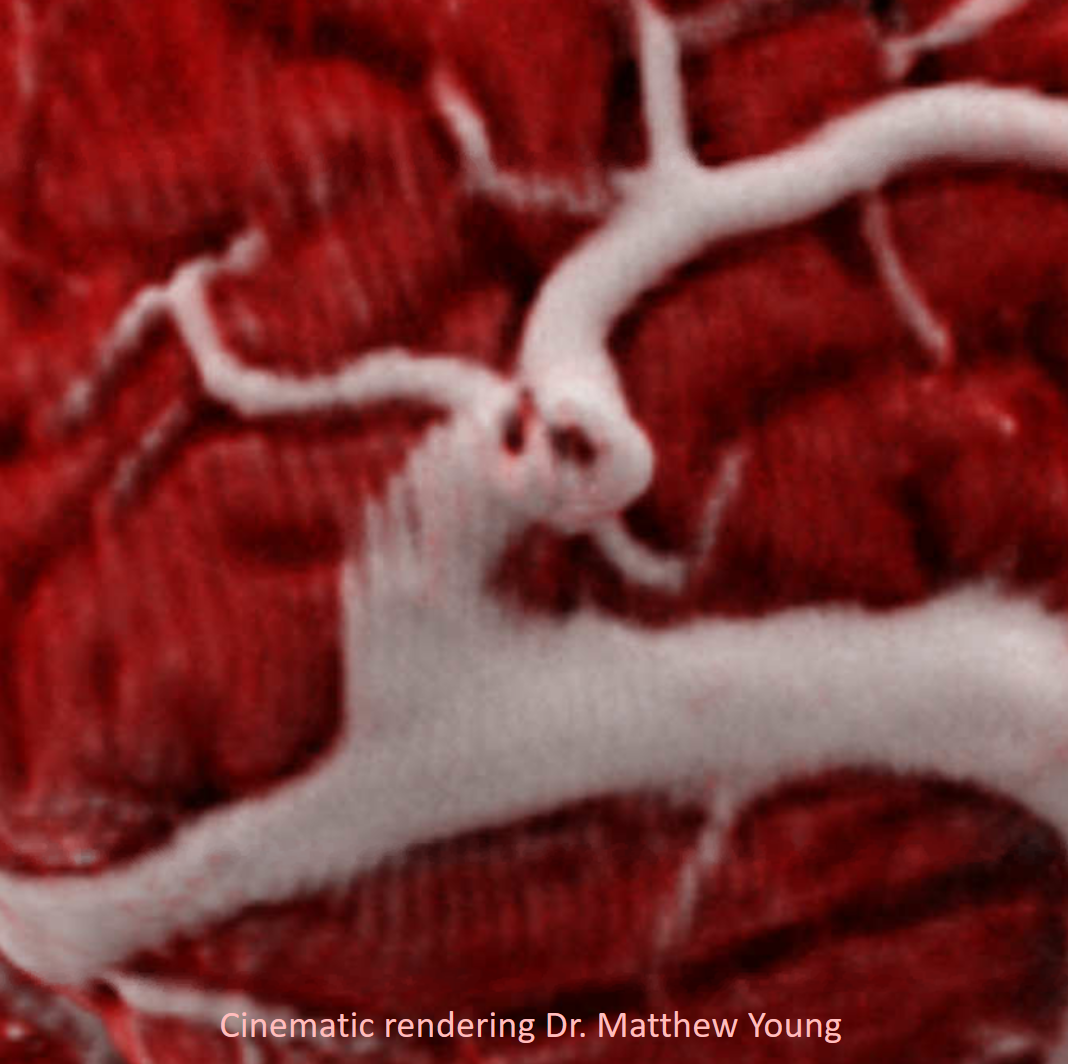
Another granulation, with beautiful cinematic rendering images by Dr. Matthew Young
Angio
MRI
Below is another one, in a thrombectomy patient. No other imaging thats good to show this available.
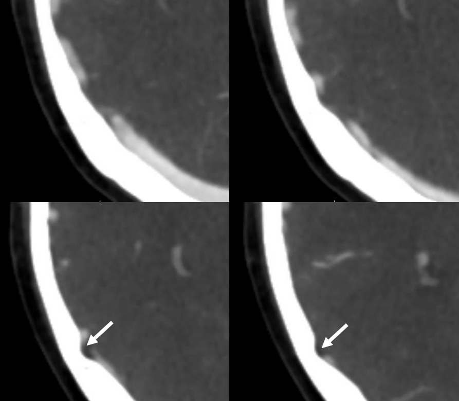
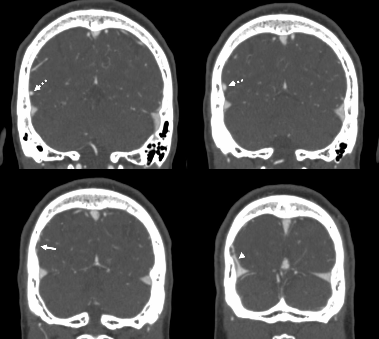
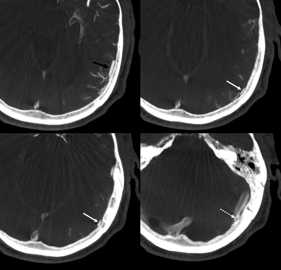
Finally, best for last — largest to date granulation (arrow) in venous sinus channel (arrowhead) — courtesy Dr. Erez Nossek
CTA of same case — not as obvious. Notice hypodensity of granulation itself (arrow) and scalloping of the skull
Subtle Granulation — these are more common than meets the eye — here is looking at angio and MRI together, carefully… There is a subtle lucency (arrows) at the confluence of 3 cortical veins into a dural venous channel
Careful look at MRI shows the granulation (multiple sequences, follows fluid surrounded by blood, scalloping of inner table etc)
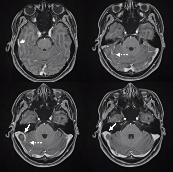
Yet another one below — just in case anyone still need convincing. Granulation — arrow; labbe — dashed arrow; convexity occipital veins (ball arrows); dural channel — arrowhead
stereos
CTA
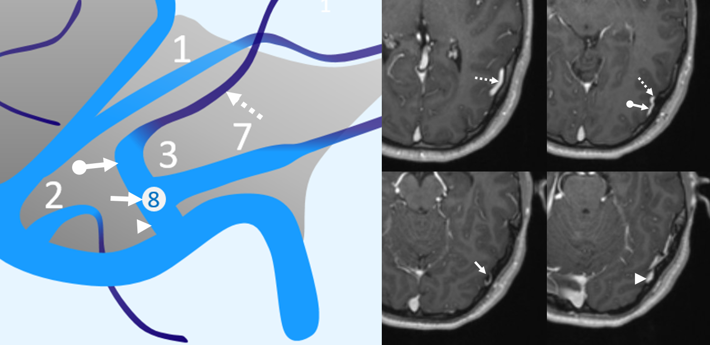
Last one (for now — it really matters to show lots of them to convince the unbelievers that these are not just flattened cortical veins — they are really intradural). Sometimes its easiest to see them on T1 post volumetric images or on T2s — once you get the hang of it. The same arrows point on diagram and images to corresponding areas. The granulation (8) is marked by solid arrow
More cases…
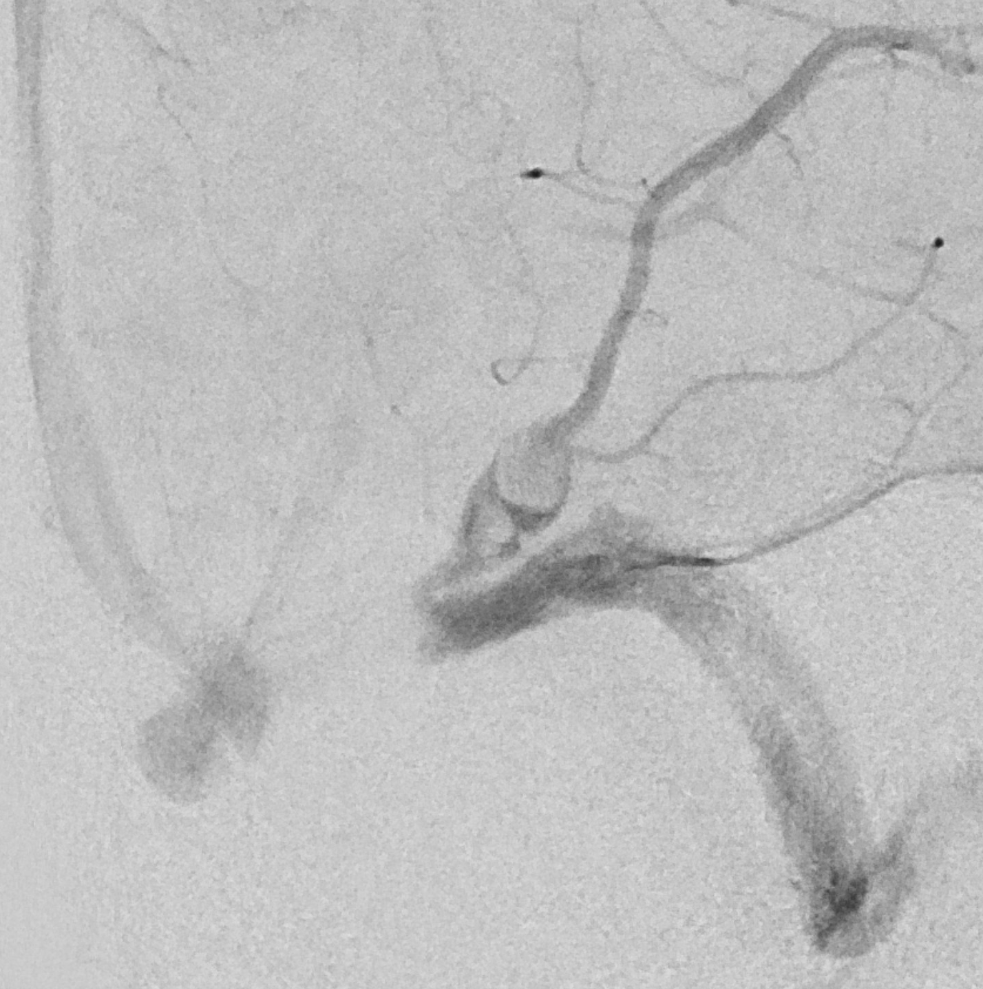
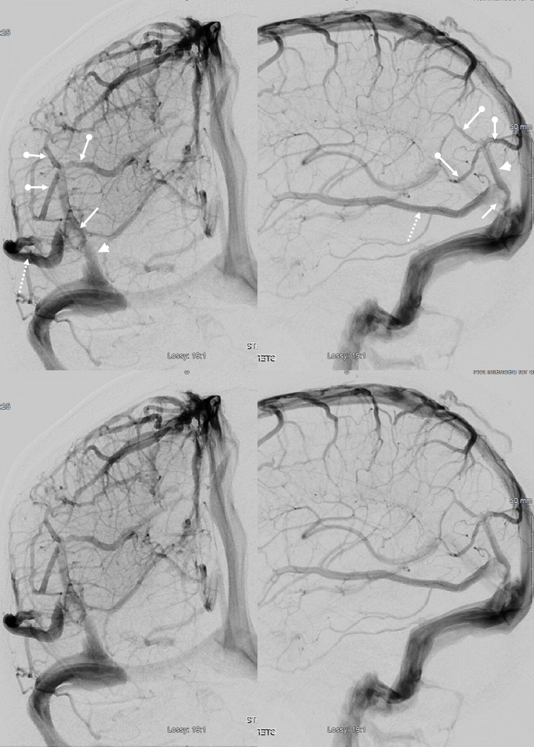
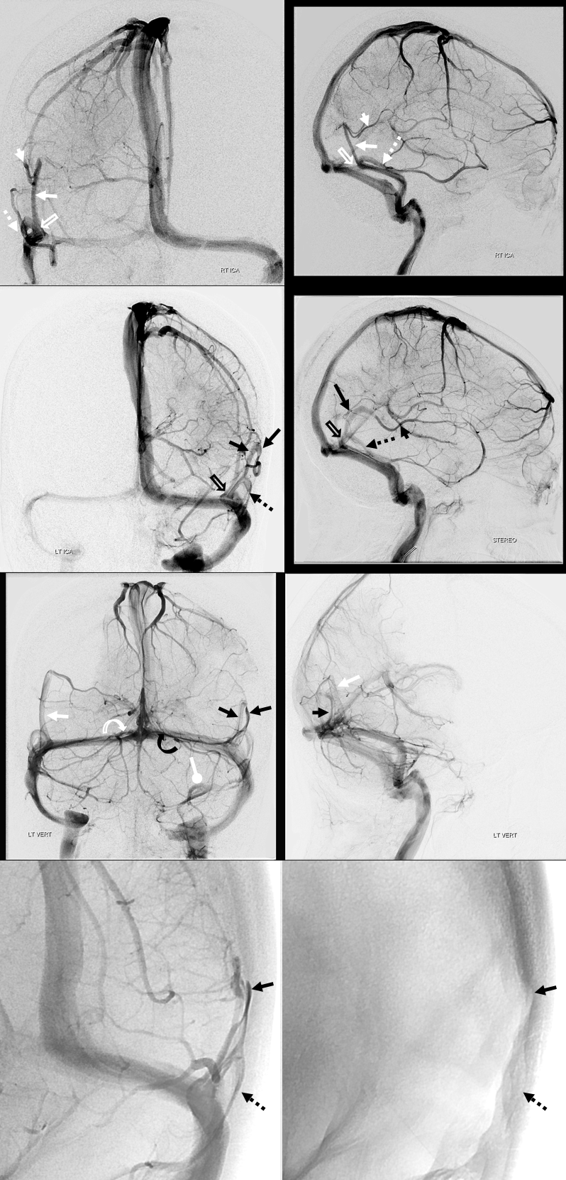
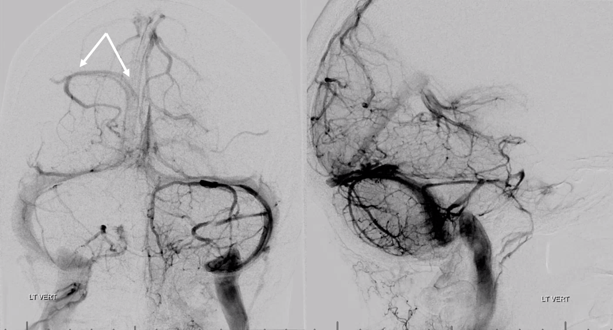
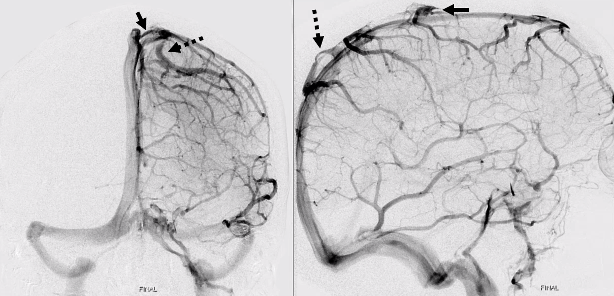
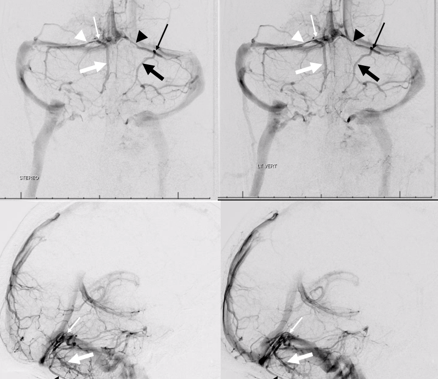
On right a small Labbe-like vein (white arrowhead) drains into a dural channel (white arrow). An inferior temporal vein drains into its own dural channel (dotted white arrow). Both dural channels converge on a common dural channel (open white arrow) that drains into the sigmoid sinus.
On the left, the situation is similar. A Labbe (black arrowhead) drains into a dual venous channel (black arrow) and inferior temporal vein drains into its own separate dural channel (black doted arrow). Both channels converge on a common dural channel (open black arrow) draining into the sigmoid sinus.
Third row from top (vertebral injection venous phase) opacifies the same dural channels because they are receiving inflow from temporo-occipital veins.
Also present are several tentorial sinuses (dural venous channels in the tentorium cerebelli – curved white arrow, curved black arrow, white arrow with ball point)
Bottom row shows optimal left anterior oblique projection that shows dural channels (black arrow, dotted black arrow) lateral to hemispheric surface, and corresponding indentations of the skull
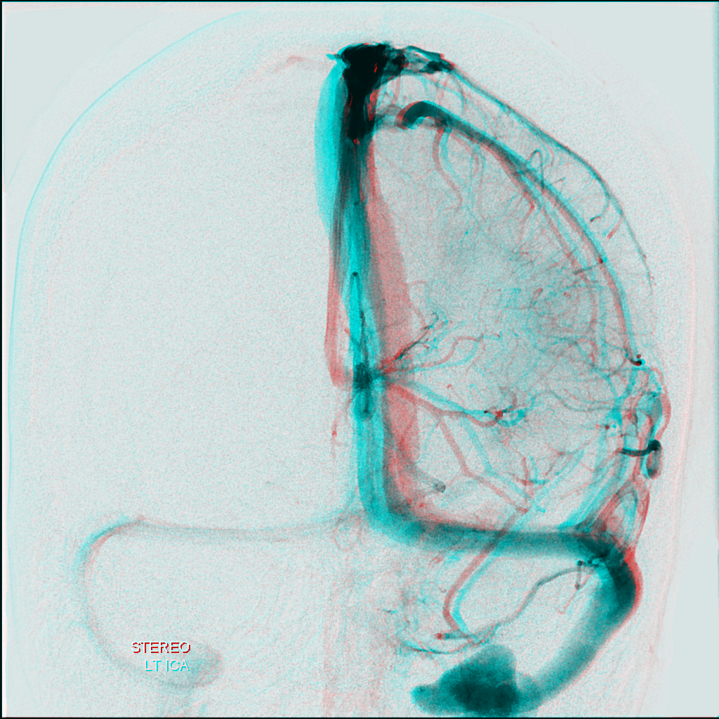
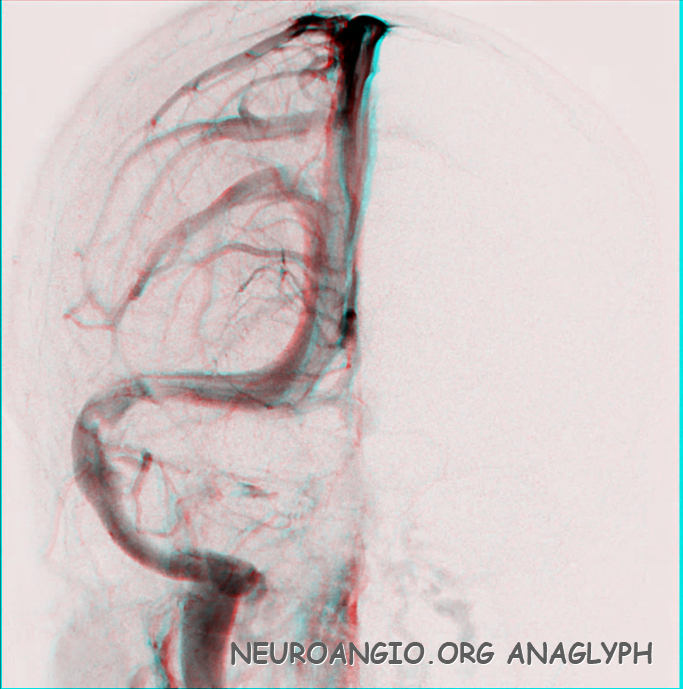
Anaglyph stereos of same case
Another case. Anaglyph images on bottom
Same case MRI shows angulation at point of transition from hemispheric to dural segment (upper right image) on axial source images. Not easy to spot unless u know what to look for. Same case volume rendered reconstruction MRI (upper left) and DYNA CT reconstructions as well. Lower left is anaglyph
DYNA CT Anaglyph reconstruction — bigger picture
Parieto-Occipital Region
Here are some weirder ones — notice how the channel projects posterior to the SSS, because it is running in the dural fold just before it enters the sinus

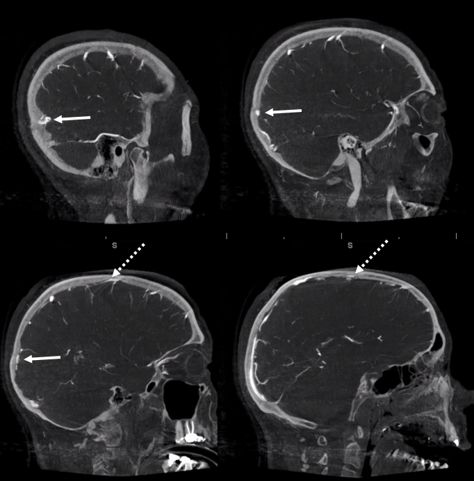
MRI of same case — black arrow points to the dural channel, dashed arrow to the tributary veins. Not as obvious, right?
Another example. Also another one (dashed arrows) near vertex (see “Venous Lakes” below)
Cross-eye stereo occipital one
Anaglyph stereos

DYNA CT of same patient. Harder to appreciate
Yet another two — one parietal, one occipital. Notice how these channels extend posterior to the contour of the superior sagittal sinus
Anaglyphs

Status Channeloticus — a predisposition by some individuals to have tons of channels
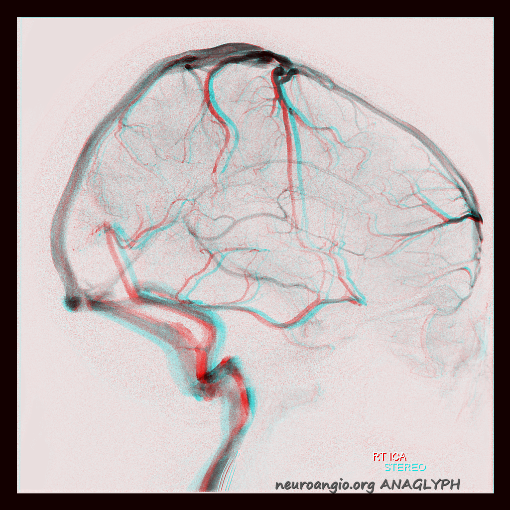
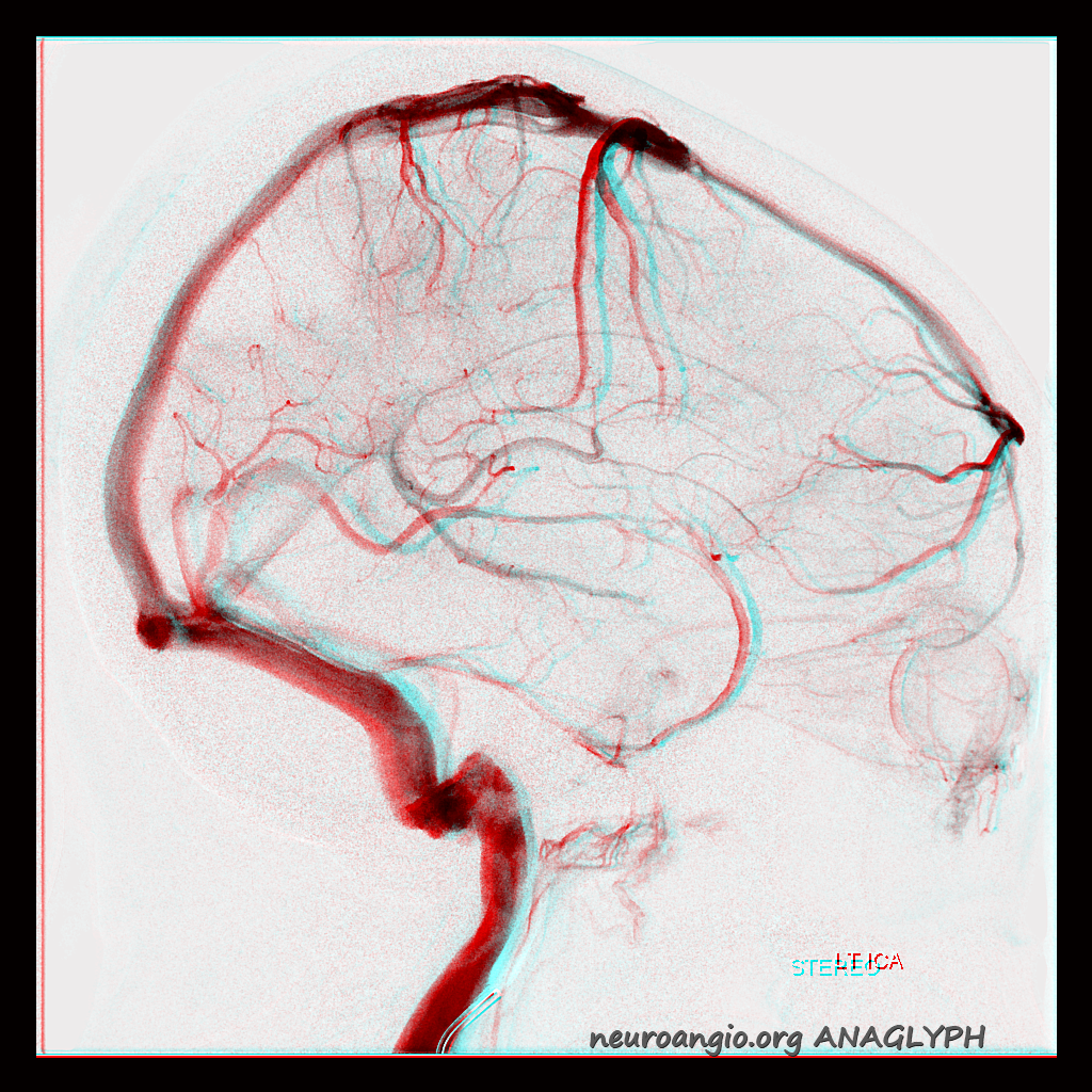
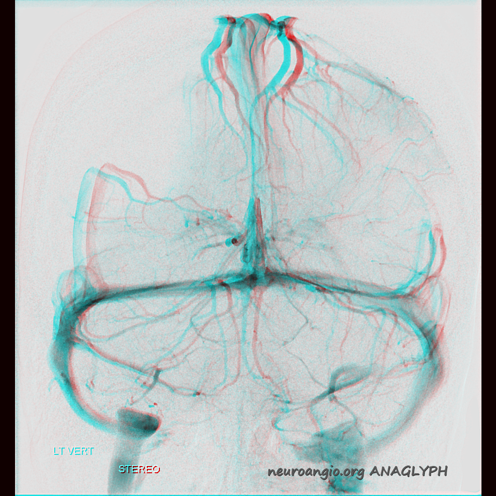
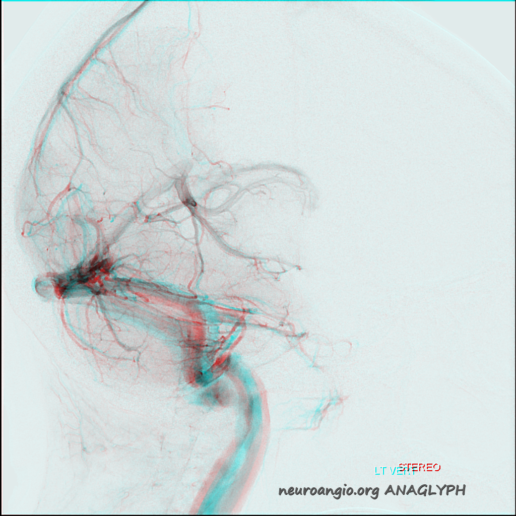
Left vert
Right ICA — white arrows point to same channel as seen from vert injection. Black arrows and arrowhead to two separate additional channels.
Right ICA of same patient
Altogether
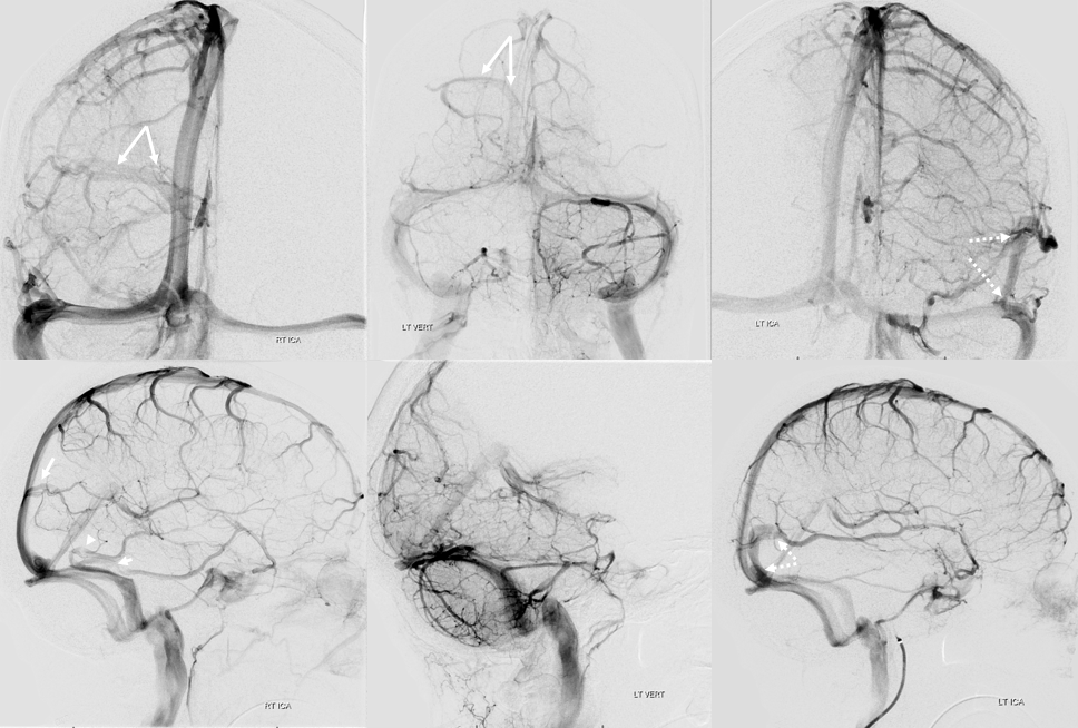
Stereos
CTA of same patient — venous contamination. Labbe – black arrow. Dural channel — white arrows
DYNA CT — better than CTA. A small arachnoid granulation seems to be present (dashed arrow) — have better examples here
Anaglyph stereo

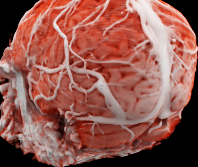
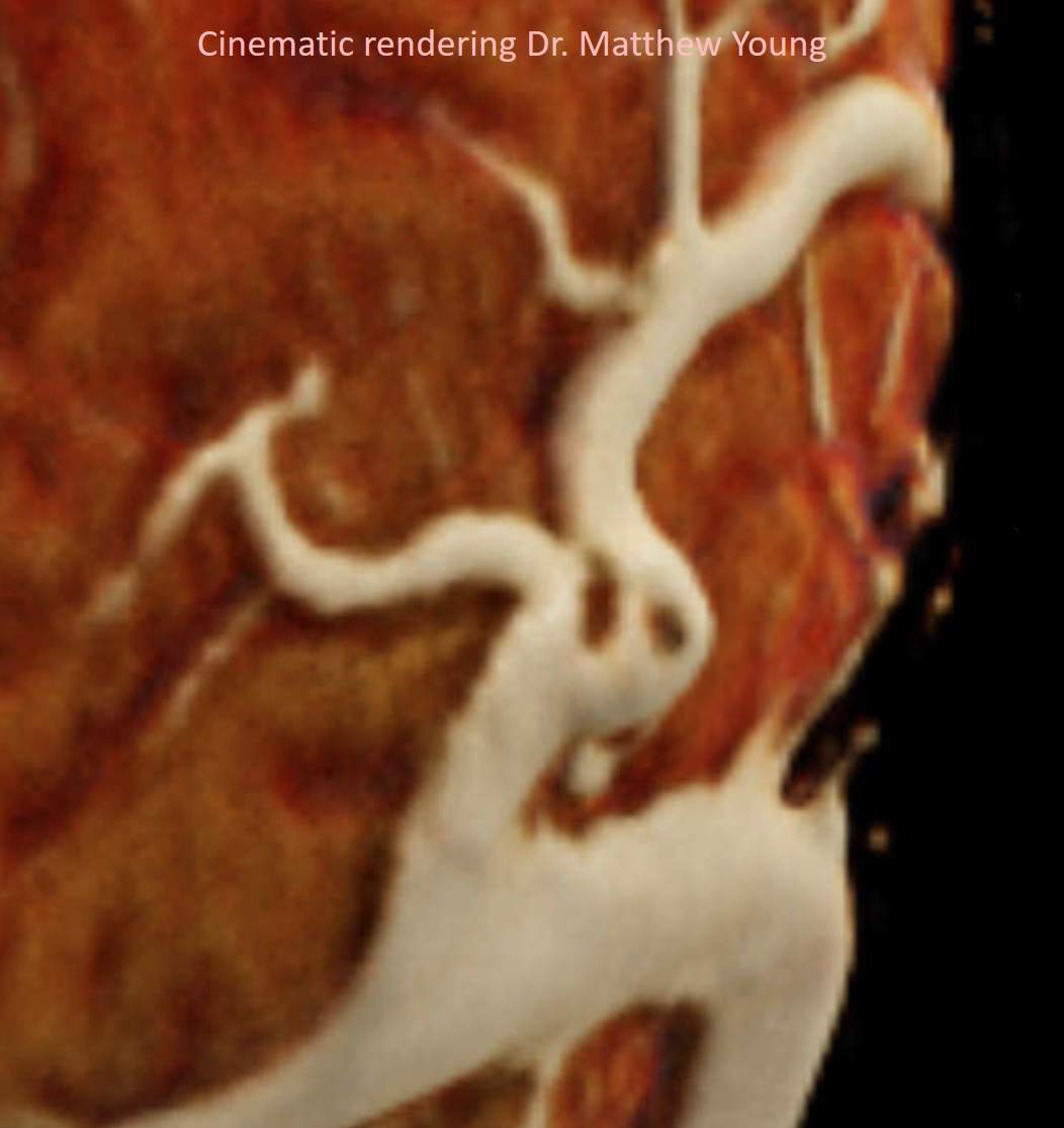
Finally an awesome cinematic rendering by Dr. Matthew Young
Falx Cerebri
Parieto-occipital Falx channel receiving mesial veins
Example of typical falx cerebri dural sinus channel (black arrows) that is opacified from both carotid and vertebral injections
These are best seen on vessel wall imaging — white arrowheads (shown above already)
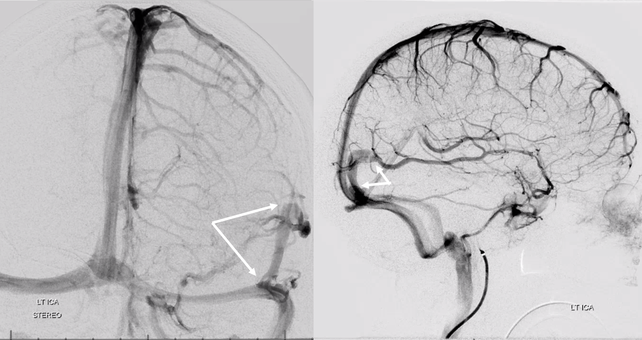
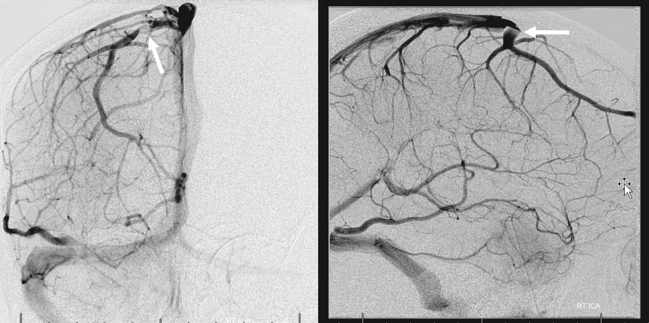
Here is another stereo example of a parietal convexity vein draining into a falx cerebri dural channel (arrows)
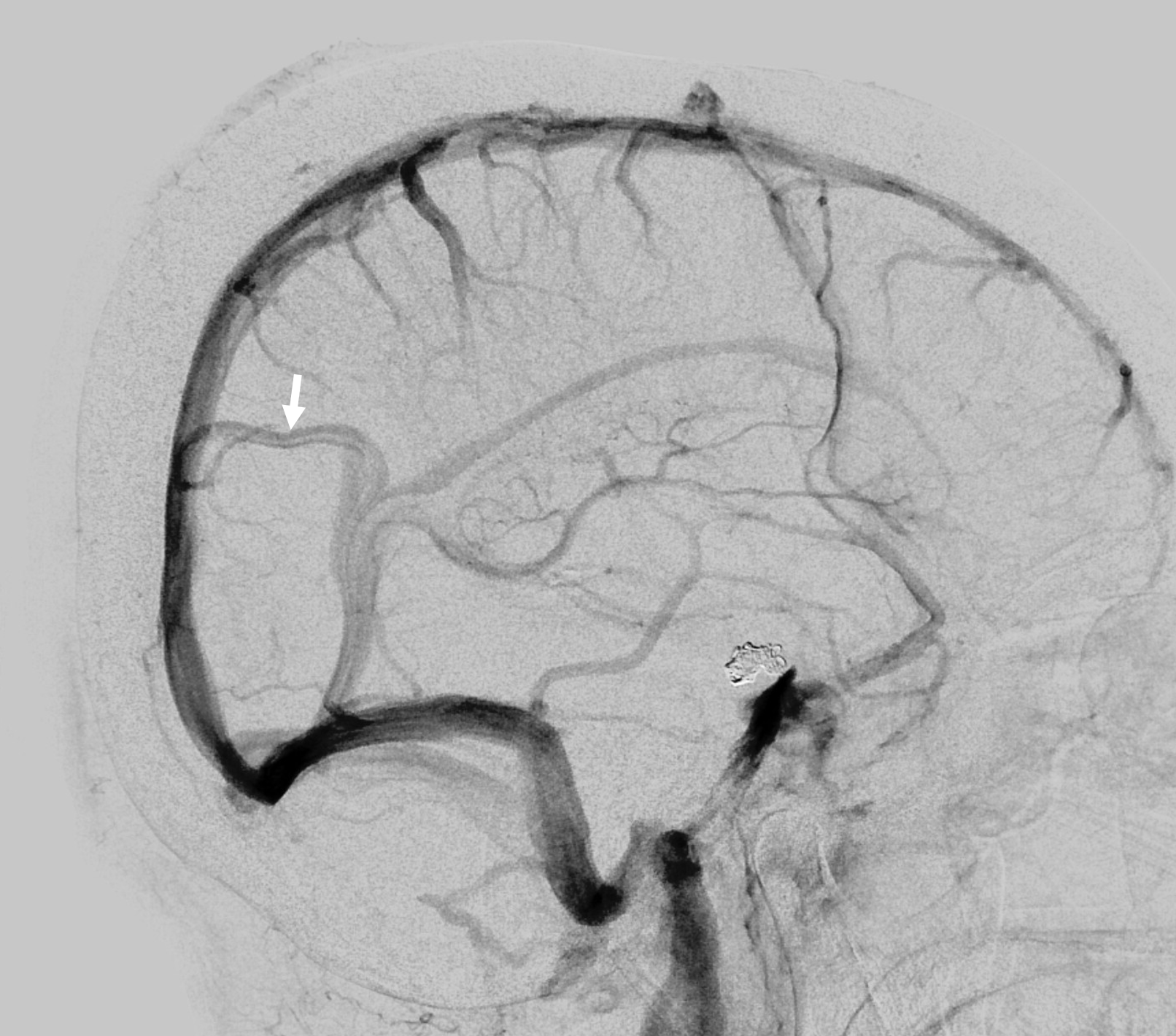
“Venous Lakes” — these are essentially dural channels / lakes adjacent to the superior sagittal sinus that receive the larger convexity veins such as Trolard or Rolando. They can be a surgical pain in the a.. when operating on parasagittal lesions esp. meningiomas. Not easy to appreciate on axial imaging because it is in plane with the lakes. Coronals are good for that. On angio they look like flattened portions of veins just adjacent to the sinus. Sometimes, the flattened portion projects above the sinus — because that’s how the dura is (see below)
Another one
Same patient on coronal MRI — dotted arrow is cortical vein, solid is dural lake part
The same channel on axial images — not obvious at all
Another example
Anaglyph

Dural Venous Lake — another example
VR image below — see transition from round convexity vein to flattened lake and junction with SSS
MIP of same case
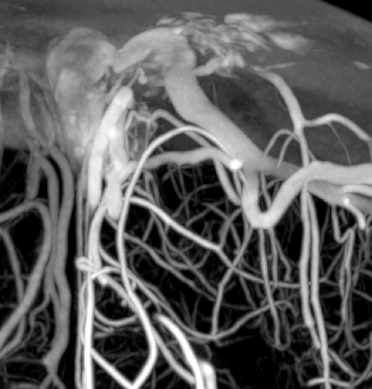
Temporal — middle cranial fossa dural channel
Rare, but again – -any part of dura can be a channel. Here is a big one (white arrows) draining superficial sylvian veins — look at the acute turn the venous channel takes when cortical sylvian veins enter the dura — ultimately draining via the ovale (arrowhead) into the pterygopalatine venous plexus
It is not convincing where this channel is on these images — however — look at the movie below — it is perfect. Stop movie to scroll individual frames and see
Tentorial Sinus (dural channel in tentorium cerebelli)
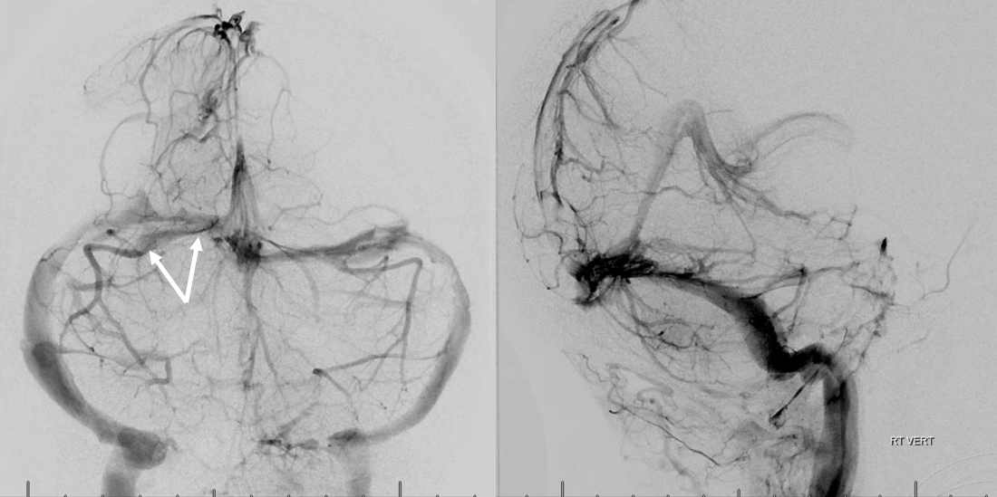
Tentorial sinus (dural channel in tentorium cerebelli, white arrows) draining the basal vein (black arrows)
Another case on T1-weighted volumetric MRI post. Tentorial dural venous channel (white arrow) receives superficial temporal (white arrowhead) and superior cerebellar (dashed arrow) veins
Some beautiful venous phase rotational angiography images of another big basal vein one
Anaglyph
The one below receives both large Labbe and several other temporal veins
DSA
Movie of venous 3D-DSA — pause to scroll thru individual frames
Cross-sectional evaluation
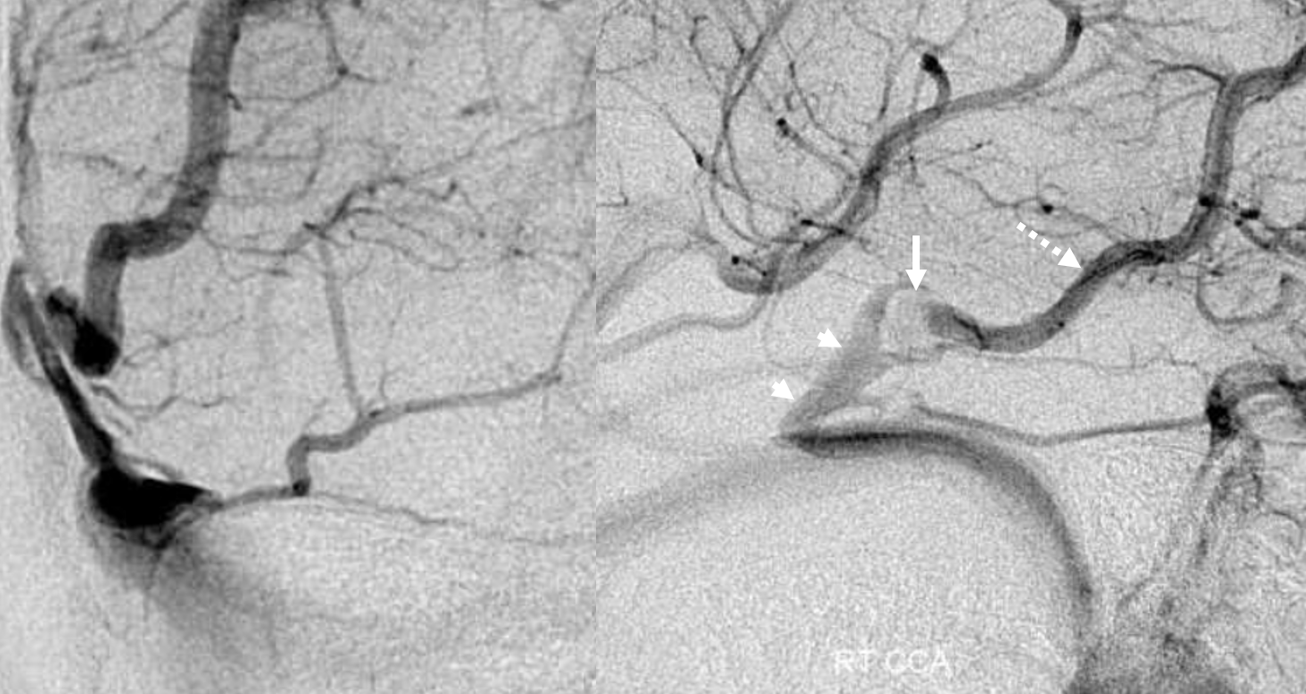
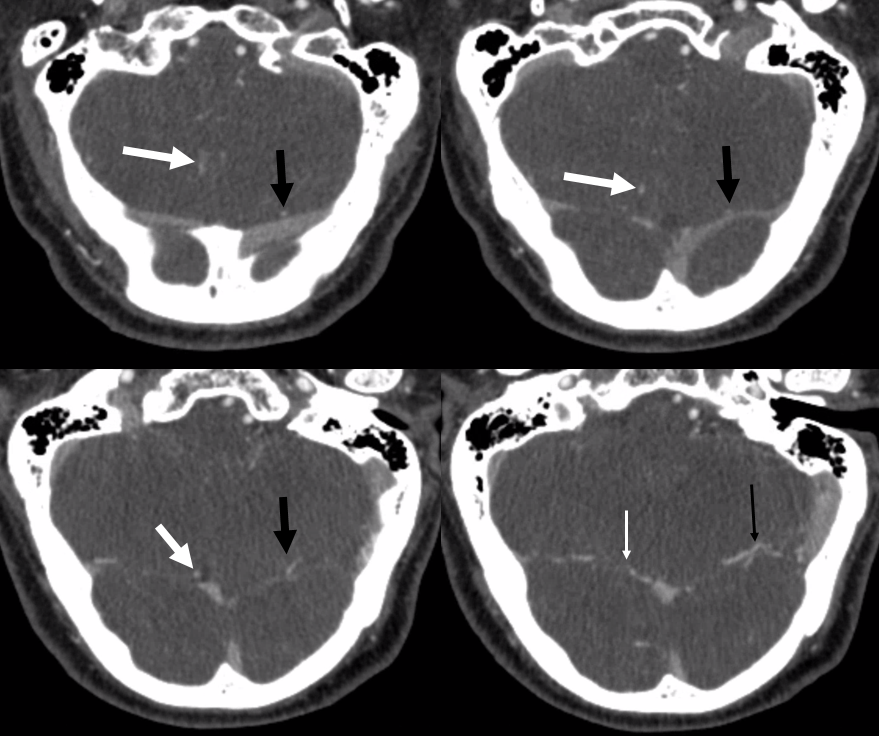
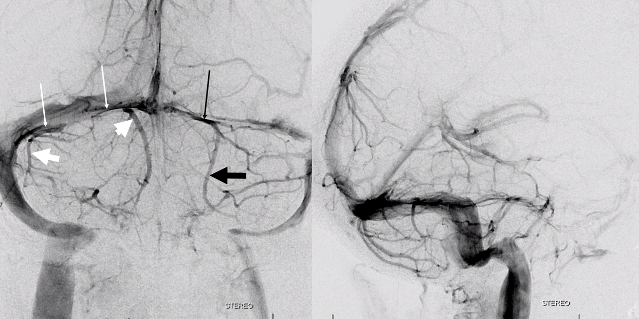
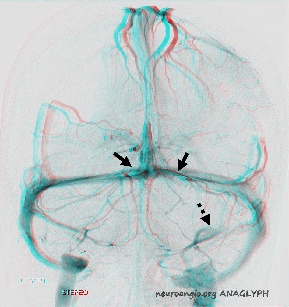
If you look at it carefully, most inferior cerebellar veins (inferior vermian ones also) actually insert into a tentorial sinus. When looking at axial images, of either MR or venous contaminated CT, you can trace these veins to insertion point along the dura, ventral / anterior to the transverse sinus/torcular. Here is a typical example, on angio (cross eye stereo views). White arrow — inferior vermian vein. White thin arrow — insertion point on tentorial sinus (white arrowhead) — the sinus has a typical line-like appearance when viewed edge-on. In this case it projects above the transverse sinus. Black arrow — inferior cerebellar vein. Black thin arrow — insertion on tentorial sinus, which drains medially (black arrowhead) — another edge-on look. On lateral views, the inferior vermian vein is deeper. Both veins curve anteriorly to insert on the dural sinus way anterior/ventral (white thin arrow)
CT of the same case
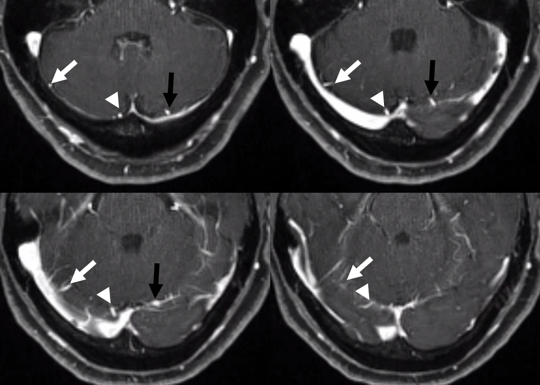
Another case with MR correlation. Inferior cerebellar veins are marked with thick arrows, corresponding tentorial venous channels with thin ones.
MRI
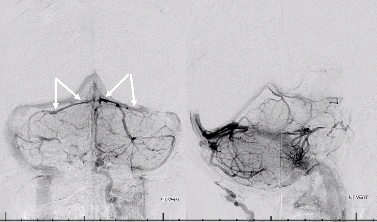
According to some publications, more than half of tentoria cerebelli have at least one dural channel. But, they are harder to spot on angio. Their appearance is usually of a dense line projecting over the transverse sinus in a Townes kind of view (arrows). Lateral is unhelpful. Here is an example
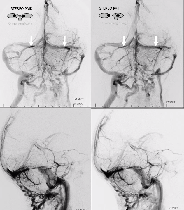
Another one
Another example
Here is an anaglyph stereo example also showing a channel connecting to the superior petrosal sinus (dotted arrow)
Clinical significance?
Coming up soon