Precentral Cerebellar Vein (C), a.k.a. Vein of the Cerebellomesencephalic Fissure, is an unpaired vein which runs in the caudocranial direction within the quadrigeminal plate cistern and serves as a key landmark for structures of the superior cerebellar vermis and dorsal midbrain.
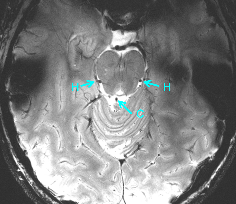
Knowledge of this landmark vein is absolutely essential. It helps establish key anatomical relationships and, even in today’s era of cross-sectional imaging, helps identify posterior fossa mass lesions by its displacement behavior.
Even though practically everyone calls it the Precentral vein, Rhoton has to be different and names it the “Vein of the Cerebellomesencephalic Fissure” because that’s his name for the precentral fissure. Personally, I prefer Rhoton’s classification because it more uniformly and intuitively names the veins according to their location with respect to the brain. As mentioned in so many places here, knowledge of vein location is more important than nomenclature, especially given the tremendous variability seen with veins. However, with big veins like the Precentral Vein, a different name, though anatomically appropriate, may not end up so catchy. Only the giants, like Galen and Rosenthal, seem secure…
The precentral vein is usually an unpaired midline vein which runs vertically along the posterior aspect of the upper brainstem, thus serving as a key landmark. The vein is most commonly formed by union of two brachial tributaries (D) which are found on the upper outer surface of the superior cerebellar peducles. (Appropriately, Rhoton calls these tributaries “Veins of the Superior Cerebellar Peduncle”) It then courses in the precentral fissure upward into the quadrigeminal plate cistern, where it is a nice landmark for that plate. Usually it empties into the Common Cerebral Vein, which is my new name for the Great Vein of Galen (A). Just kidding…
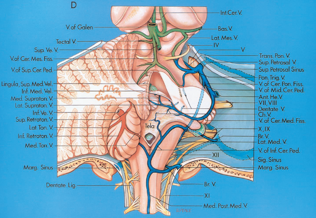
From Rhoton, The Posterior Fossa Veins. Neurosurgery 2000 supplement, Volume 47 (3), S69-92; Vein of the Cerebellomesencephalic Sulcus (Precentral Vein) being formed by paired Veins of the Superior Cerebellar Peduncle (Brachial Tributaries).
Yung Peng Huang and Bernard Wolf. Veins of the Posterior Fossa. Chapter 75, in Newton and Potts Radiology of the Skull and Brain. Volume 2, book 3. 1974 Precentral Vein (C) formed by union of the Brachial Tributaries (D)
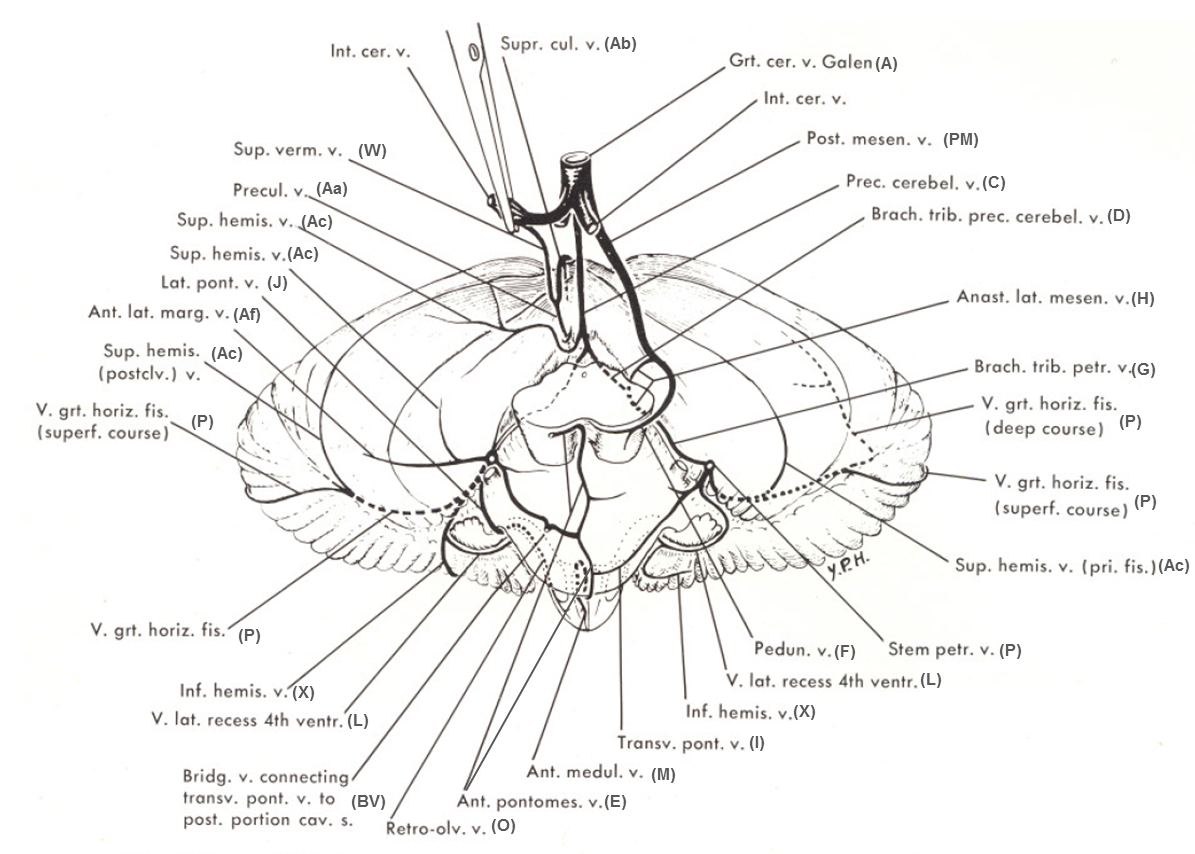
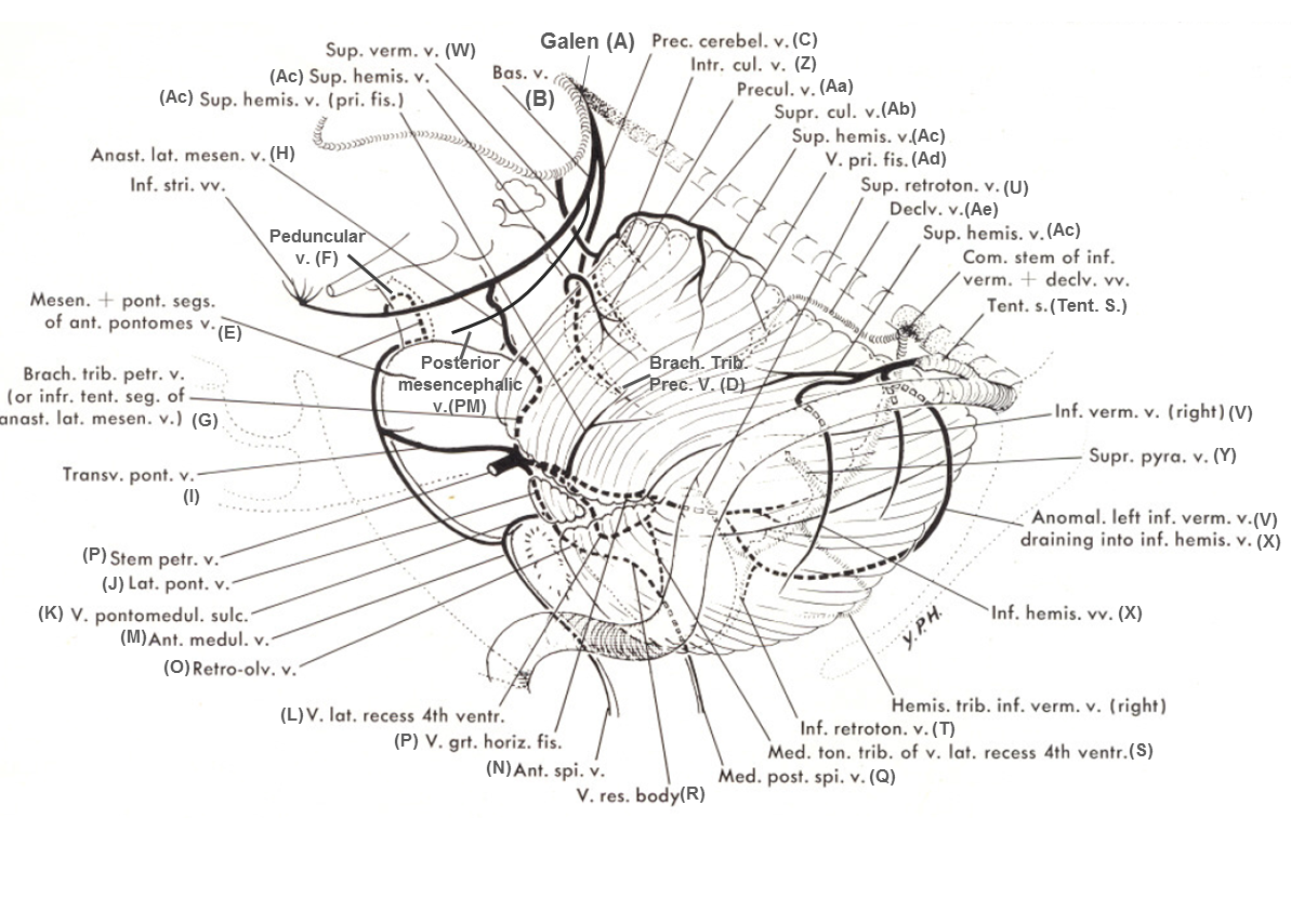
A table of correlational nomenclature is included here for reference, to the best of my ability to read minds of both Drs. Huang and Rhoton. It is not an exact science.
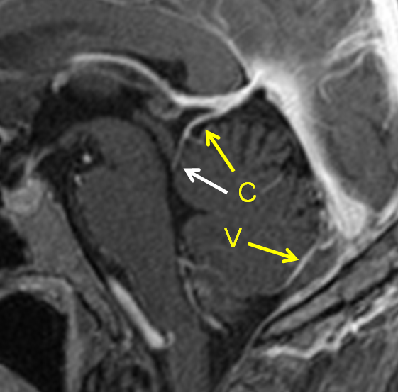
Midsagittal postcontrast T1 MRI of the brain, showing typical precentral vein (C) running within the Precentral Fissure (a.k.a. the Cerebellomesencephalic Fissure) and the quadrigeminal plate cistern. Also well seen is the inferior Vermian vein (V).
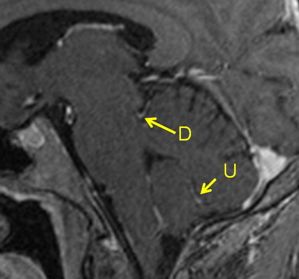
A parasagittal section of the same case, where the Brachial Tributary (D) a.k.a. the Vein of the Superior Cerebellar Peduncle links to the Precentral Vein. Also well seen is the Superior Retrotonsillar (U) tributary of the Vermian vein.
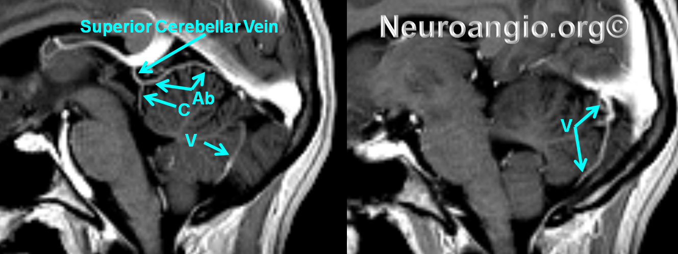
The superior vermian vein (W) and the precentral vein may connect to the Galen (A) separately, or come together into whats known as the “Superior Cerebellar Vein” before joining the Galen, as seen in this example. The upper part of the Superior Vermian Vein is named Superior Culmen Vein (Ab)
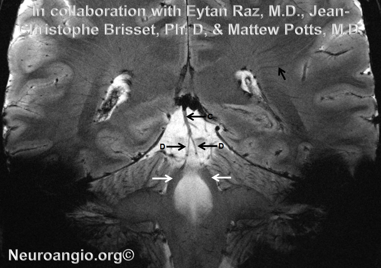
Stunning 7T MRI Coronal Gradient Echo T2-weighted images of the precentral (C) and brachial (D) veins along the superior cerebellar peduncles (white arrows). The transmedullary veins are shown with an unlabeled black arrow
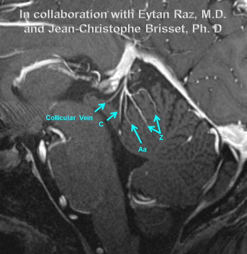
7T post-contrast MP-RAGE amazing demonstration of the precentral, preculmenary, and collicular veins, in collaboration with Eytan Raz, M.D. and Jean-Christophe Brisset, Ph. D
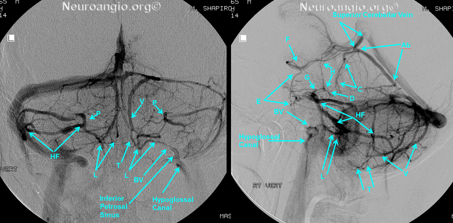
The veins of the superior cerebellar peducle (brachial tributaries [D] of the precentral vein [C]) are also connected to the lateral mesencephalic vein (H), a.k.a. the Pontotrigeminal vein in Rhoton. More often then drain into the precentral vein (C), but not always, and instead could empty into the Lateral Mesencephalic ystem. This is why the lateral mesencephalic vein is sometimes hypoplastic. The following example shows veins of the superior cerebellar peduncle (D) draining into the lateral mesencephalic vein (H). Notice also how the Superior Culminate Vein (Ab) portion of the superior vermian vein unites with the Precentral vein, forming what’s called the “Superior Cerebellar Vein”. Other times the Superior Vermian vein and the Lateral Mesencephalic drain separately.
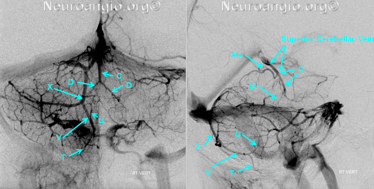
The opposite is seen here, with “classic” large brachial tributaries (D), a.k.a. veins of the superior cerebellar peduncle (D). There is a characteristic inverted “V” configuration which helps identify these veins. Also seen is variant connection of the inferior vermian vein (V) into an inferior hemispheric vein (X), with the characteristically more superficial course of (X) in the lateral projection than would be expected for the classic inferior vermian vein, which more closely hugs the vermis. Well seen are superior (U) and inferior (T) retro-tonsillar veins, defining the posterior border of the tonsil.
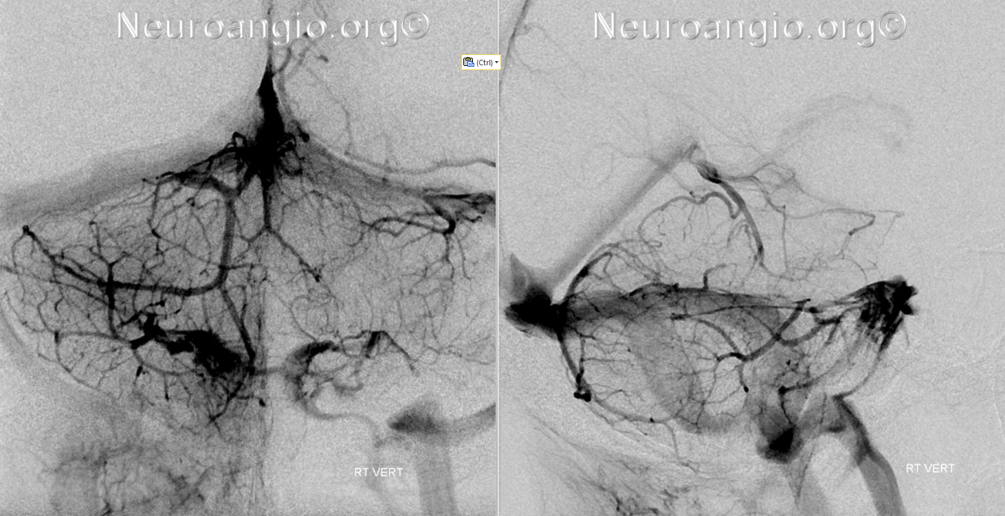
The same images, without overlapping labels:
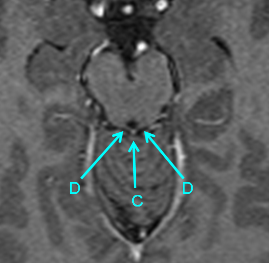
Post-contrast volumetric T1-weighted images are excellent for venous anatomy. This MRI shows brachial tributaries (D), a.k.a. veins of the superior cerebellar peduncle converging to form the Precentral Vein (C). Notice how (D) are less well-developed on the left, where the lateral mesencephalic vein (H) is more prominent, and vice versa on the right.
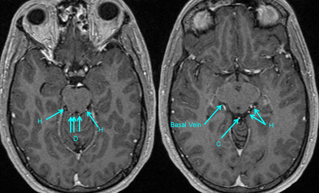
The convergence of paired (D) to form (C) is well-seen here:
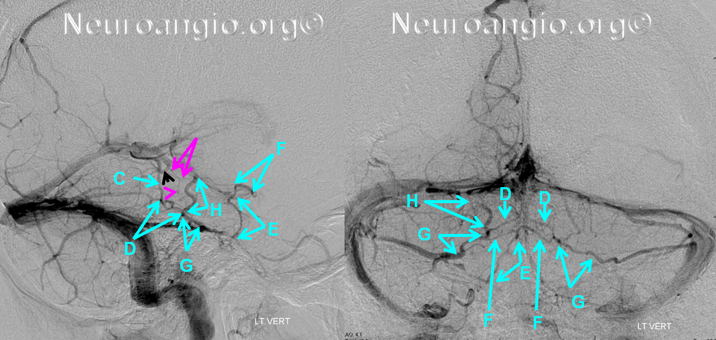
Another example, with well-seen anastomosis between the Precentral vein (C) and the lateral mesencephalic vein (H), a.k.a. Pontotrigeminal Vein in Rhoton via the Brachial Tributaries of the Precentral vein (D), a.k.a. Veins of the Superior Cerebellar Peduncle, especially on the right. Notice separate entry of the precentral vein (C) and superior culmen vein (black arrow) into the Galen. Tectal plate veins are also present (pink)
Same picture, with brainstem outline. This is a key concept — the precentral vein defines the posterior portion of the mesencephalon — the tectal plate. Below the precentral vein, at the level of the pons, is going to be the 4th ventricle. Remember that the aqueduct is located in front of (ventral to) the tectal plate. Immediately below the plate the aqueduct opens into the 4th ventricle.
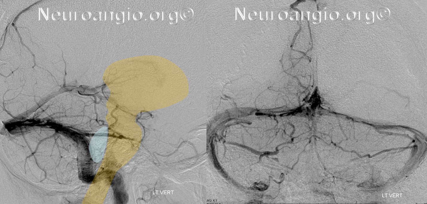
MRI already shown above, illustrates the above concepts. The precentral vein defines the posterior portion of the mesencephalon — the tectal plate. Below the precentral vein, at the level of the pons, is going to be the 4th ventricle. Remember that the aqueduct is located in front of (ventral to) the tectal plate. Immediately below the plate the aqueduct opens into the 4th ventricle.

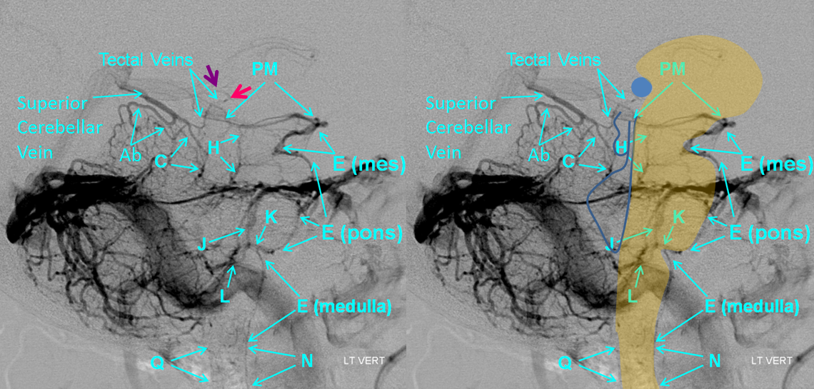
Another image, with the added convenience of seeing the infrequently visualized Tectal Veins. Notice even how the upper part of the Tectal Vein curves anteriorly (pink arrow) and then posteriorly again (purple arrow), outlining the posterior portion of the Pineal Gland (blue circle). The Superior Cerebellar Vein is formed by union of the Precentral (C) and Superior Culmen Veins (Ab). The Superior Culmen vein is really the superior vermian vein (runs on top of the vermis)
References:
Yung Peng Huang and Bernard Wolf. Veins of the Posterior Fossa. Chapter 75, in Newton and Potts Radiology of the Skull and Brain. Volume 2, book 3. 1974
Yung Peng Huang et. al. The Veins of The Posterior Fossa — Anterior or Petrosal Draining Group. September 1968. American Journal of Radiology. For a full text PDF click here.
Yung Peng Huang et. al. The Veins of The Posterior Fossa — Anterior or Petrosal Draining Group. September 1968. American Journal of Radiology. For a full text PDF click here.
Albert Rhoton, The Posterior Fossa Veins, Neurosurgery. 2000 Sep;47(3 Suppl):S69-92.
Albert Rhoton, The Cerebellopontine Angle and Posterior Fossa Cranial Nerves by the Retrosigmoid Approach. Neurosurgery. 2000 Sep;47(3 Suppl):S93-129.

