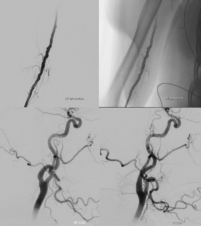
Another radial case with a small twist of pretty obvious FMD in the brachial artery. Not something femoral access is likely to encounter. Of course there is carotid and vert FMD but brachial is not usually imaged. Here is a ruptured ACOM case. Typical beading of FMD seen on radial roadmap imaging
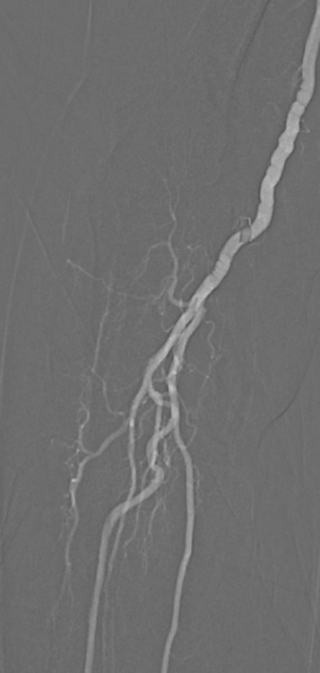
Brachial imaging. We always go up radial and brachial under imaging guidance
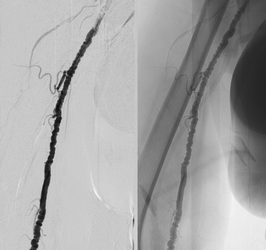
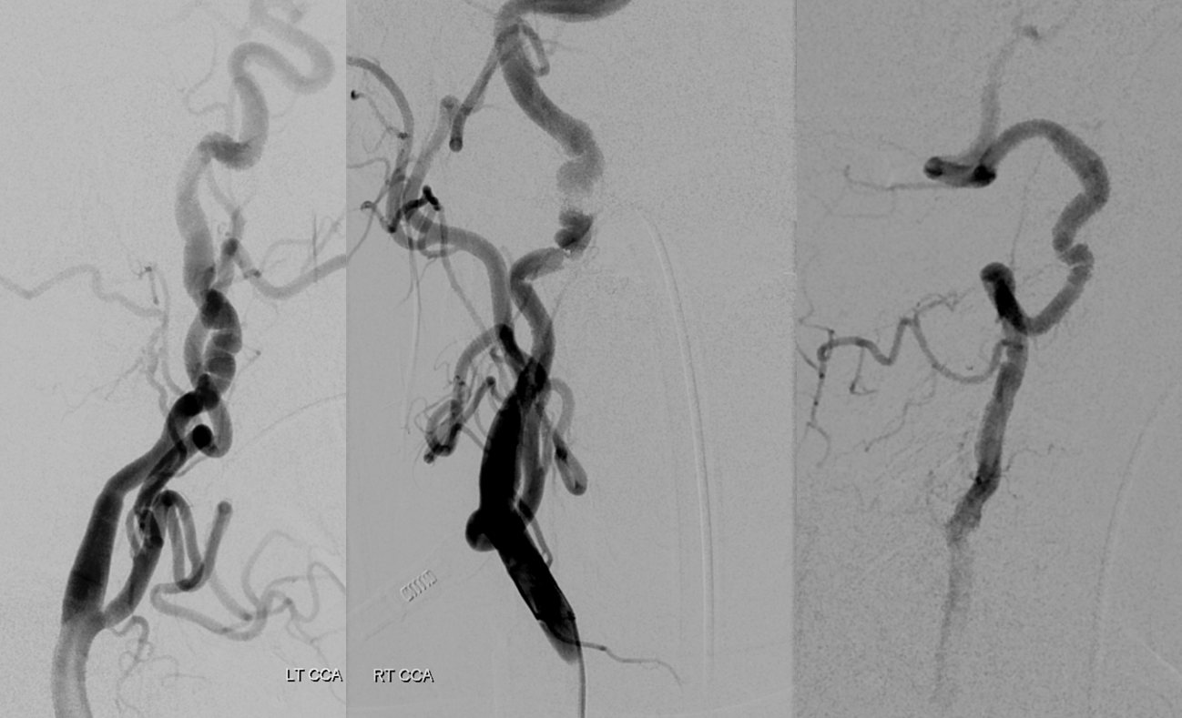
Ultimately we had a 6F glidesheath and 6F Envoy (ID=070) there with no issues in right extremity perfusion. Here is typical bilateral ICA and right vert FMD
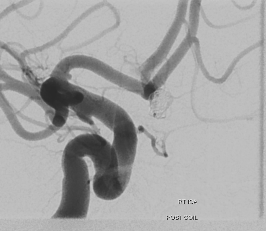
ACOM aneurysm working projection
Post coil
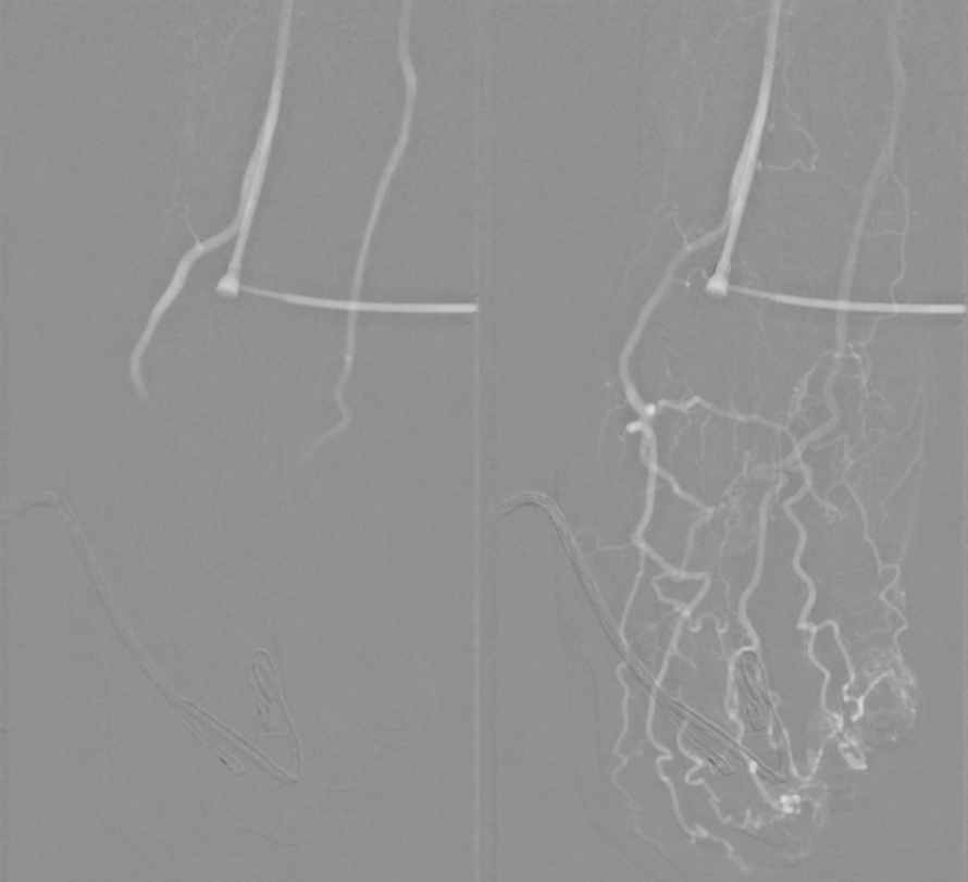
Post procedure check shows antegrade flow in right radial and nice palmar arch. We inject with enough forward flow to visualize the ulnar as well.
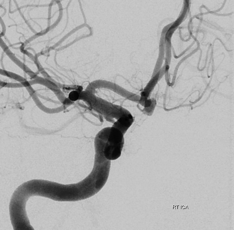
Brachial FMD need not be associated with other sites. In the patient below, there is no carotid involvement. One vertebral was injected and was clean also. A small left ophthalmic aneurysm is there however — a known but not very strong association
Conclusion: FMD something to keep in mind for radial access; another reason to use imaging when going up that arm
