
A sad case showing how bad webs can be…
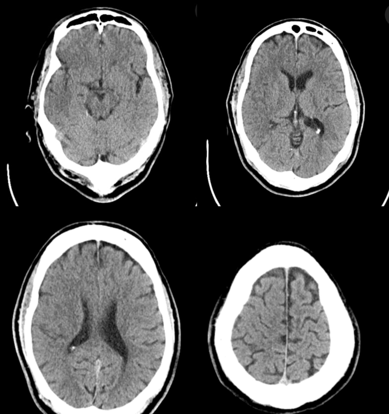
Presentation with right hemisphere large vessel occlusion. NCHCT
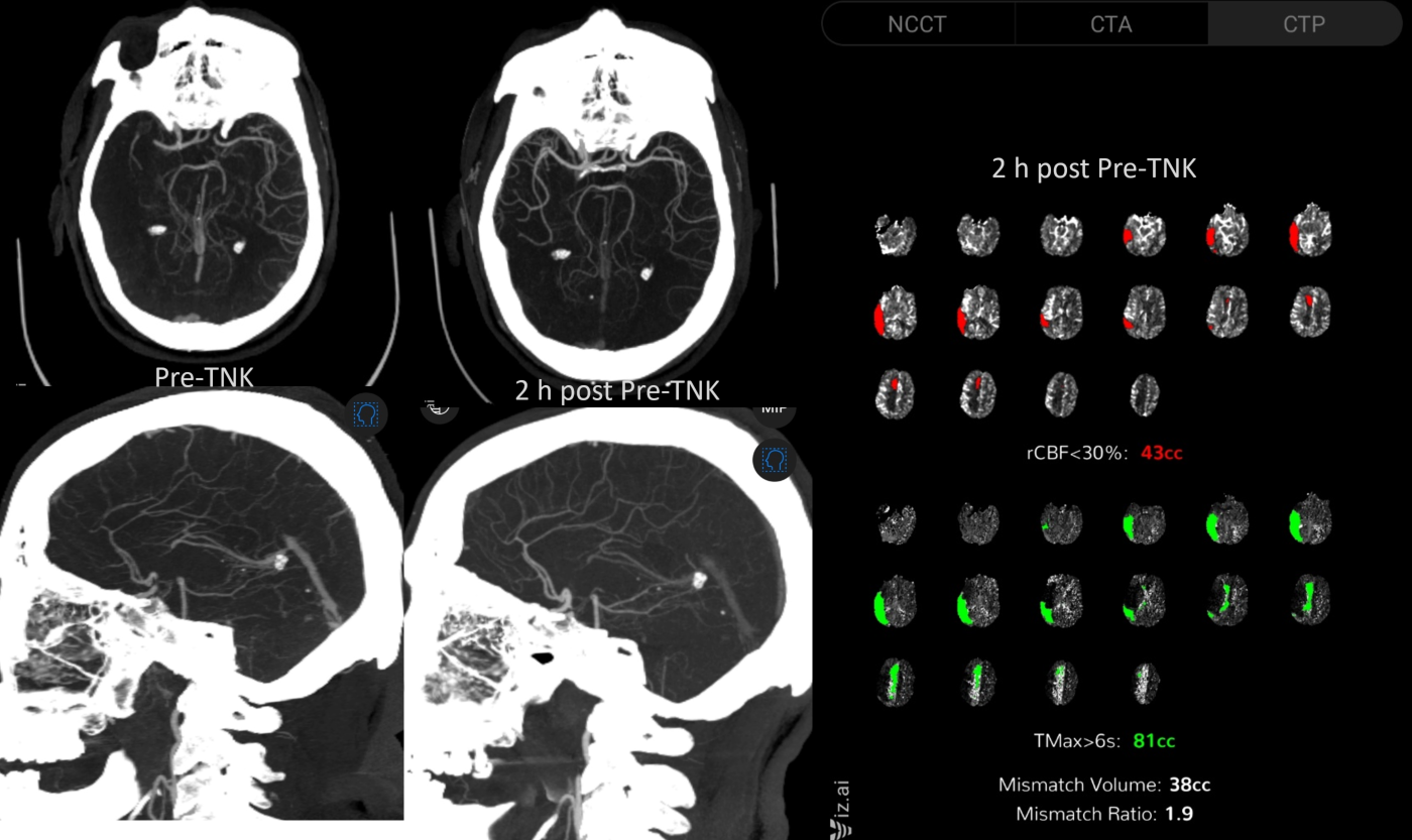
CTA shows right M1 near-complete and right A2 complete occlusions. TNK is given. Transferred to thrombectomy center. Two hours later, the subocclusive M1 improved. A2 remains closed. Perfusion does not reach convexity and shows residual M3 level occlusion
CT shows extensive early ischemic changes of the superior convexity. The Middle and Anterior cerebral tandem emboli are especially bad, since each derives the other of one’s natural collateral. Note also sparing of basal ganglia given distal nature of M1 occlusion, beyond the lenticulostriates
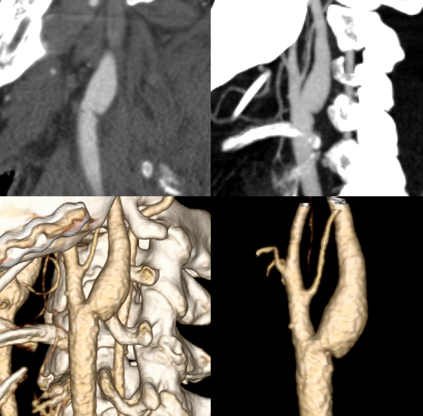
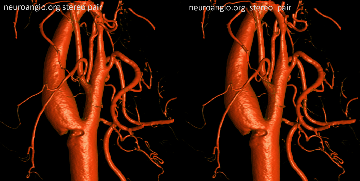
CTA of the web. See carotid web page for more info
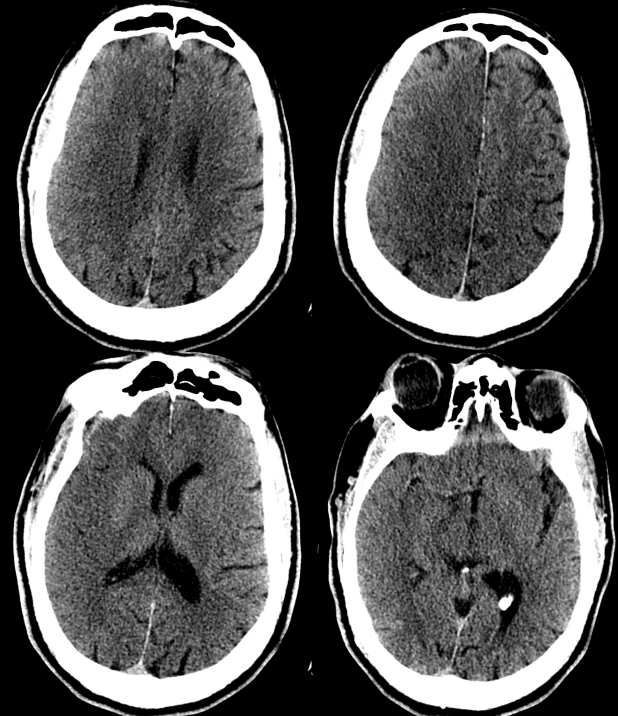
Thrombectomy offered due to time of presentation and overall preponderance of evidence.
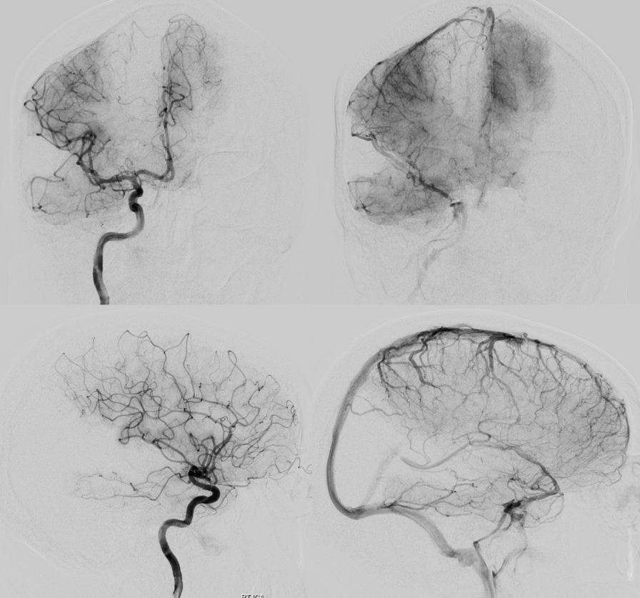
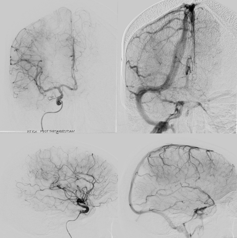
Post Zoom 55 aspiration of the residual M3 and the A2.
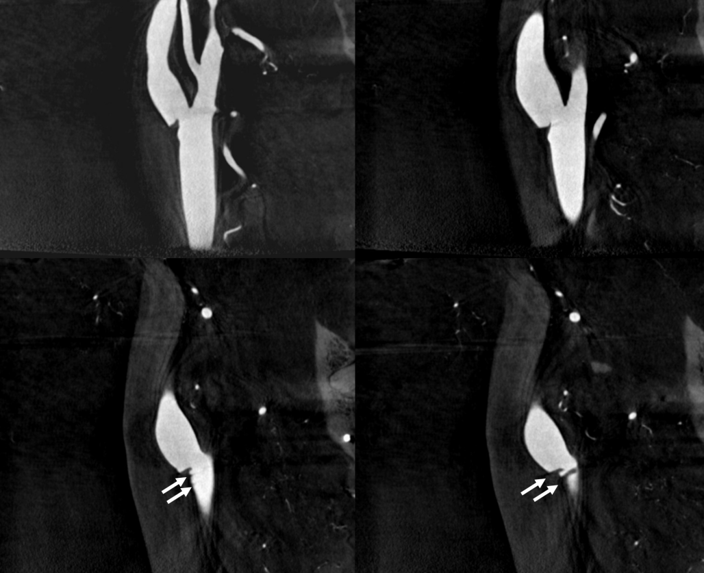
DYNA CT of the web
Note how well multiple connective tissue shelves can be resolved on the MIP images
See other web cases on Case Archives Page