
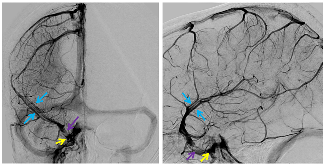
Knowledge of regional venous anatomy is critical for optimal surgical outcomes. Interruption of dominant cerebral veins, intentional or not, may lead to hemorrhagic venous infarcts in the distribution of the compromised vein. In this case, preoperative angiography demonstates several dominant superficial sylvian veins (blue) draining the frontotemporal operculum and adjacent convexities into the cavernous sinus (yellow) via the sphenoparietal sinus (purple)
Pterional craniotomy was perfromed iwth careful attention to preserving these veins. One of the veins could not be saved despite full recognition of its significance, which can still be tolerated in many patients. Here, however, the result is a typical venous infarction, with hemorrhagic foci within a larger hypodense, swollen territory. This particular event was well-tolerated.
