Case courtesy Dr. Peter Kim Nelson
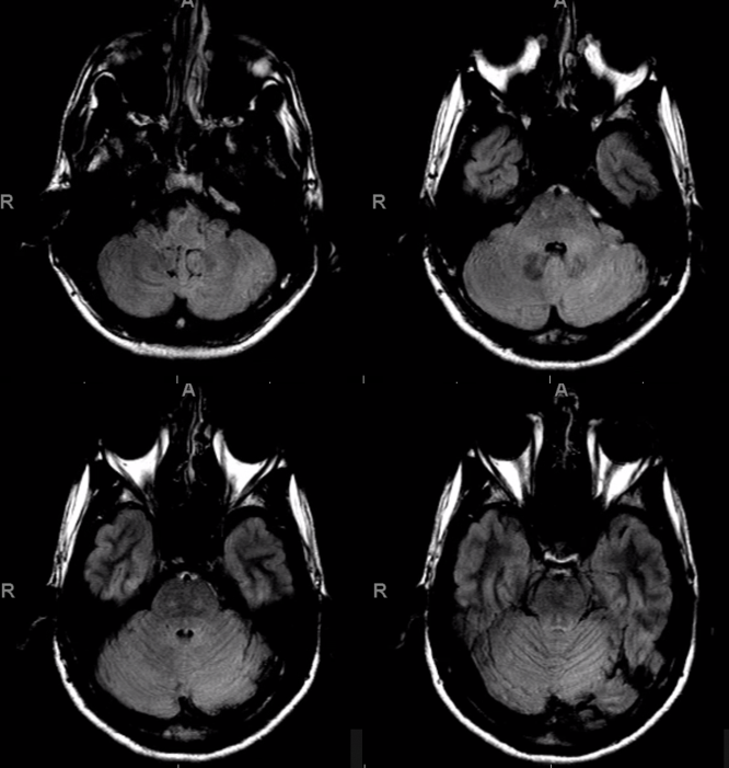
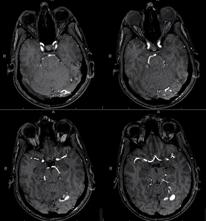
Another example of tentorium cerebelli fistula — a fistula located on the tentorium, not directly associated with a venous sinus, instead draining directly into cerebellar veins
FLAIR shows some congestion of the left cerebellum
MRA
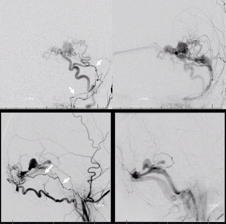
Angio. Supply from artery of tenorium cerebelli — part of posterior meningeal group. Notice fistula is not primarily involving the transverse sinus
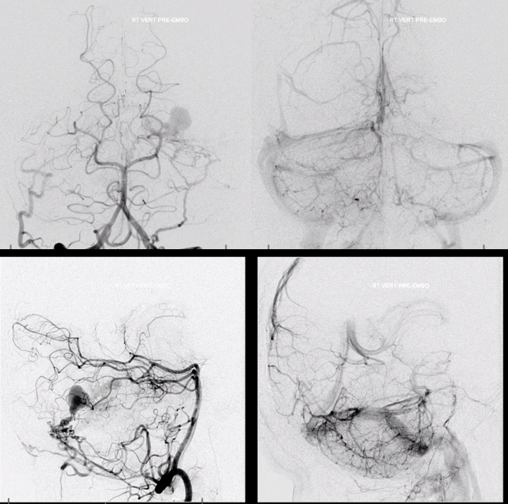
Right vert
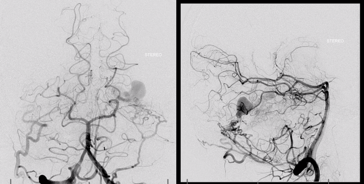
Typical supply from MMA, transosseous occipital, and ascending pharyngeal jugular division contributing to artery of the sigmoid sinus (artery in wall of sigmoid sinus, white arrows)
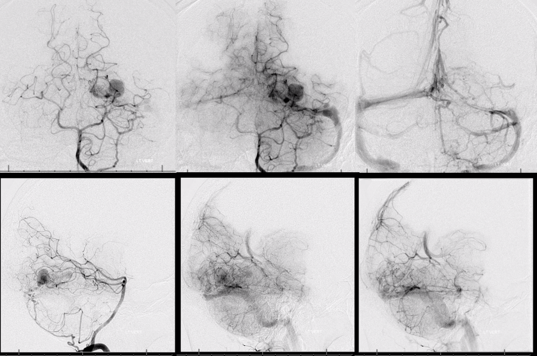
Left vert injection. Important to understand arterial supply (left), fistula venous drainage (center) and brain venous drainage (right) phases
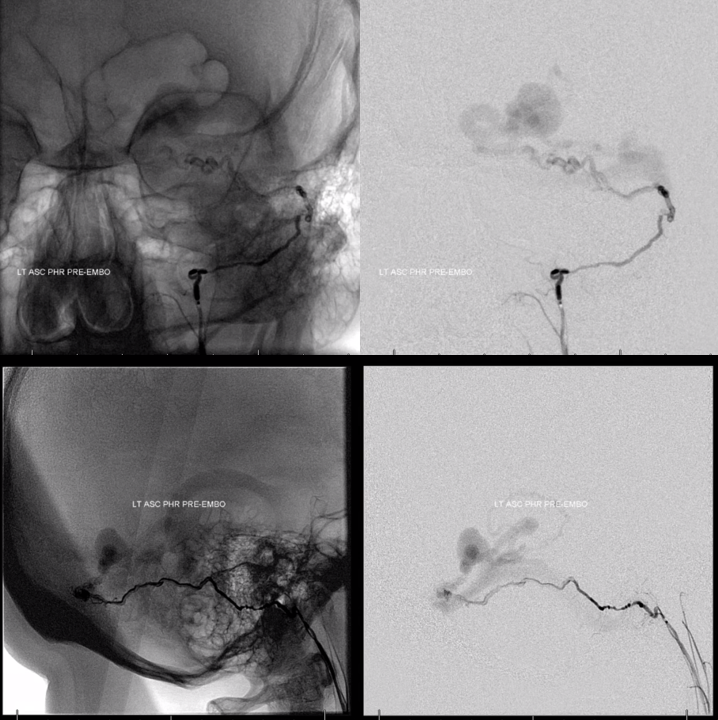
Superselective injection — ascending pharyngeal . Not a good embolization position (CN IX, X, XI)
MMA is a much better choice
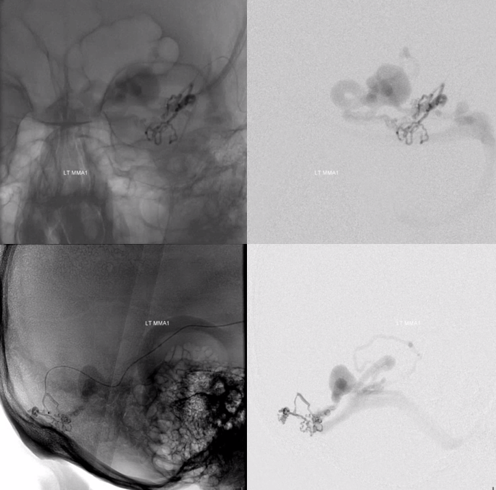
Despite excellent position, nBCA does not penetrate (this is pre scepter and pressure cooker era)
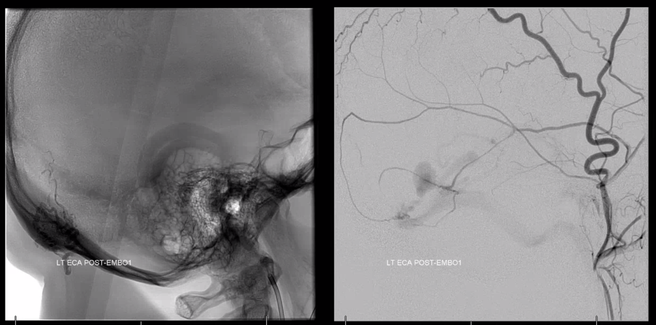
Going back to the MMA
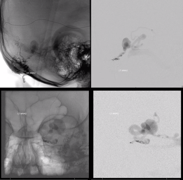
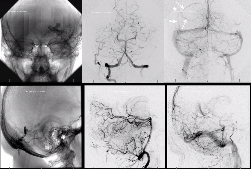
Now there is plenty of glue in the vein. No more fistula. Notice a supratentorial dural venous channel (upper right image) — collecting several temporo-occipital veins (dashed arrows) into the dural sinus channel (white arrow). Transition point characterized by acute angulation is white arrowhead
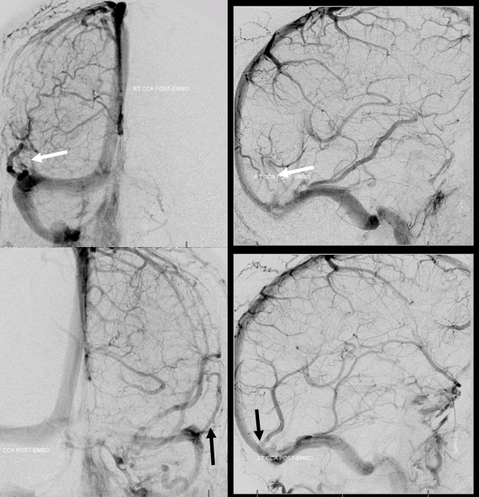
Right and left ICA injections. Notice dural venous channels in the supratentorial dura also (arrows). The one on right is the same one that was seen in the above vert injection
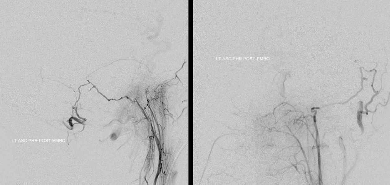
Control ascending pharyngeal injection, with beautiful reflux into occipital and vert
For more, see additional dural venous channel cases on “Cases” page
