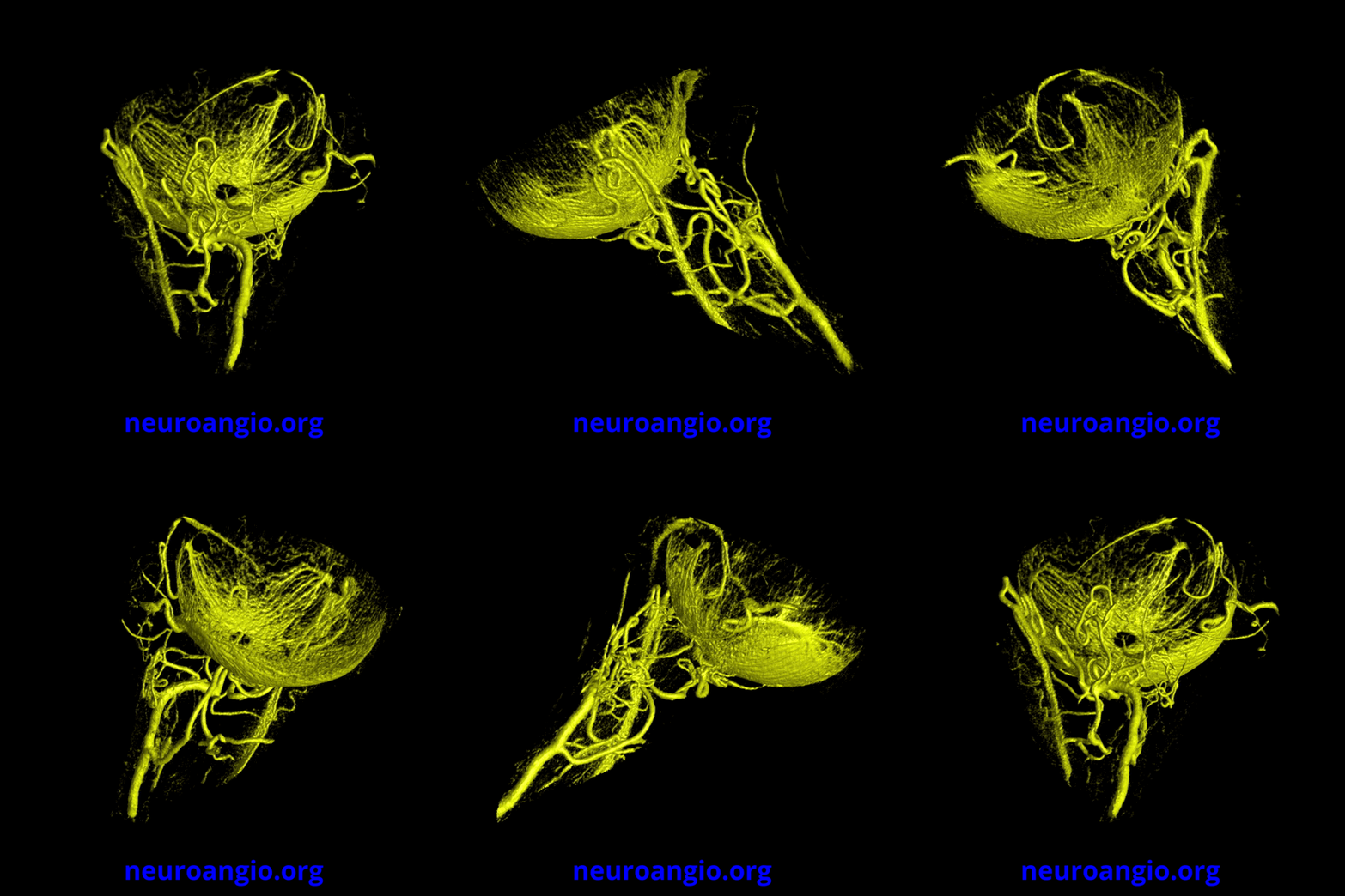
Visualization of Central Retinal Artery is routinely possible with Cone Beam imaging, opening window into seeing disease states previously not directly observed. See Ophthalmic Artery background page for more info. In this case of posterior ischemic optic neuropathy, a diseased central retinal artery is seen (thready and irregular). Treatment remains medical, as artery is too small for mechanical intervention.
Note how the choroid remains unaffected and prominently visible, with large corresponding vorticose veins.
This is very much on the leading edge of medical imaging and many questions remain. However, this is how advances are made.
This is contrasted with normal appearance of central retinal artery below

