by Drs. Erez Nossek and Cen Zhang
Anatomy knowledge matters. Here is an acute completed PCA infarct
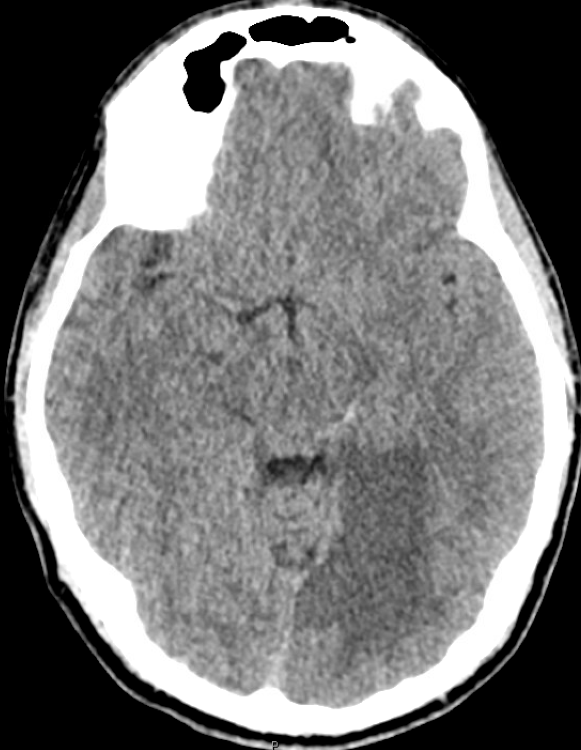
MP4 of CTA — stop and scroll — see something?
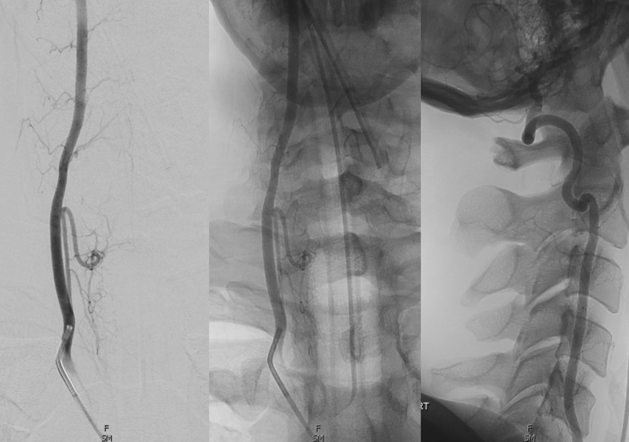
Angio — look at transition in vertebral artery caliber. Why?
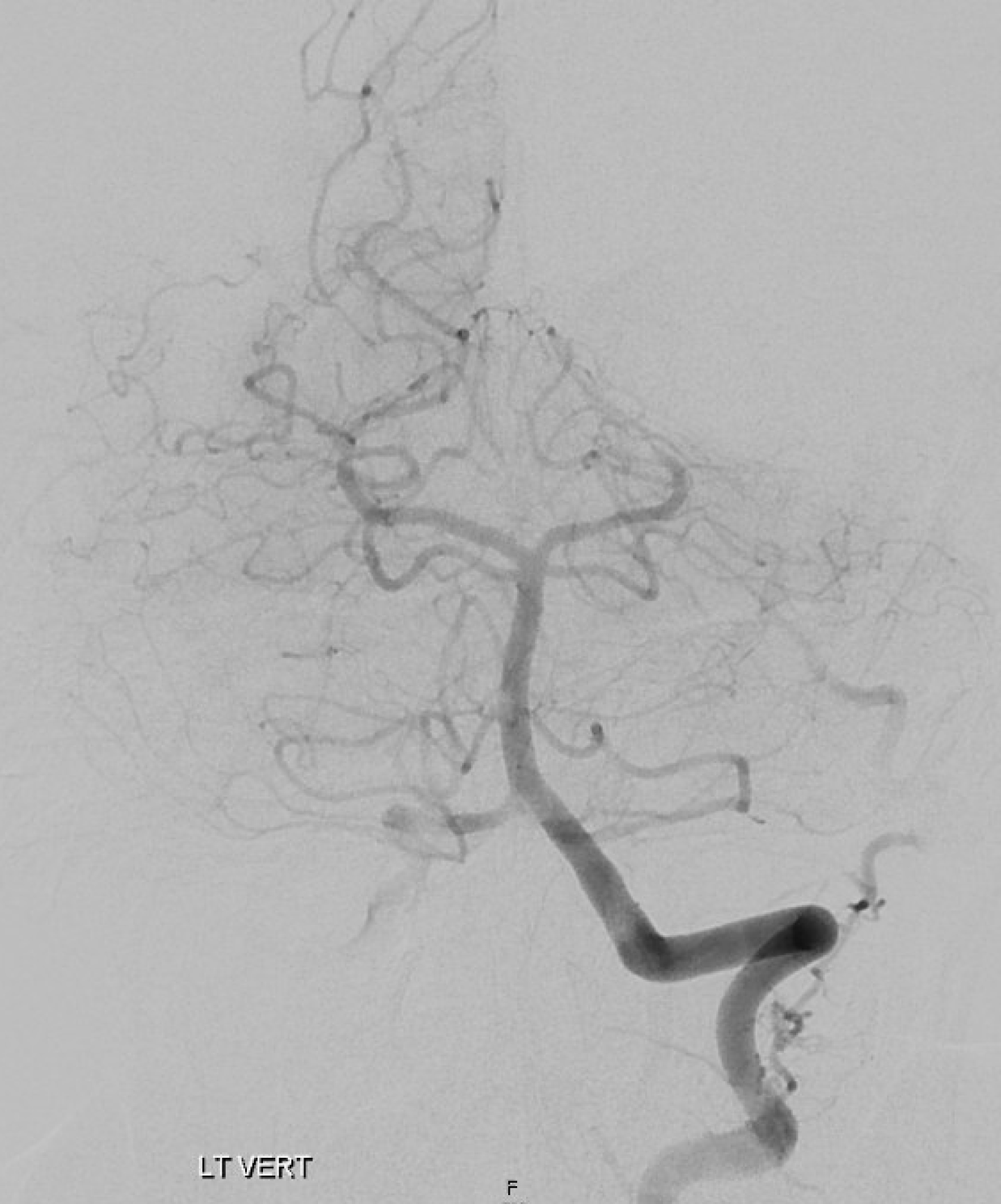
Selective vert injection
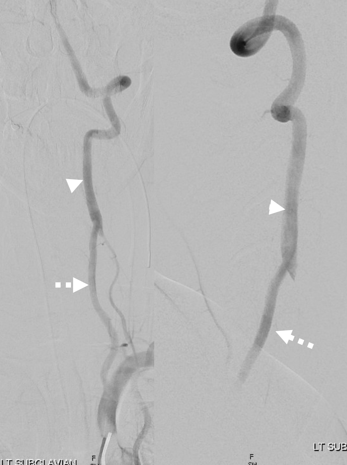
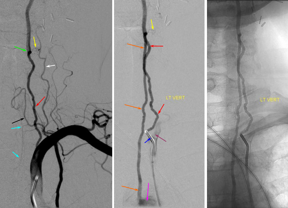
What is this? Answer is knowledge of anatomy. There is a variant of proximal “duplicated” vert — where direct aortic origin vert and “classic” subclavian origin vert origins co-exist, usually fusing somewhere in the lower cervical segment into a single vessel. Below is a different case of this anatomical disposition. See Vertebral Artery page for much more on this, including key to the arrows
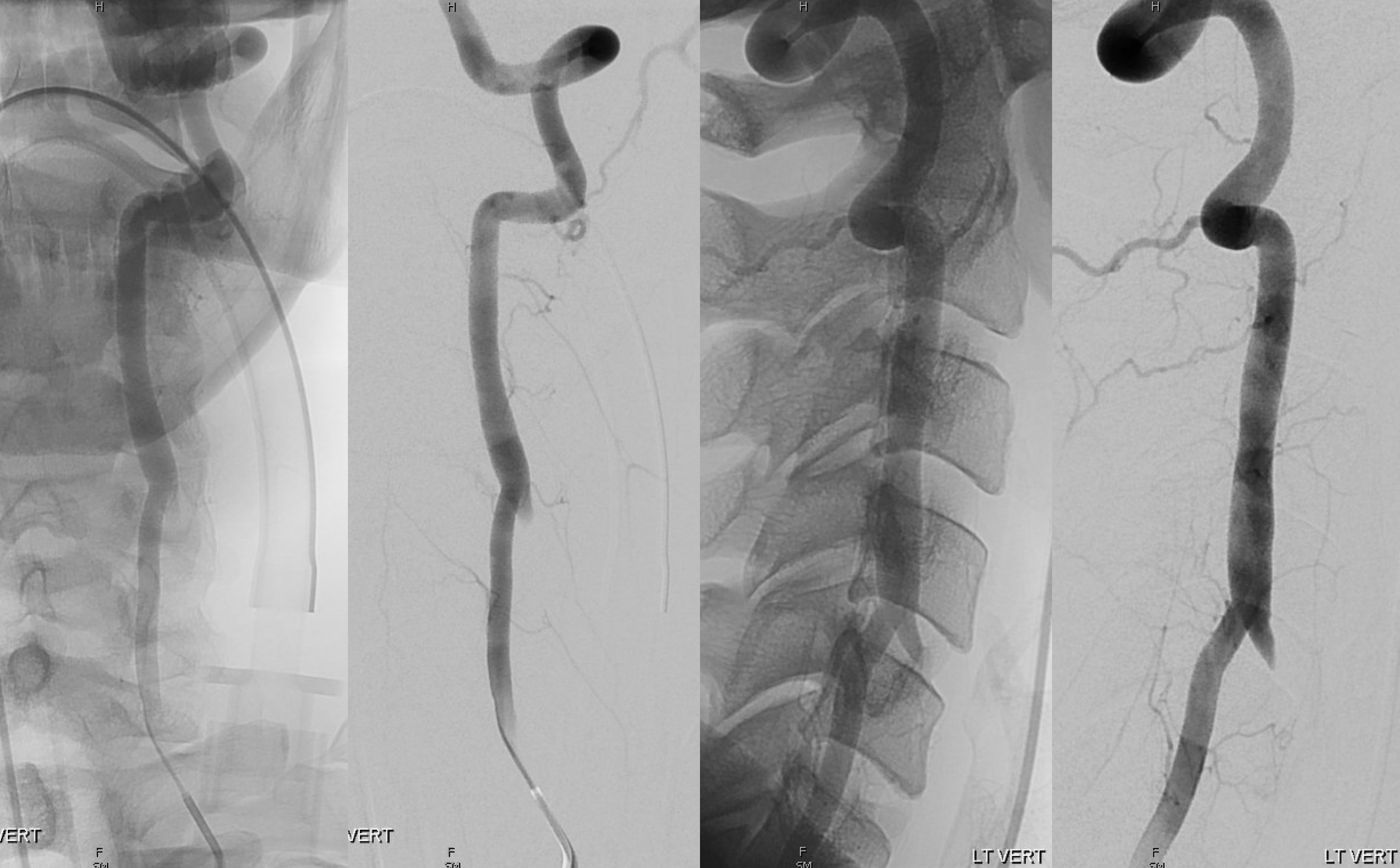
Knowledge of this prompts us to look for another vert origin — and here it is — direct origin off aortic arch — with the associated occlusion. Also note branch to the anterior neck
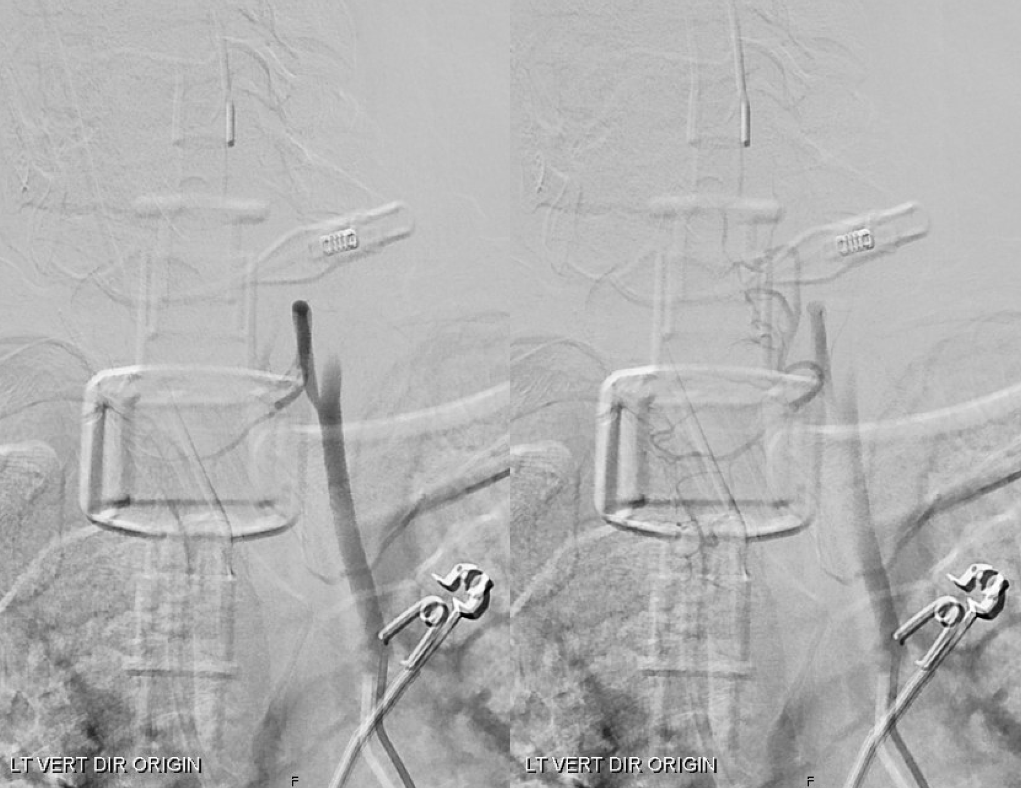
This is not just anatomical satisfaction — it establishes stroke etiology as artery-to-artery embolism due to dissection. Mystery solved
Cranial view of PCA occlusion
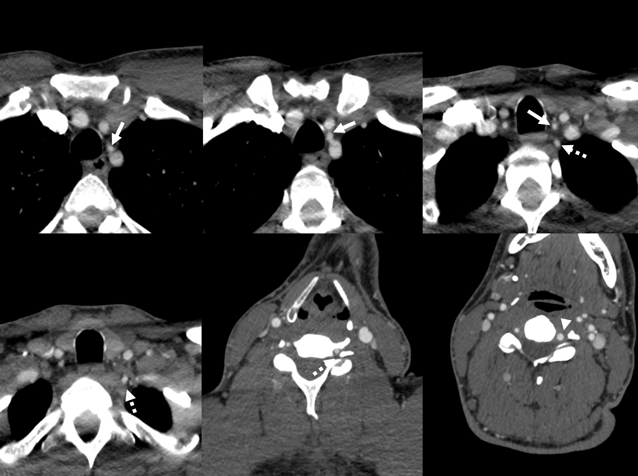
Retrospective review of CTA shows the same — direct origin vert = solid arrows; subclavian origin vert = dashed arrows; larger caliber vert distal to fusion of the two origin verts = arrowhead;
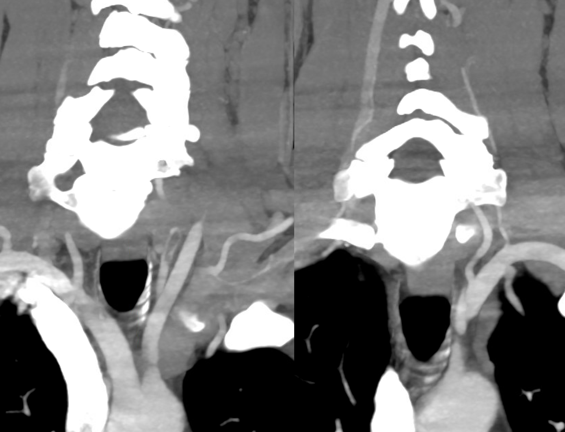
MIP coronal CTA — image on left shows nicely the dissected segment of the direct origin vert
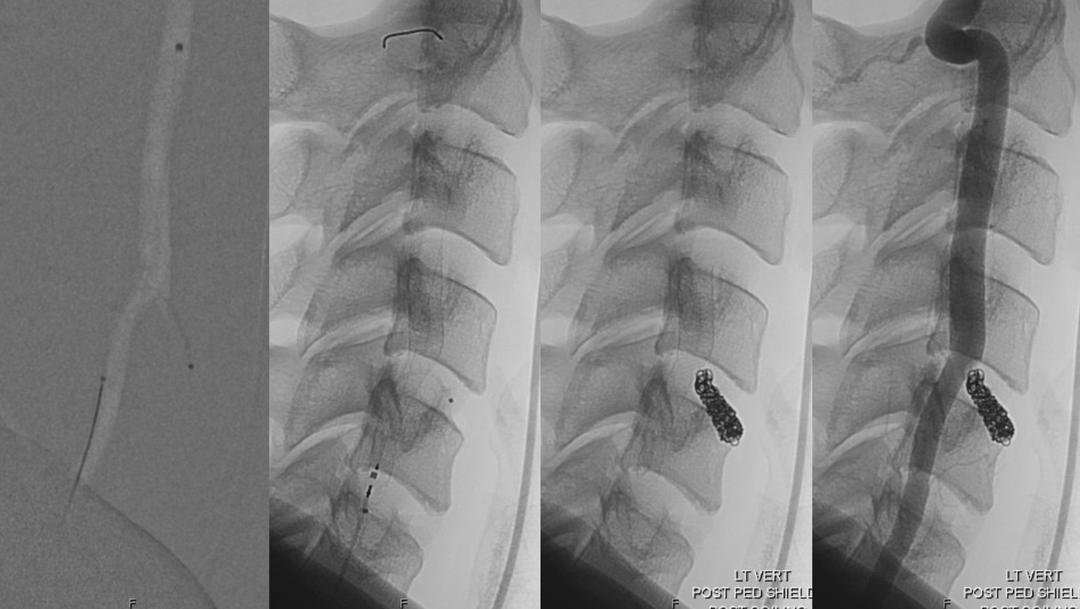
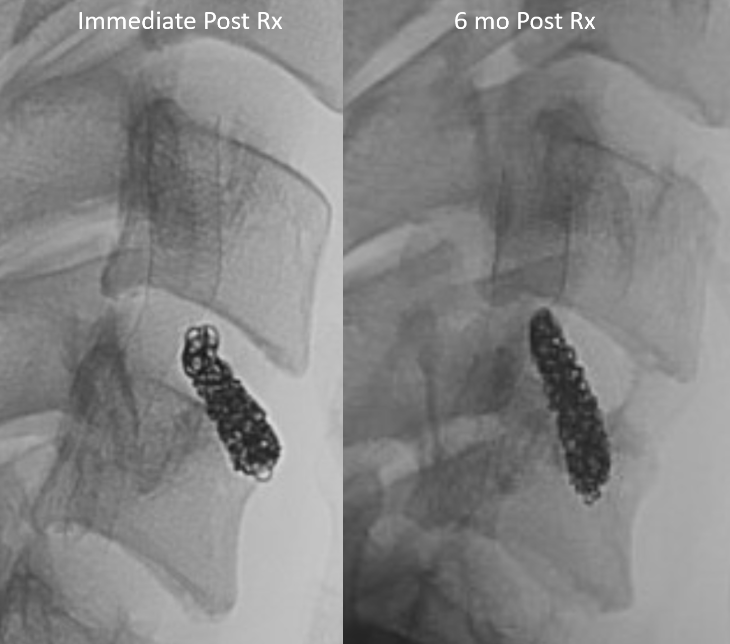
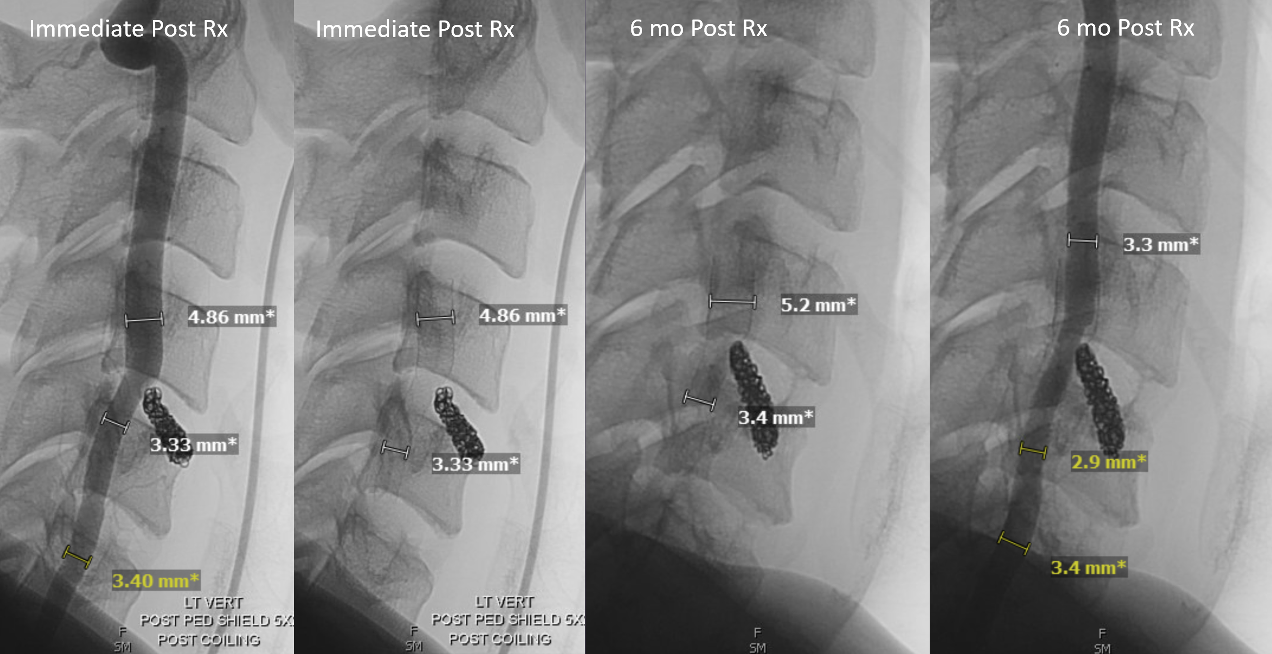
What to do? Post vert Pipe and coil of direct origin stump
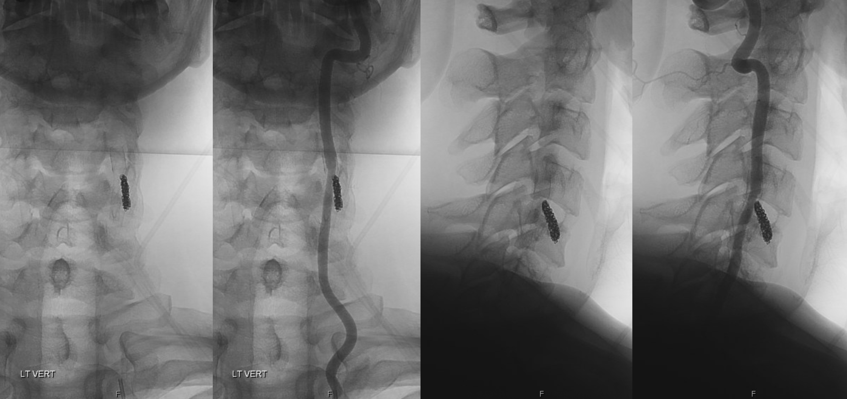
6 mo later
Direct origin stump supply now exclusively to inferior thyroid artery
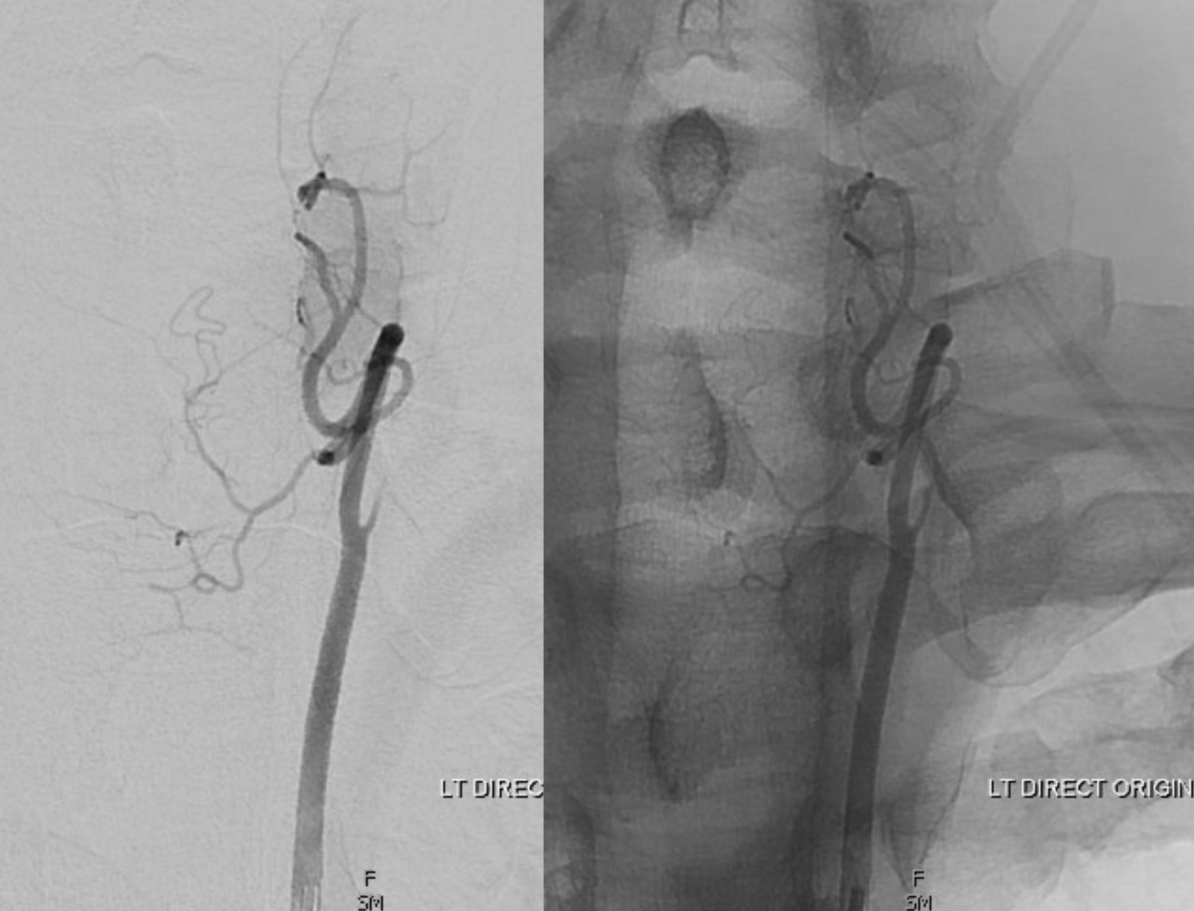
Comparison has some interesting points
And, finally, back to anatomy. The same common origin inferior thyroid and vert on the right — the homology between vertebral and deep cervical arteries
