Posterior Communicating Artery (under very slow constuction)
Embryology: The posterior communicating artery, embryologically, belongs to the anterior circulation, representing the “caudal ramus” of the internal carotid artery. In most “higher” species, the anterior circulation (ICA) is responsible for supply of the entire supratentorial brain, as well as the bulk of brainstem structures which, in the human, are the property of the vertebrobasilar (posterior) circulation. This is a very recent change in cerebral vascular arrangement, and as such may just as well be regarded as a deviation, rather than forward “evolution”. Regardless, the origin of PCOM as an ICA vessel is very much alive in the ubiquitous “fetal PCOM”, estimated at 25% prevalence in the human. For a detailed discussion of PCOM and other embryology/phylogeny, please see Neurovascular Evolution and Vascular Neuroembyology sections.
PCOM Infundibulum
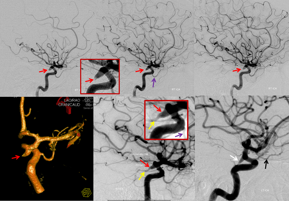
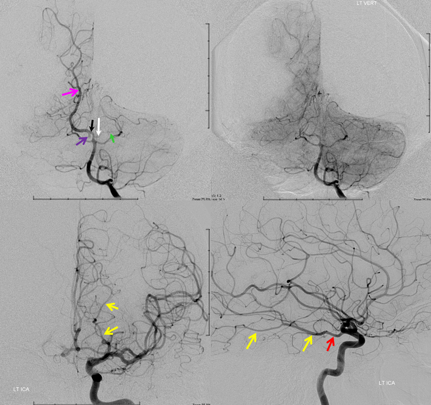
Fetal PCOM
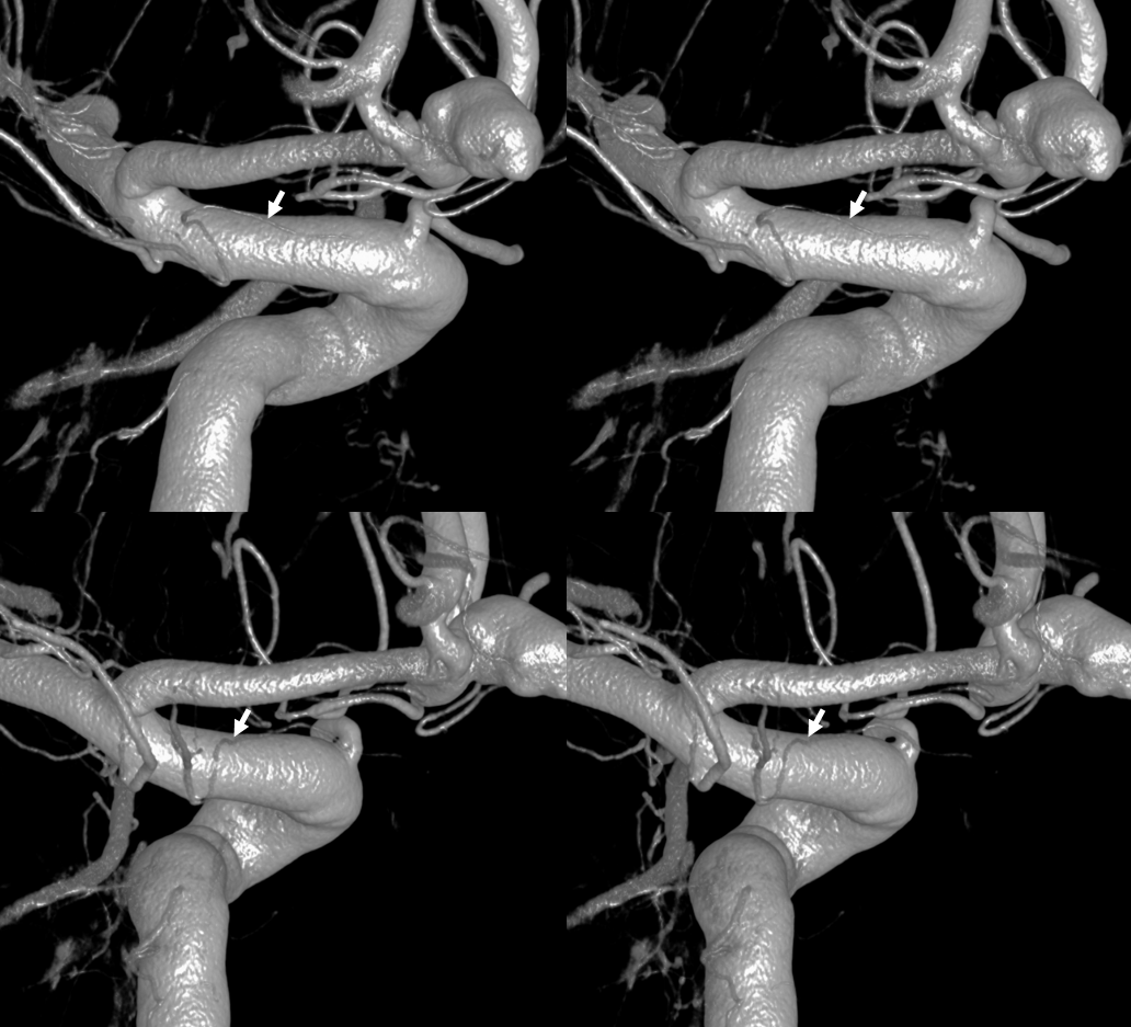
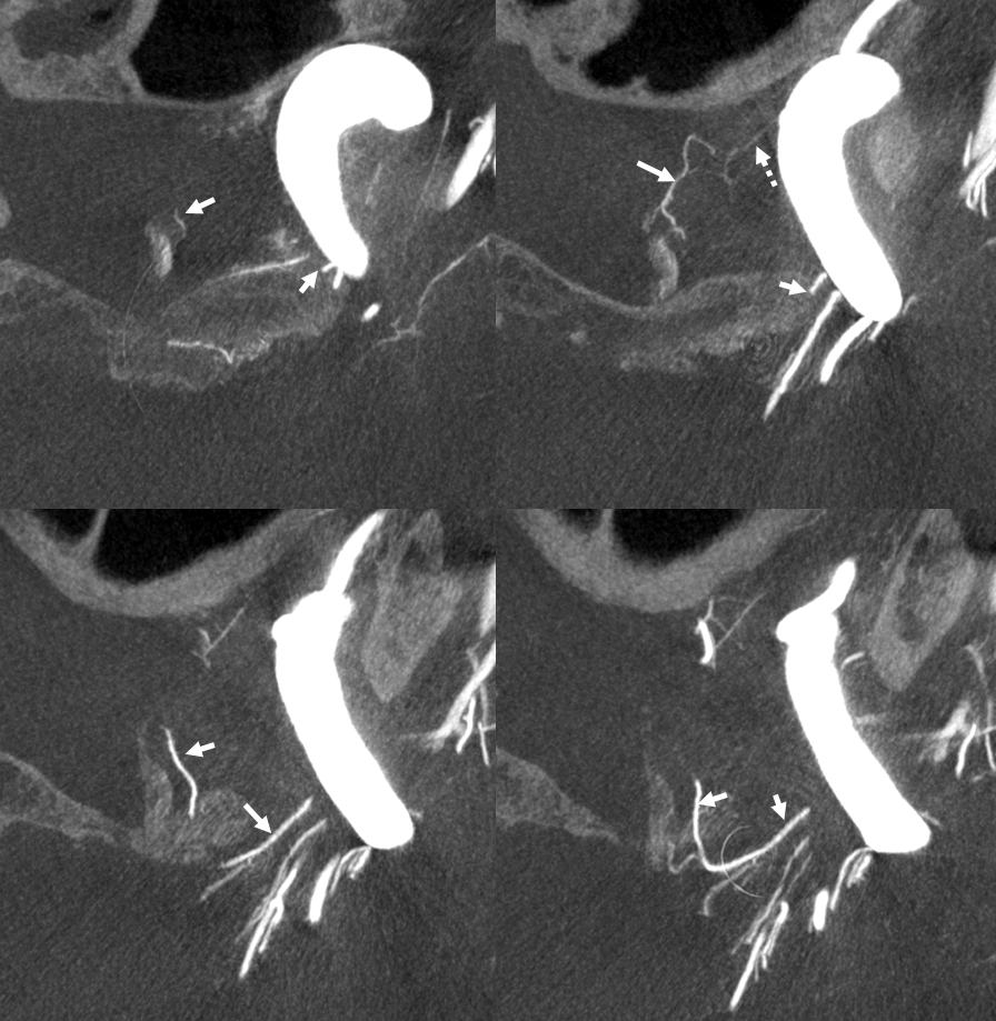
PCOM / Superior Hypophyseal Artery Balance
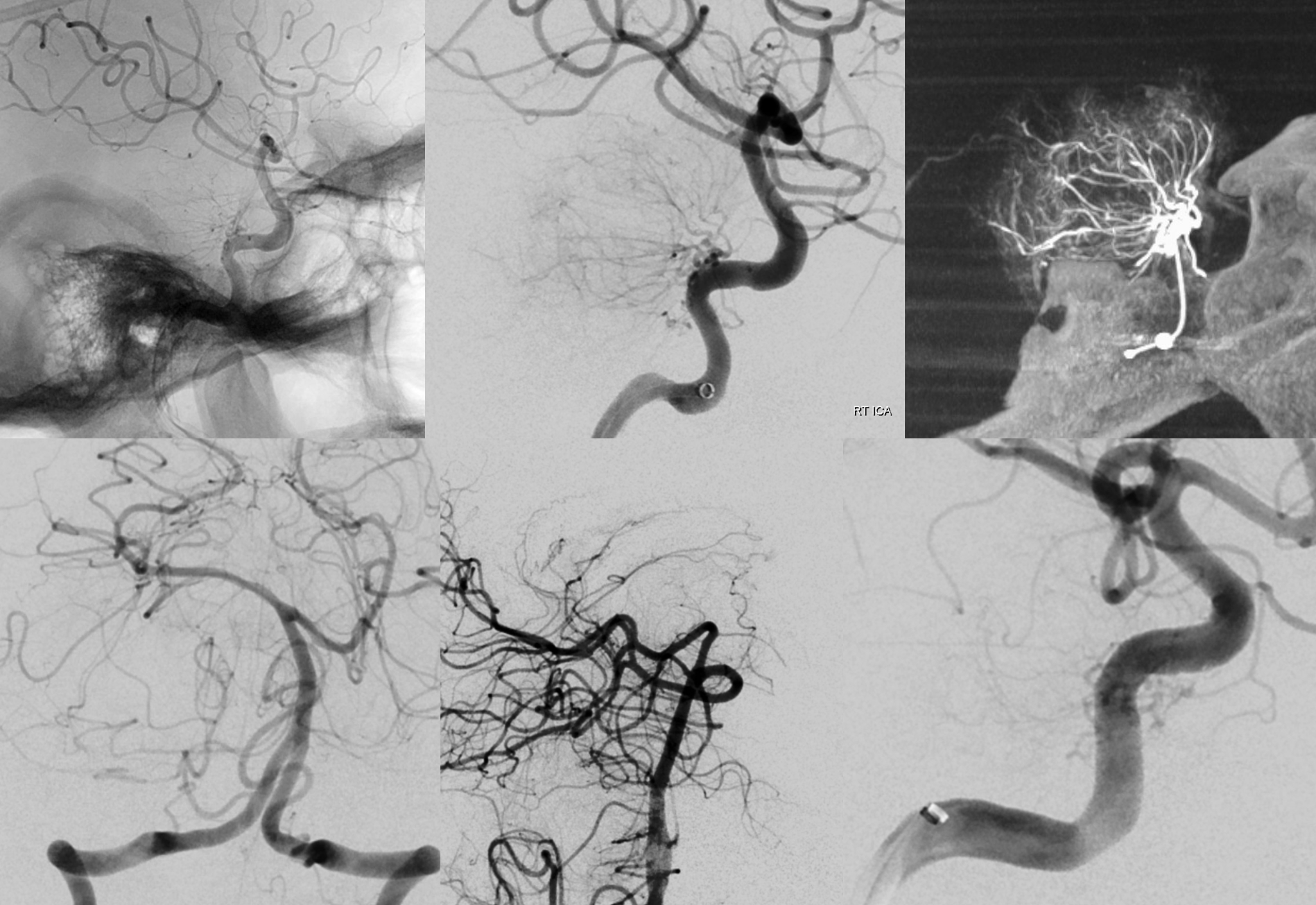
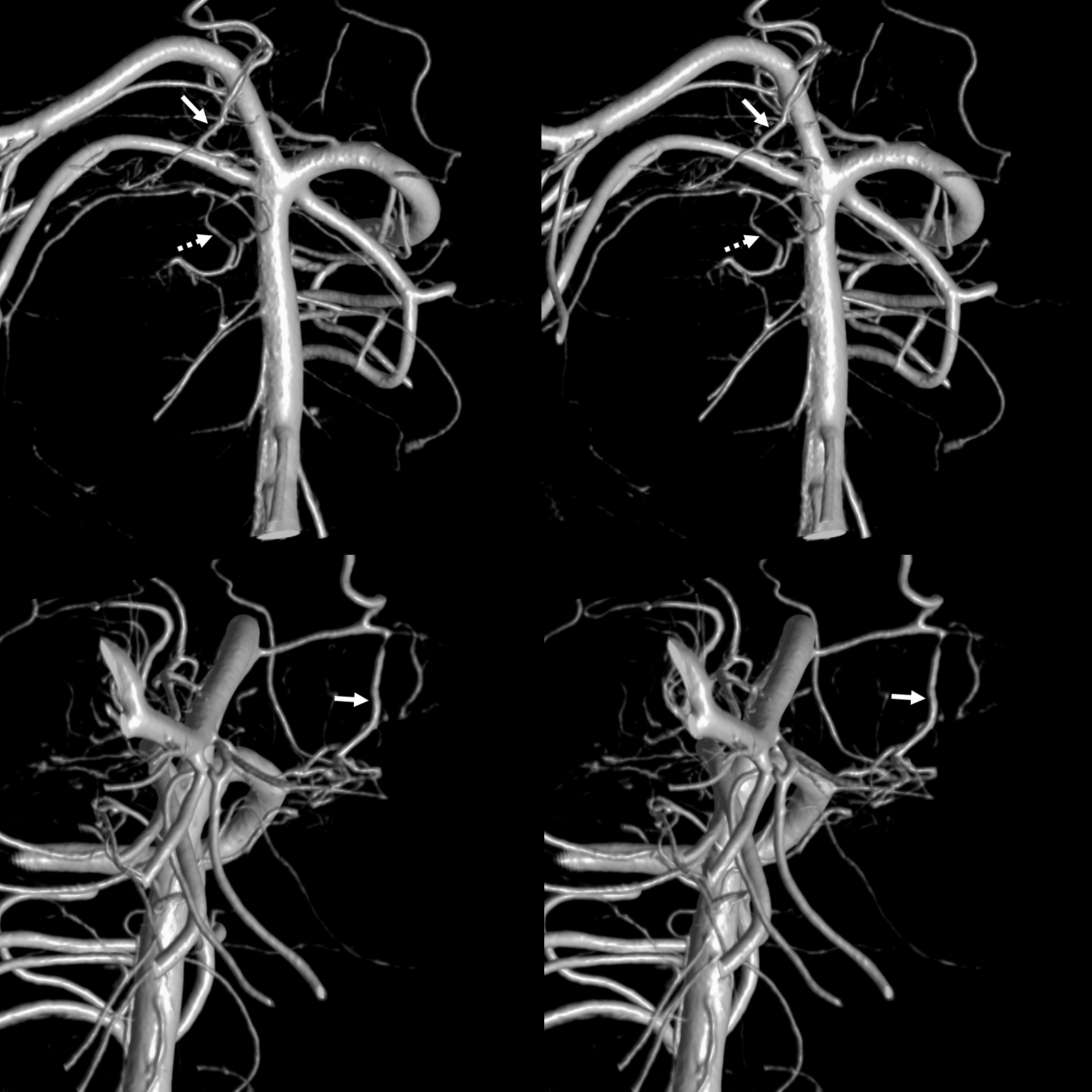
There is a balance between these two that is rarely discussed. Here, the superior hypophyseal artery is hypoplastic — very tiny actually. The supply of its territory is from a branch of the PCOM (arrows)
STEREO PAIRS
AXIAL DYNA IMAGES — the hypoplastic superior hypophyseal is dashed arrow. The PCOM collateral is arrrows.
Note pituitary stalk
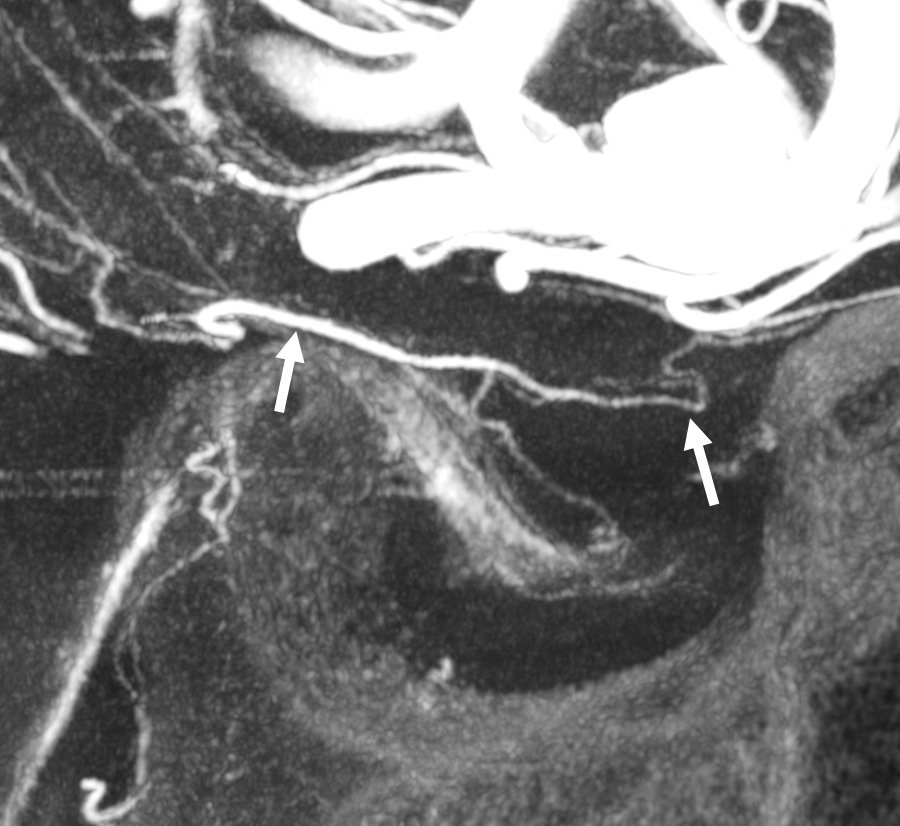
PCOM Pituitary supply — a lot
Aneurysm — perhaps a superior hypophyseal one. Note relatively distal origin of the ophthalmic. The PCOM supplies large portion of the pituitary (arrows). The classic superior hypophyseal branch is very small
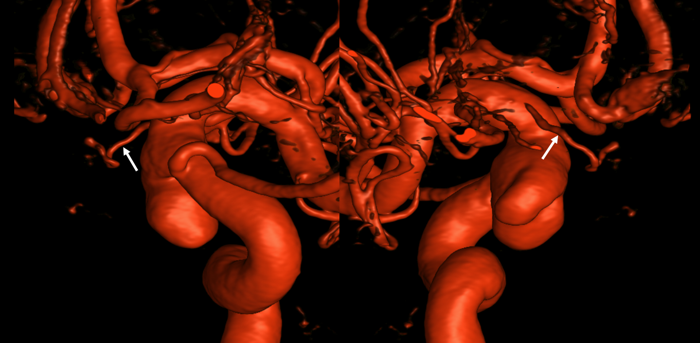
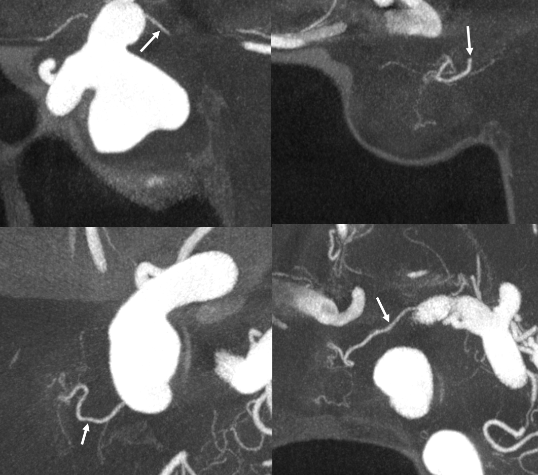
Small superior hypophyseal (dashed arrows) and large PCOM origin superior hypophyseal to the pituitary

PCOM Origin Piodural Anastomosis (PCOM Dural Branch)
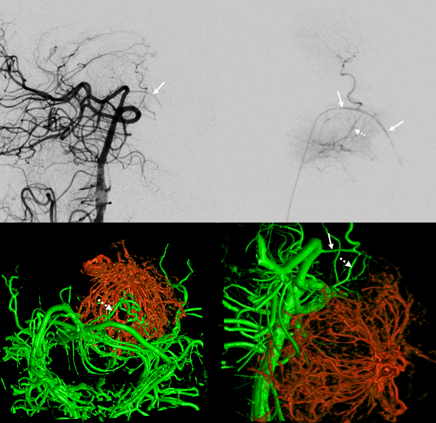
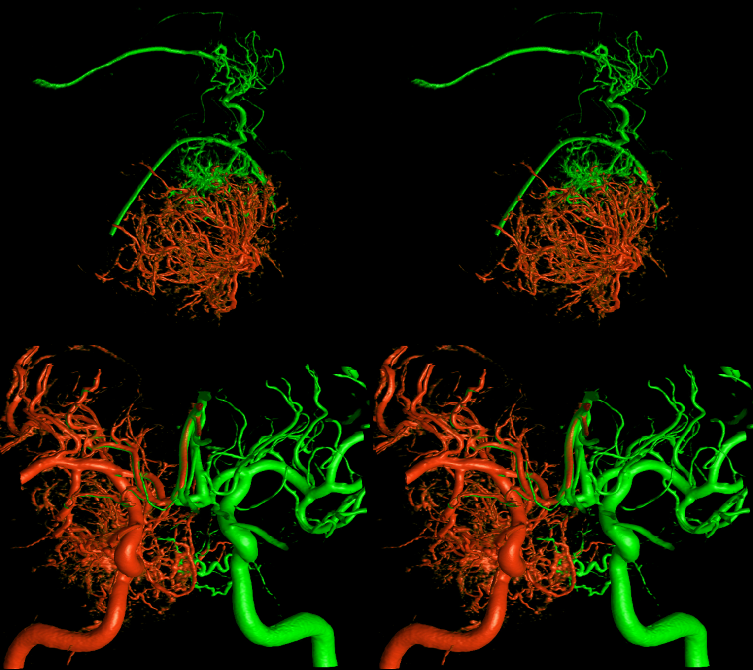
Never say never. Favored places for piodural connections include the PCA (Davidoff-Schechter), the SCA (Wollschleger and Wollschleger), the AICA (subarcuate artery), the PICA (as origin of the posterior meningeal artery — one can argue about all of that). PCOM origin — have only seen one. Patient with a very bad meningioma
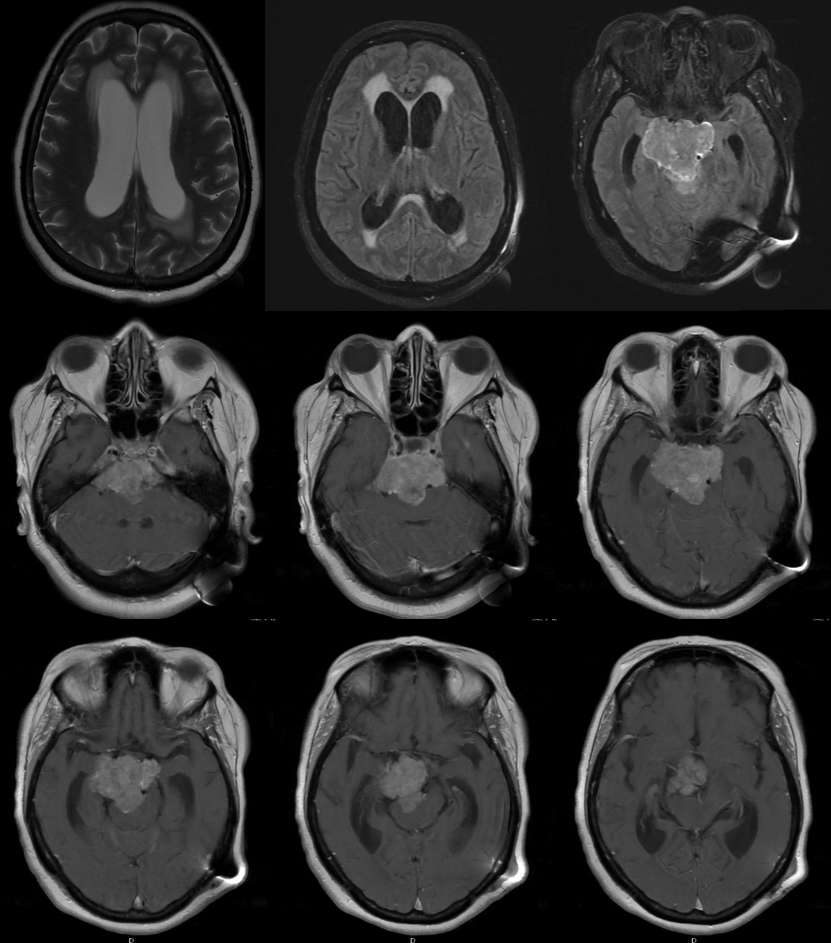
The various supplies and mass effects are shown:
Stereo DYNA of the vertebrobasilar injection — the PCOM origin dural branch supplying the top of tumor is marked by arrows. Another piodural supply from a large basilar perforator is dashed arrows.
Different arrows below — PCOM is solid arrows, its dural branch is dashed arrows. Bottom row are fusion images of the right MHT injection (red) and vertebral (green). Left lower view is from top, right lower from side
Stereo images — top are selective PCOM injection (from posterior circulation) and bottom are ICA fusions.
Full case is here
