Spinal vessels are very small. Bellies can be very big. This is a good test of your angiographic equipment. Many older machines are not able to image in beyond a certain Body Mass Index at all. Getting good images is of course essential to find the fistula and avoid problems — spinal arteries.
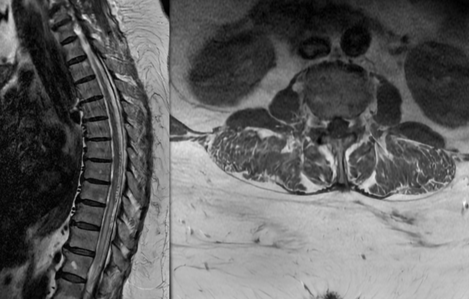
This one presents with several days of near-paralysis

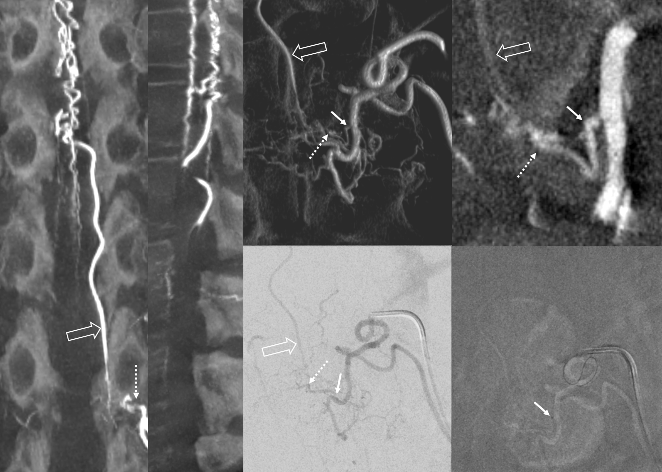
Visualization is excellent, especially for DYNA CT / CONE BEAM CT — no binning is essential. No veins are seen — they are congested by the fistula. If one is searching for a fistula, lack of venous visualization after injection of Adamkiewicz is a sure sign of congestion.

Amazing images of left L1 fistula. The venous system is incredibly congested — with no radicular or bridging vein outflow until c-spine. Lack of outflow is as important in pathology as the fistula itself.

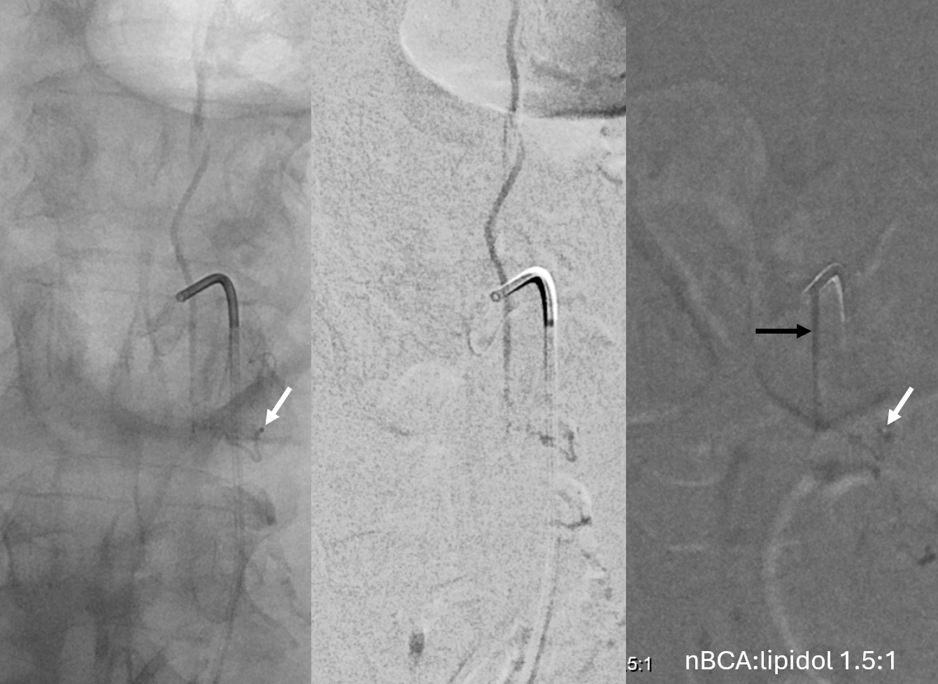
Visualization is key in understanding microangioarchitecture and succeeding in treatment. Cone Beam CT / DYNA of the fistula is good quality. The fistula (dashed arrows) is supplied by pedicle (arrows) off the segmental artery. Radicular vein is open arrow. Lower right image shows catheter tip at the origin of the “feeding pedicle”

Micro injection and nBCA cast

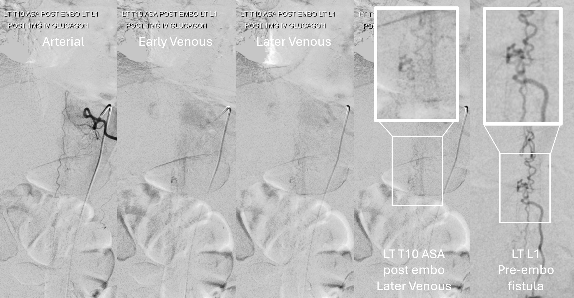
Post-embo injection of Adamkiewicz now opacifies the same spinal veins as were previously parasitized by the fistula (boxes)

It is important to understand that the venous system “returned” to the cord post embo is profoundly abnormal. Especially in patients like this one, with very few outflow options left, venous congestion persists, and may be in part responsible for incomplete recovery. Nothing can be done except to place patients on anticoagulation to prevent thrombosis of the remaining system.
