Case in conjunction with Dr. Eytan Raz
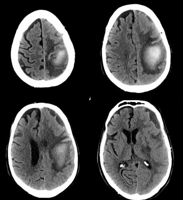
Another case illustrating how high grade dural fistulas adjacent to but not directly involving a venous sinus can do a lot of damage. Clinical presentation is with right weakness.
Pattern looks like venous congestion infarct. There is also a subdural.
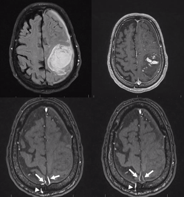
MRI with scattered enhancement and enlarged arteries in wall of the superior sagittal sinus (white arrows). Also well seen is a transosseous branch of the superficial temporal artery (arrowhead). See meningeal vessels page for more info on arteries in walls of sinuses
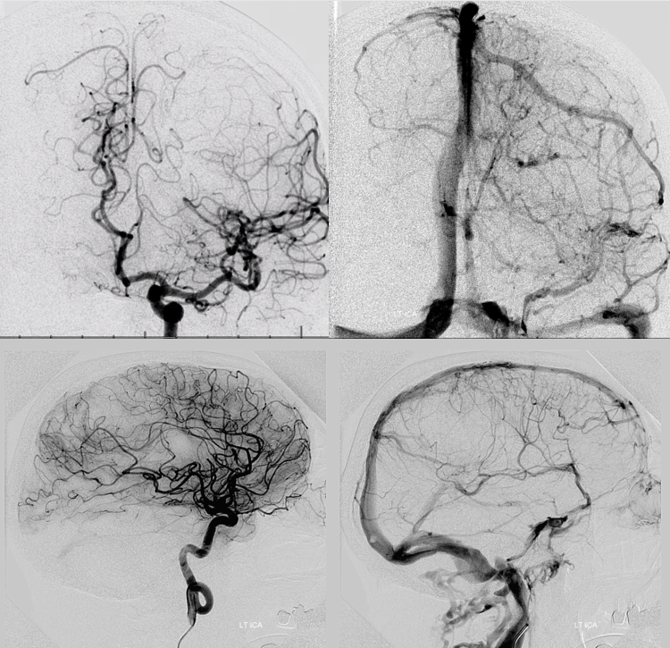
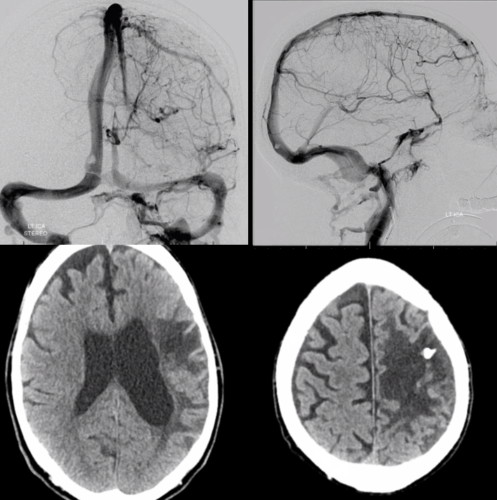
Left ICA — extensive venous congestion of the posterior frontal/parietal convexity. Drainage via medullary veins into the internal cerebral venous system
Cross-eye stereo views of bilateral MMA injection — parasagittal dural fistula drains directly into an ectatic Trolard-family vein
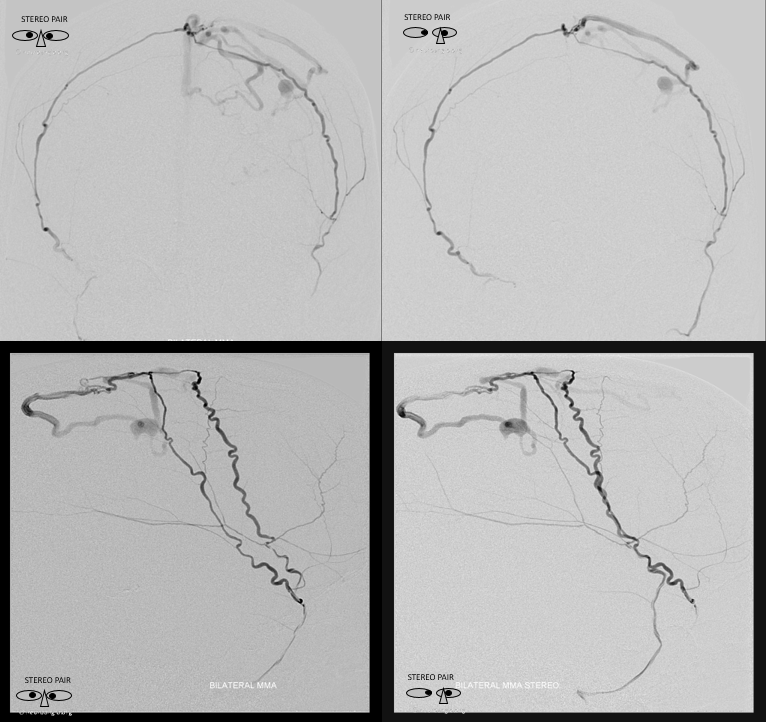
Anaglyph Stereos
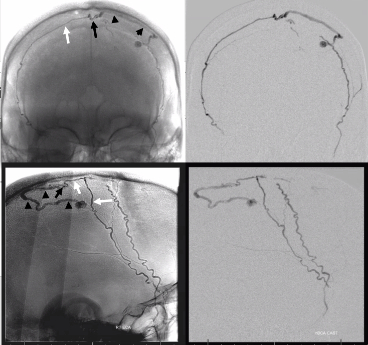
Glue cast. Bilateral simultaneous MMA nBCA injections from as distal positions as was possible. Far from fistula but both were “wedge positions”, right side reached fistula. MMA– white arrow, superior sagittal sinus artery – white arrowhead, fistula site – black arrow, draining Trolard-family vein black arrowheads
Post embo left ICA showing somewhat improved congestion. Bottom row – delayed CT with glue in venous aneurysm
See “Dural Venous Channel” Page for more info
