Another example of what happens when skull breaks. See companion MMA traumatic fistula case. In this one, man found down with extensive intracranial subarachnoid and subdural hemorrhages.
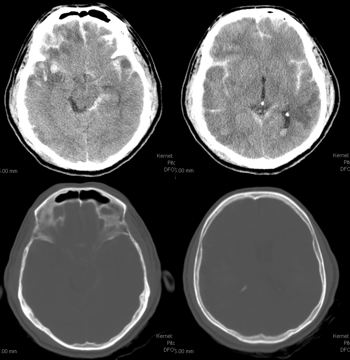
Lateral projection of single plane angio shows an arteriovenous shunt (red arrow). Also see parietal skull fracture. Pooling of blood is present on the late arterial and venous phase images over the pterion and overlying the proximal basal vein (not lableled)
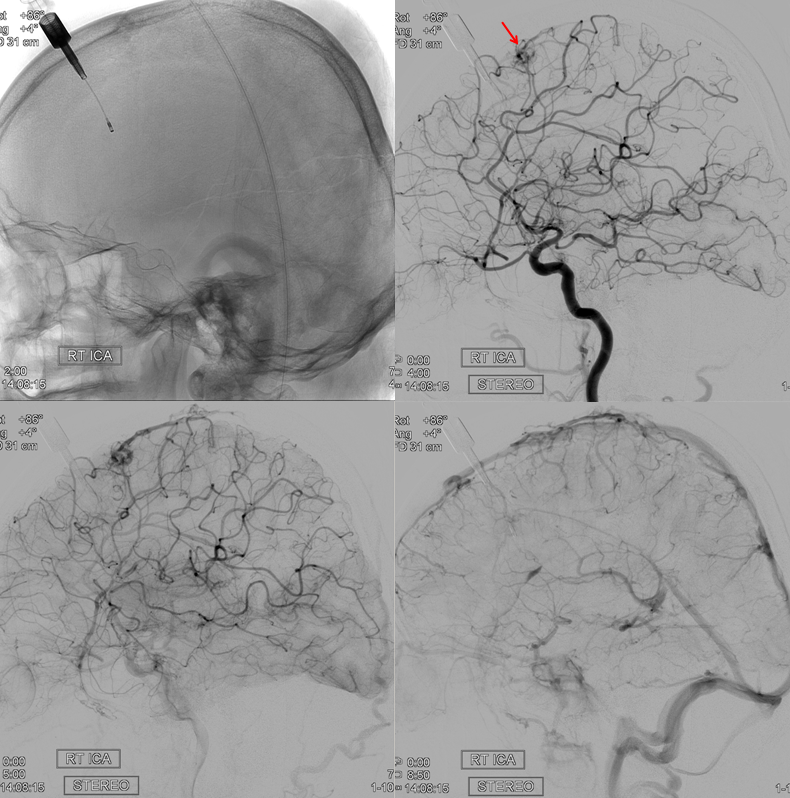
Did a brain AVM rupture? Not really. The shunt marked by the red arrow is a traumatic fistula. Notice a large vessel arising from the ophthalmic artery, heading superiorly (white arrow). That’s the recurrent meningeal artery — see ophthalmic artery and MMA pages for more info. Recurrent MMA is a variant where MMA arises from the ophthalmic. The opposite is the meningo-ophthalmic variant, where the ophthalmic arises from the MMA — that’s the more important one, for MMA embo work, perhaps the best known “dangerous anastomosis” out there. The pools of contrast alluded to above are now shown by yellow arrows.
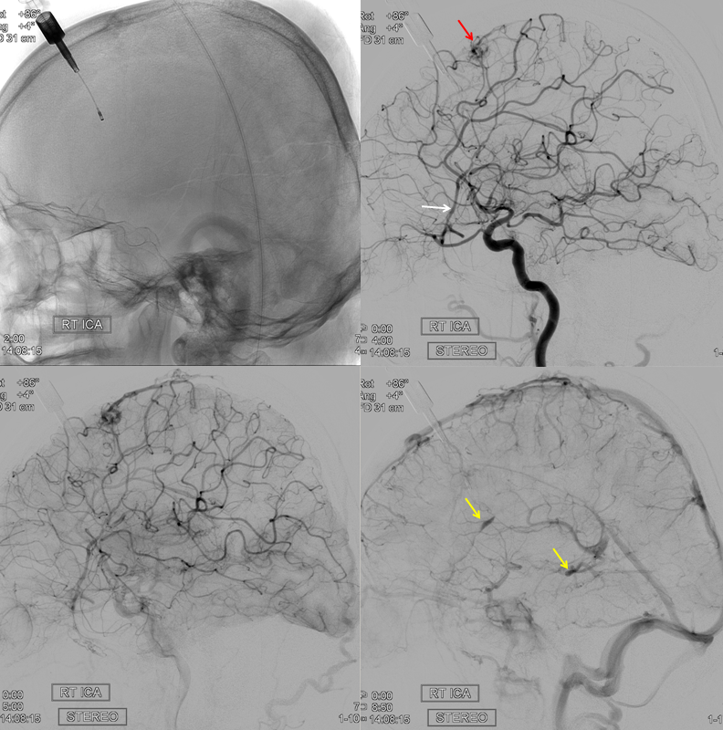
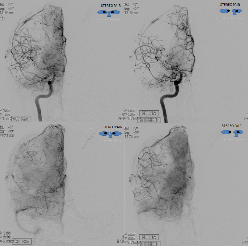
stereo pair
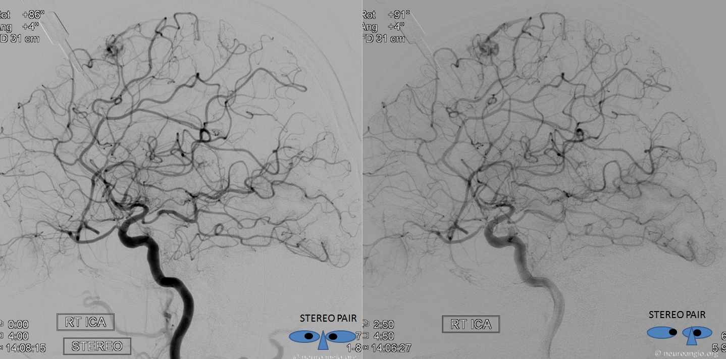
Frontal views. Notice tremendous trauma-related MCA vasospasm — something we do not appreciate enough of as a field. Could some delayed infarctions in trauma patients be due to this kind of spasm?
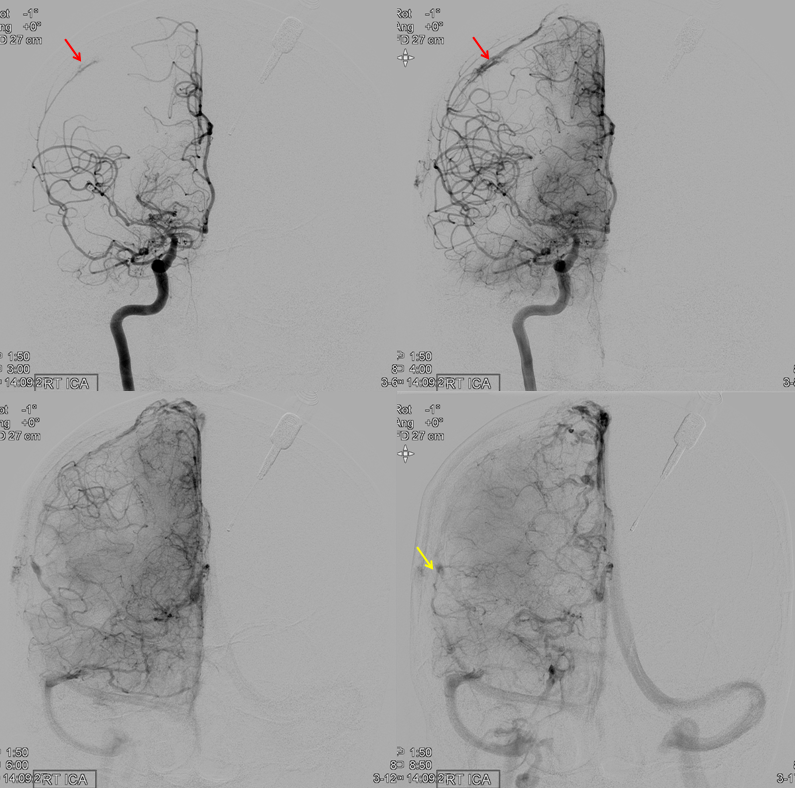
Stereos
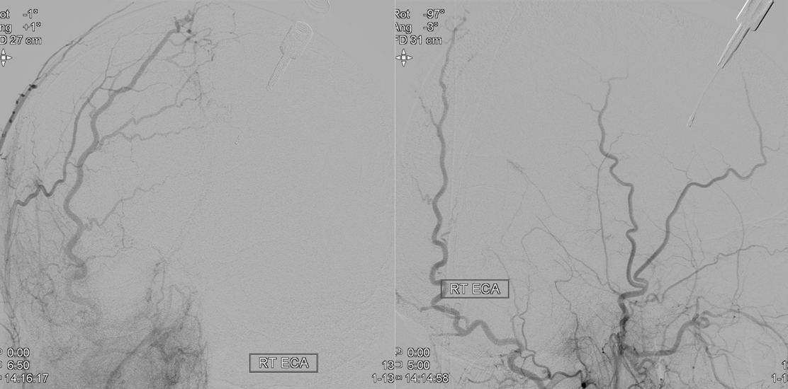
ECA injections demonstrating absence of MMA
Conservative management of fistulas was pursued. The patient did very badly due to extensive trauma
