The Anterior Pontomesencephalic Vein, as well as Anterior Medullary vein (M).
The anterior pontomesencephalic vein (E), made up of the anterior mesencephalic and anterior pontine veins, makes up a portion of the longitudinal venous system running on the anterior surface of the brainstem, extending from the mesencephalon to the cervical spinal cord. For the purposes of this discussion, we can call this group the “anterior longitudinal group”
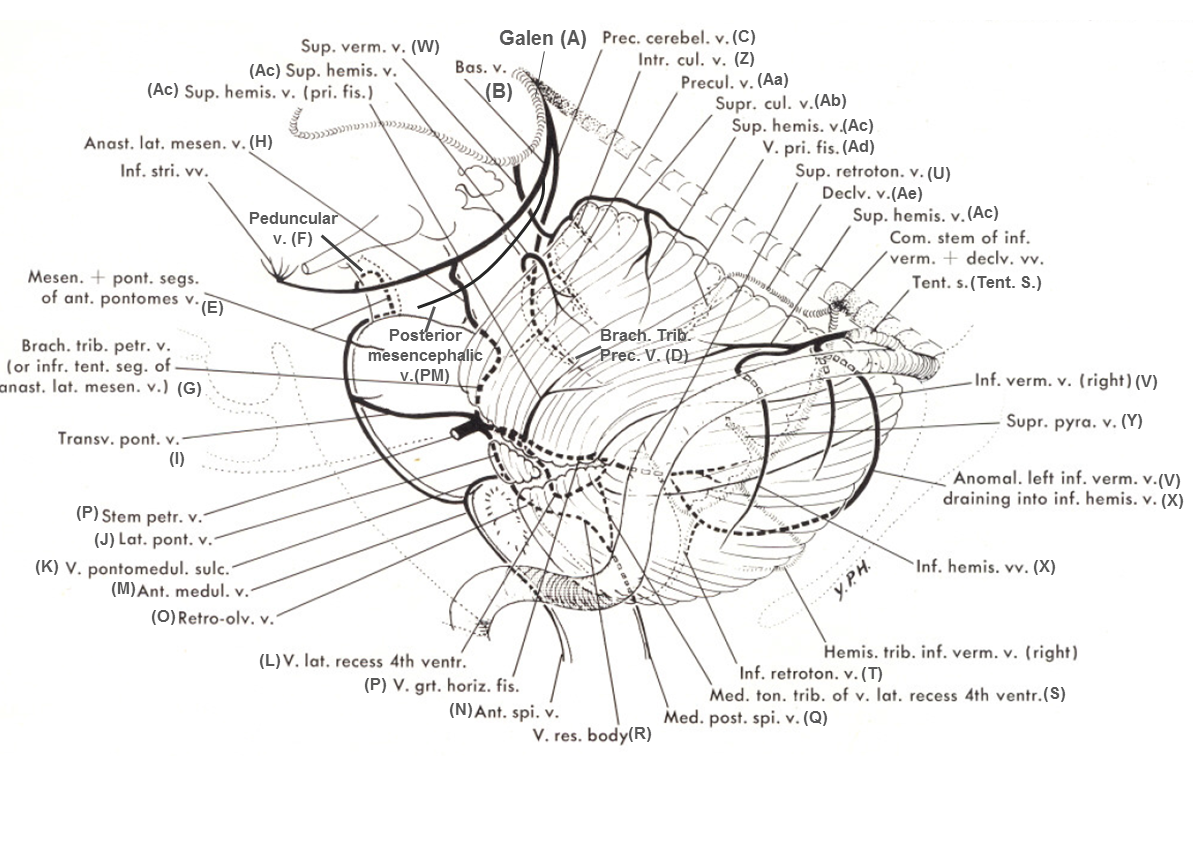
Rhoton calls the anterior pontomesencephalic vein the “Medial anterior pontomesencephalic vein”. The anterior medullary vein is named “medial anterior medullary vein”. Rhoton’s terminology is important in recognising that the anterior group is sometimes made up of “Lateral anterior pontomesencephalic vein” and “lateral anterior medullary vein” — reflecting the fact that these veins often don’t run exactly in midline, and may even be paired on the anterior surface. Again, the key concept of these pages is to show that knowledge of where the vein is in relation to the brainstem is more important than terminology.
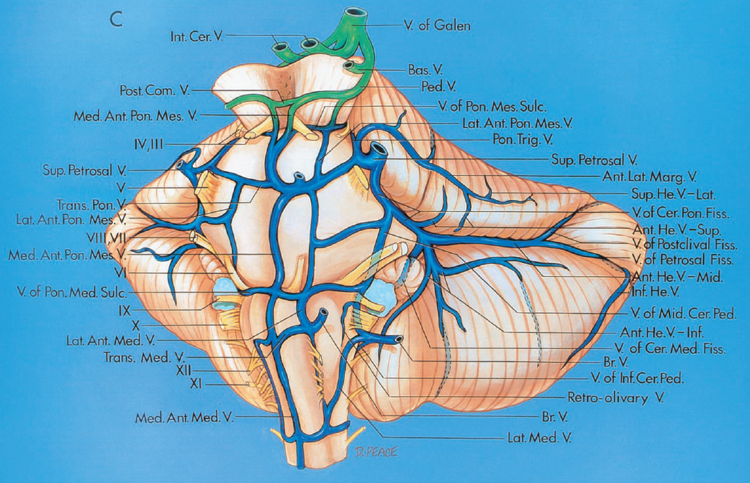
The following table serves as a correlation between the nomenclatures of Huang and Rhoton, to the best of my ability to read both of those great minds…
Rostrally, the pontomesencephalic vein usually collateralises with the peduncular veins (F), which are connected by the interpeduncular vein, also known as the posterior communicating vein. Caudally, the pontomesencephalic vein may be contiguous with the anterior medullary vein (M), which is contiguous with the anterior spinal vein (N). This is the easiest system to understand — it marks the anterior border of the brainstem in the lateral view, and as such is usually located adjacent to the basilar artery. It is a truer landmark for the brainstem than the often tortuous basilar, as seen in the following example where the whole system is particularly well-developed (the anterior medullary vein is mislabeled “E” and should be “M”).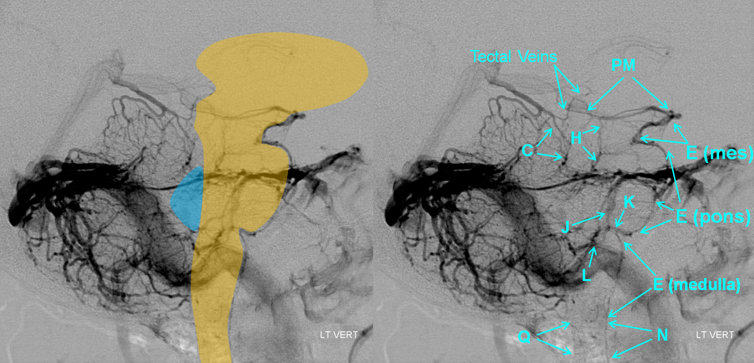
Notice how low the pons sits in repation to the jugular bulb. The 4th ventricle is located essentially behind the pons and below the precentral vein (a.k.a. the cerebellomesencephalic fissure vein in Rhoton).
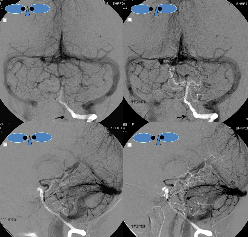
Another instance of very dominant anterior pontomesencephalic vein, draining via peduncular veins (F) into the basilar vein (B). This is particulary well seen on the Townes frontal view, where it is critical to understand the relationship of the brainstem with respect to the venous structures.
The same case, with brainstem overlay; again, the most important thing is to understand the relationship between the veins and the brain. The peduncular vein (F) outlines the cerebral peduncle, connecting to the Basal Vein (B).
An amazing stereo view of the same case:
Theory: Balance of Longitudinal and Transverse Veins
The various components of the anterior longitudinal group are typically in balance with other longitudinal brainstem veins. These are the longitudinal veins at various levels:
Mesencephalon: Anterior Mesencephalic (E), Anterior Lateral Mesencephalic (ALM), Lateral Mesencephalic (H), Precentral (C)
Pons: Anterior Pontine (E) or Anterior Lateral Pontine,
The bottom line is that at a given level, one may see a relatively “balanced” pattern or some form of dominance of one or more longitudinal systems, with corresponding hypoplasia of the other longitudinal channels at the same level. For example, dominant lateral mesencaphalic vein usually implies hypoplasia of the anterior pontomesencephalic and precentral veins. This is no different from the situation in the supratentorial compartment, where for example dominant Labbe means hypoplastic Trolard, etc.
In the above example, the anterior group his highly dominant, and therefore the precentral vein (C) and the lateral mesencephalic vein (H) are hypoplastic. In the example below, the lateral mesencephalic (H) group is dominant, and the anterior group is correspondingly hypoplastic.
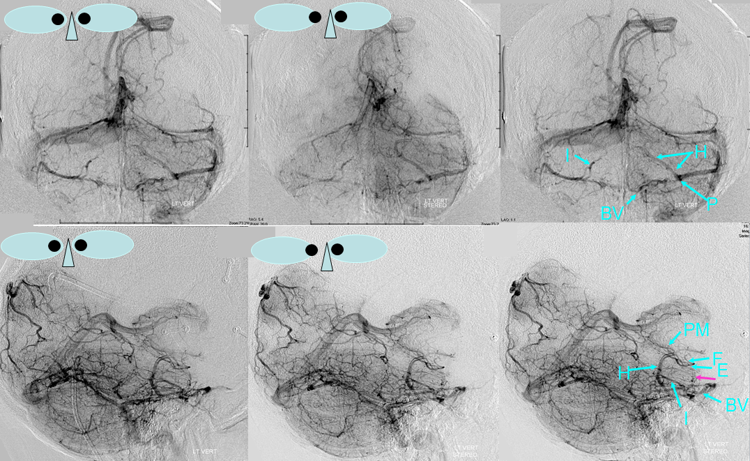
Same case in better detail:
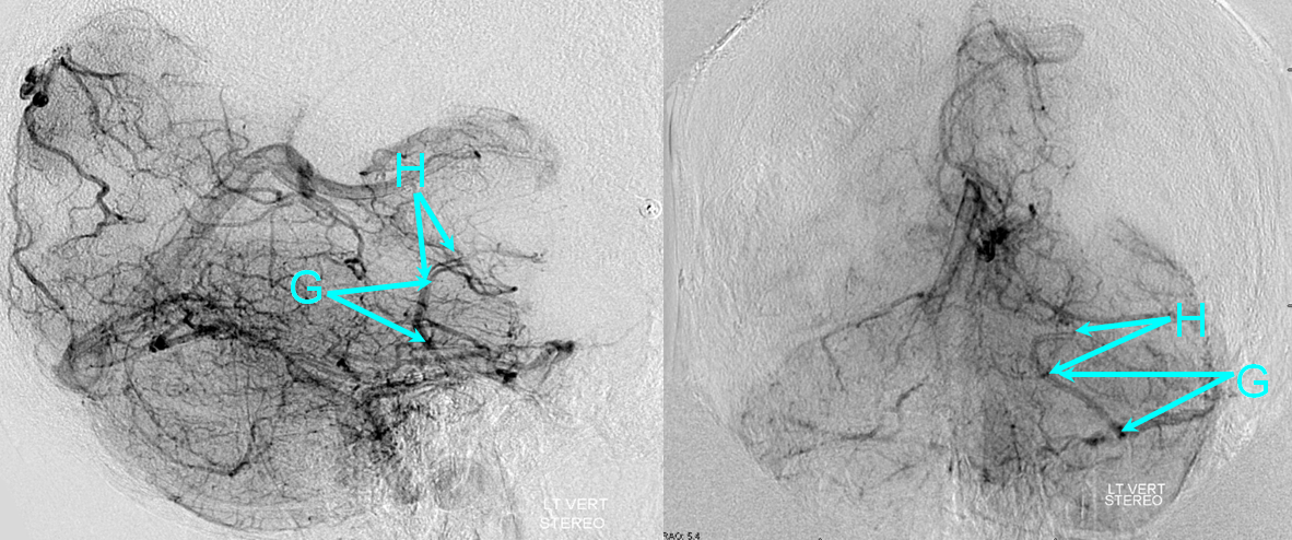
Finally, in this patient the precentral vein (C) and its brachial tributaries (D), a.k.a. veins of the superior cerebellar peduncle according to Rhoton, are dominant, and therefore both the anterior pontomesencephalic (E) and lateral mesencephalic (H) veins are hypoplastic.
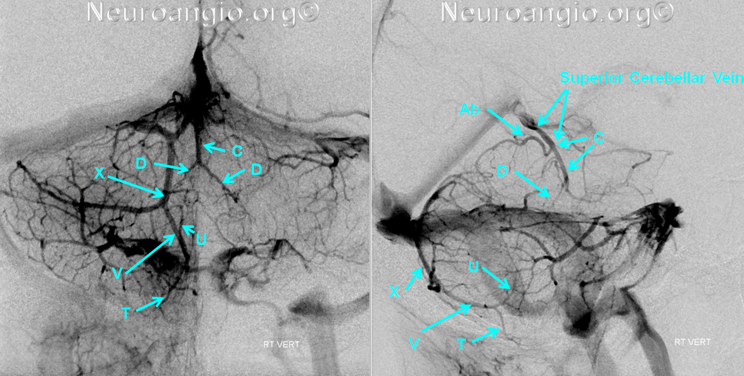
You get the idea. The same is true for the transverse group of veins, such as the pontomesencephalic sulcus, transverese ponine, and pontomedullary sulcus veins.
The anterior pontomesencephalic vein usually runs adjacent to the basilar artery, as seen in this image where the arterial phase is selected as a “mask” image, this serving as a while landmark on a gray background. It is important to understand the position of the anterior group in the frontal view. The optimal frontal view for the brainstem veins is the Townes view, which looks “down the barrel” of the brainstem somewhat. Even though the brainstem is thus foreshortened, the various cerebellar and brainstem veins overlap less and the brainstem group of veins sits lower than the cerebellar veins.
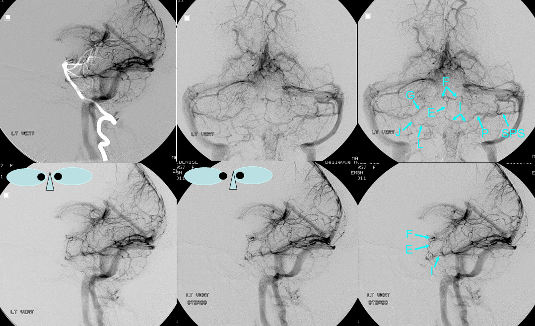
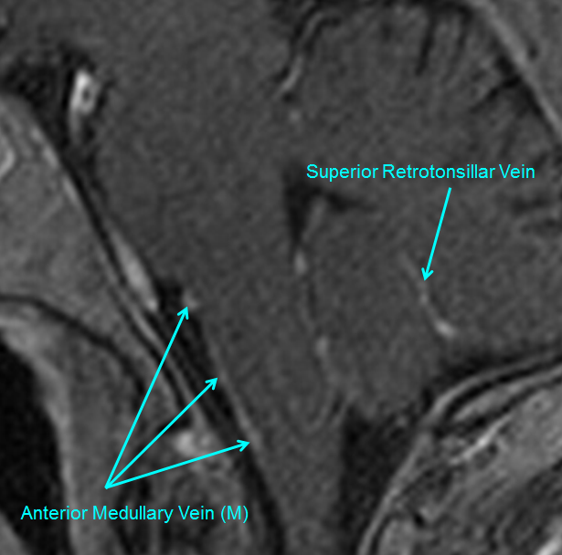
MRI is a very nice way to see these veins. The anterior pontomesencephalic and anterior medullary veins will be closely applied to the surface of the brainstem, and are a better landmark than the basilar.
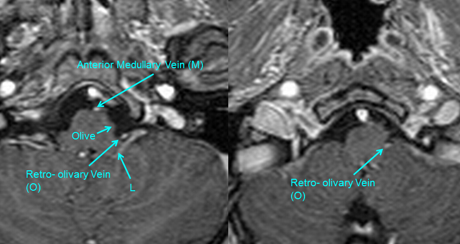
In this axial view, the anterior medullary vein is well-seen
Axial view of anterior medullary vein (M) as well as the retro-olivary vein (O)
Drainage of the anterior brainstem veins
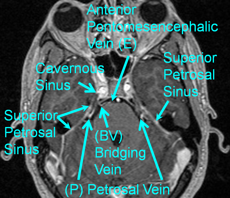
The anterior brainstem group (anterior pontomesencephalic, anterior pontomedullary, anterior medullary) of veins empty into the Petrosal vein (P), the Bridging Veins (BV) which directly connect the anterior veins to the cavernous sinus or inferior petrosal sinus, into the posterior mesencephalic (PM) veins draining into the Galen (A), or rarely via the anterior spinal vein () into the cervical spinal bridging /radicular veins and epidural venous plexus (see Spinal Venous Vasculature).
Classically, veins of the posterior fossa are organized into Anterior, Superior, and Posterior groups. The anterior group is collected by the Petrosal Vein (P), Superior group drains into the Galen (A), and Posterior Group into the Torcular and Transverse sinus. In this manner, the anterior brainstem group would belong to the anterior group draining into the Petrosal Vein, as is seen in the example below, where (E) drains into Petrosal vein (P) via the transverse pontine vein (I):

This is not always the case, however. Bridging Veins (BV) are very common, and connect the anterior brainstem veins to the posterior portion of the cavernous sinus across the prepontine cistern. Here, large bridging veins in the prepontine cistern (light blue, dark blue) are in the angio and MRI of the same patient, draining the anterior group into the posterior cavernous sinuses (pink). Notice also elegantly seen transverse pontine vein (white) and other unnamed transverse pontine veins (yellow, brown).
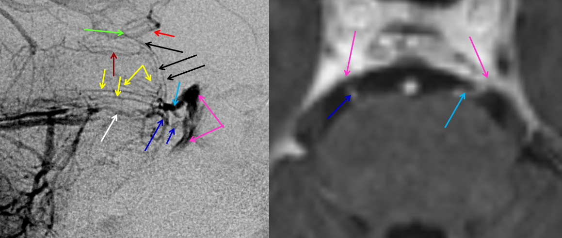
MRI demonstrating homology of the Bridging Vein (BV) draining into the Cavernous Sinus with the Petrosal vein (P) draining into the Superior Petrosal Sinus.
In this example, the anterior medullary vein (purple) drains via a radicular vein (orange) into the epidural venous plexus. Notice how the anterior pontine (blue) and medullary (purple) veins are a better landmark for the brainstem than the tortuous basilar artery: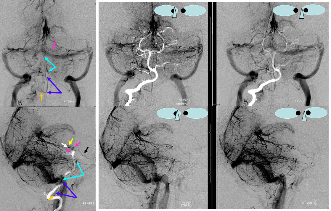
In the below, already shown example, the anterior pontomesencephalic vein drains via the peduncular vein (F) into the Basal vein (B)
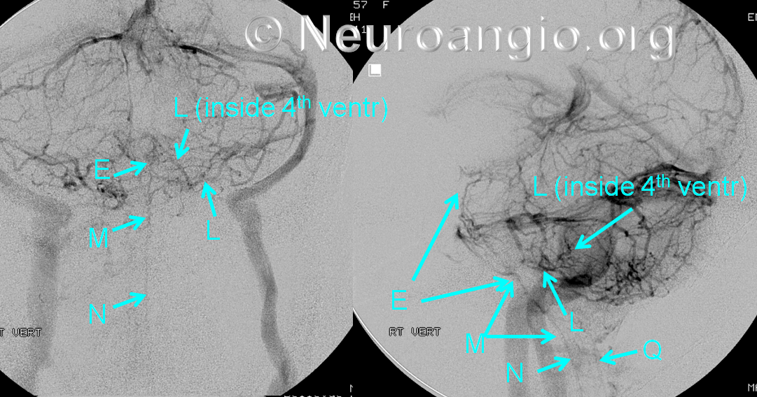
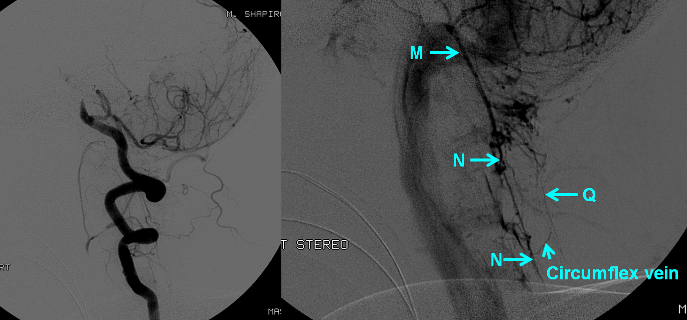
Here’s another example of a well-seen pontomesencephalic (E) and anterior medullary (M) veins. The notch between E and M marks the pontomedullary sulcus. Even the typically clandestine posterior spinal vein (Q) is a bit visible here. It is also a particularly well-developed lateral recess vein, including prominently the potion deep inside the 4th ventricle. The reason for this — the inferior vermian veins are not well-developed, such that its tonsillar territory is drained into the lateral recess vein partially (see below)
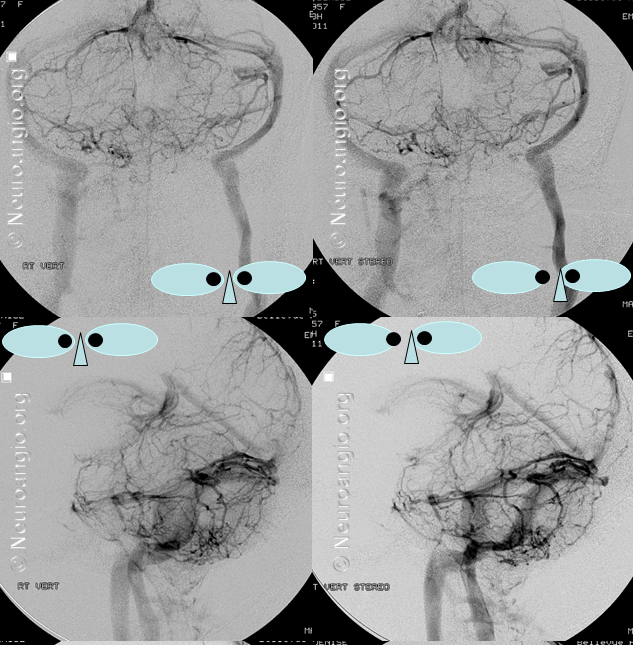
Here is a stereo of the same injection
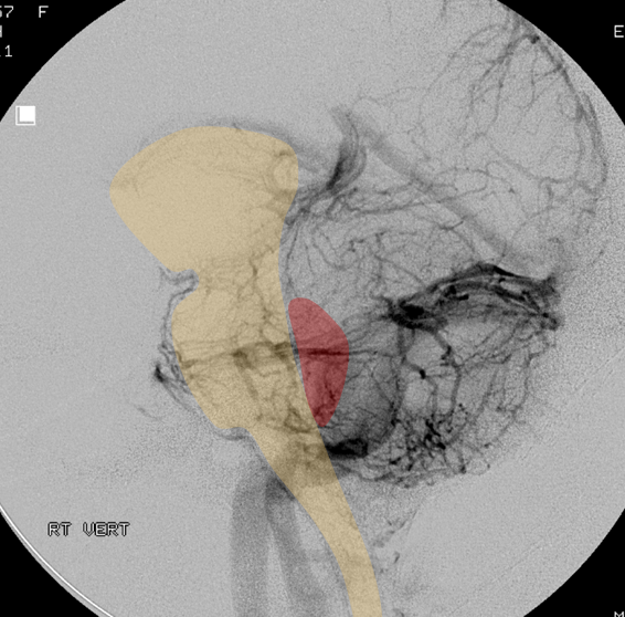
An overlay of the brainstem and the 4th for clarity
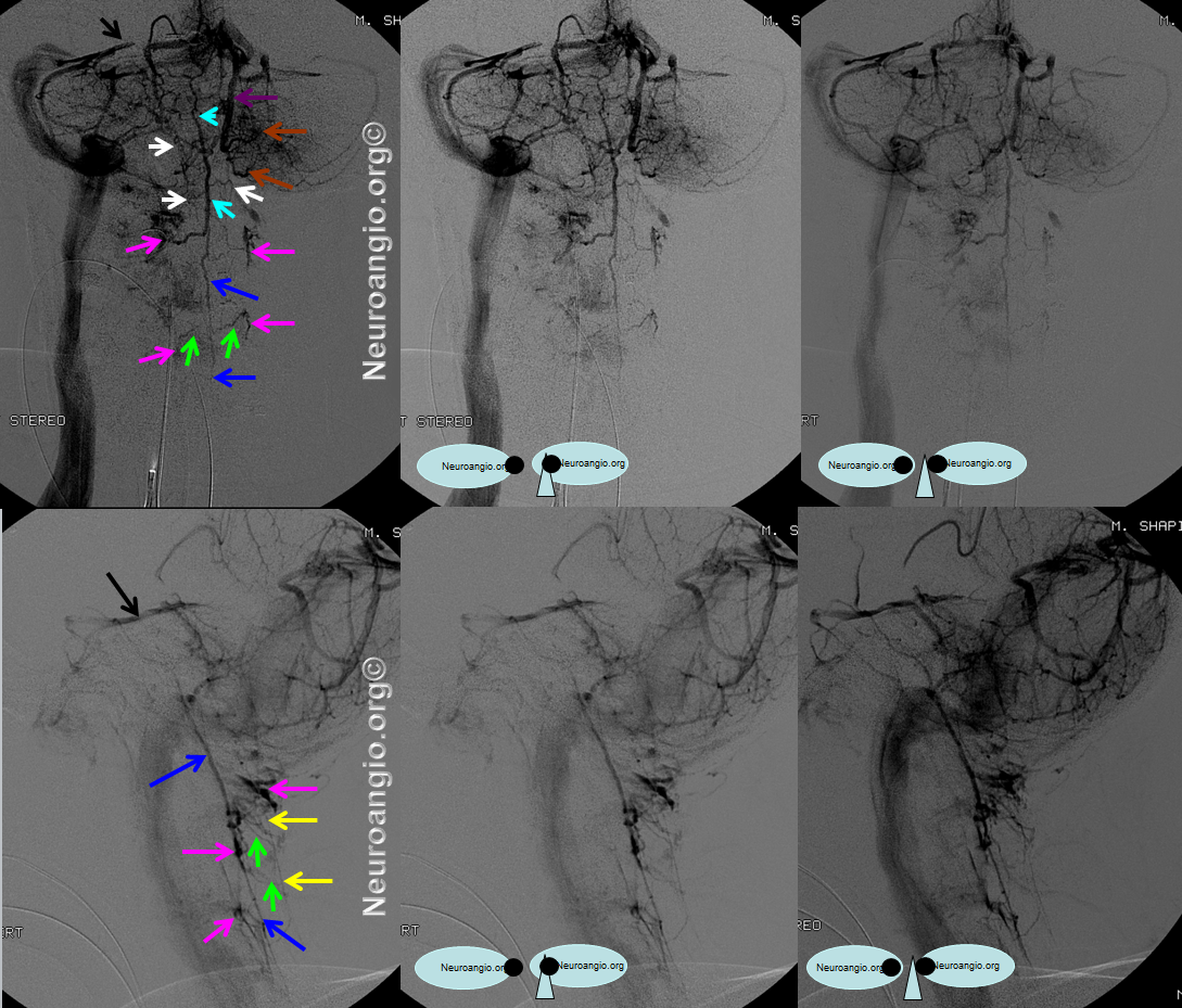
In this patient with a basilar occlusion, the outflow is prominently directed into medullary and cervical veins, which are thus seen to great advantage
The same, in stereo. The anterior medullary vein is light blue, anterior spinal dark blue, the cord tributaries of radicular veins are green, lateral spinal veins are white, and the epidural venous plexus into which the spinal veins drain is pink.
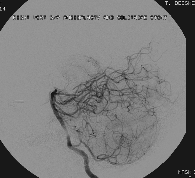
We did, for the record, reopen the basilar, leaving behind, in this case, a Solitaire FR:
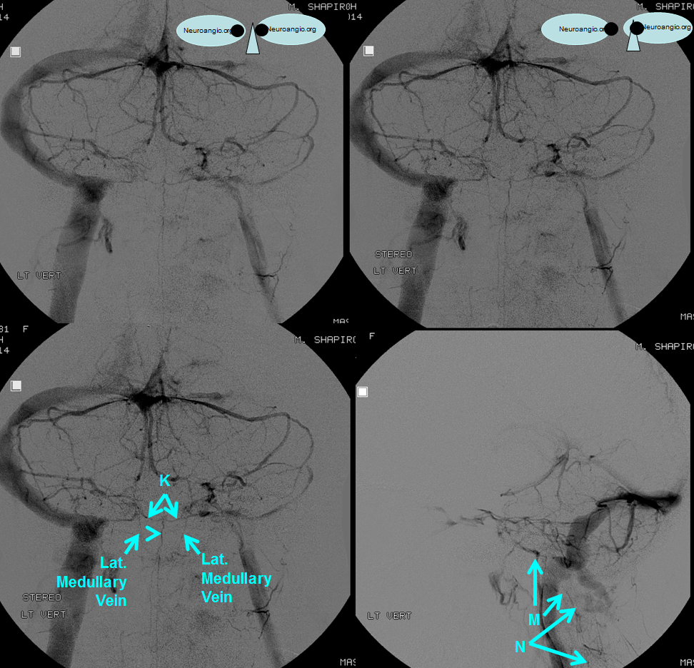
Here is an illustration of the veins of the pontomedullary sulcus (K): The stereo is from behind, to best showcase the inferior vermian veins.
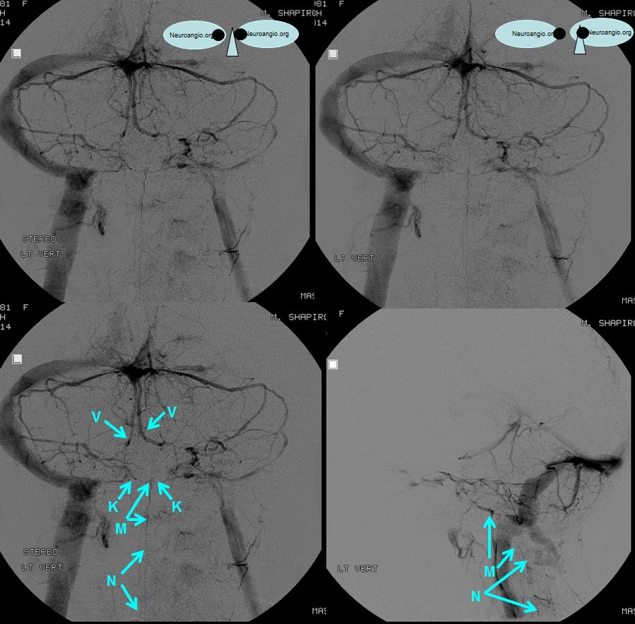
The same patient also shows well-developed lateral medullary veins (not shown on figures of Huang, but present in Rhoton and undoubtedly well-developed in many patients. The stereo is now from the front (switch right and left images)
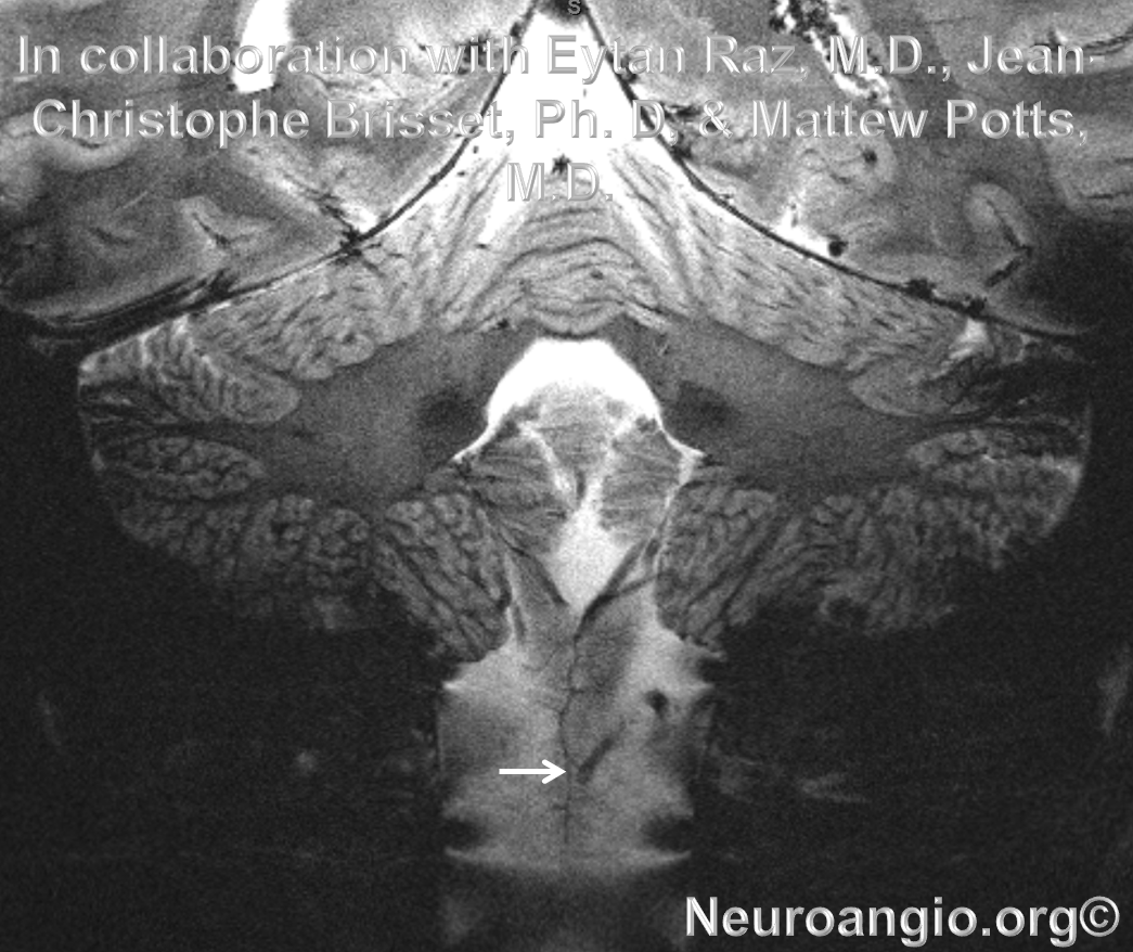
The posterior spinal vein (white arrow) is exceptionally well seen on this stunning 7T coronal plane gradient echo T2-weighted image
References:
Yung Peng Huang and Bernard Wolf. Veins of the Posterior Fossa. Chapter 75, in Newton and Potts Radiology of the Skull and Brain. Volume 2, book 3. 1974
Yung Peng Huang et. al. The Veins of The Posterior Fossa — Anterior or Petrosal Draining Group. September 1968. American Journal of Radiology. For a full text PDF click here.
Yung Peng Huang et. al. The Veins of The Posterior Fossa — Anterior or Petrosal Draining Group. September 1968. American Journal of Radiology. For a full text PDF click here.
Albert Rhoton, The Posterior Fossa Veins, Neurosurgery. 2000 Sep;47(3 Suppl):S69-92.
Albert Rhoton, The Cerebellopontine Angle and Posterior Fossa Cranial Nerves by the Retrosigmoid Approach. Neurosurgery. 2000 Sep;47(3 Suppl):S93-129.
Albert Rhoton, The Posterior Fossa Veins. Neurosurgery 2000 Sept 47(3), S69-92.
