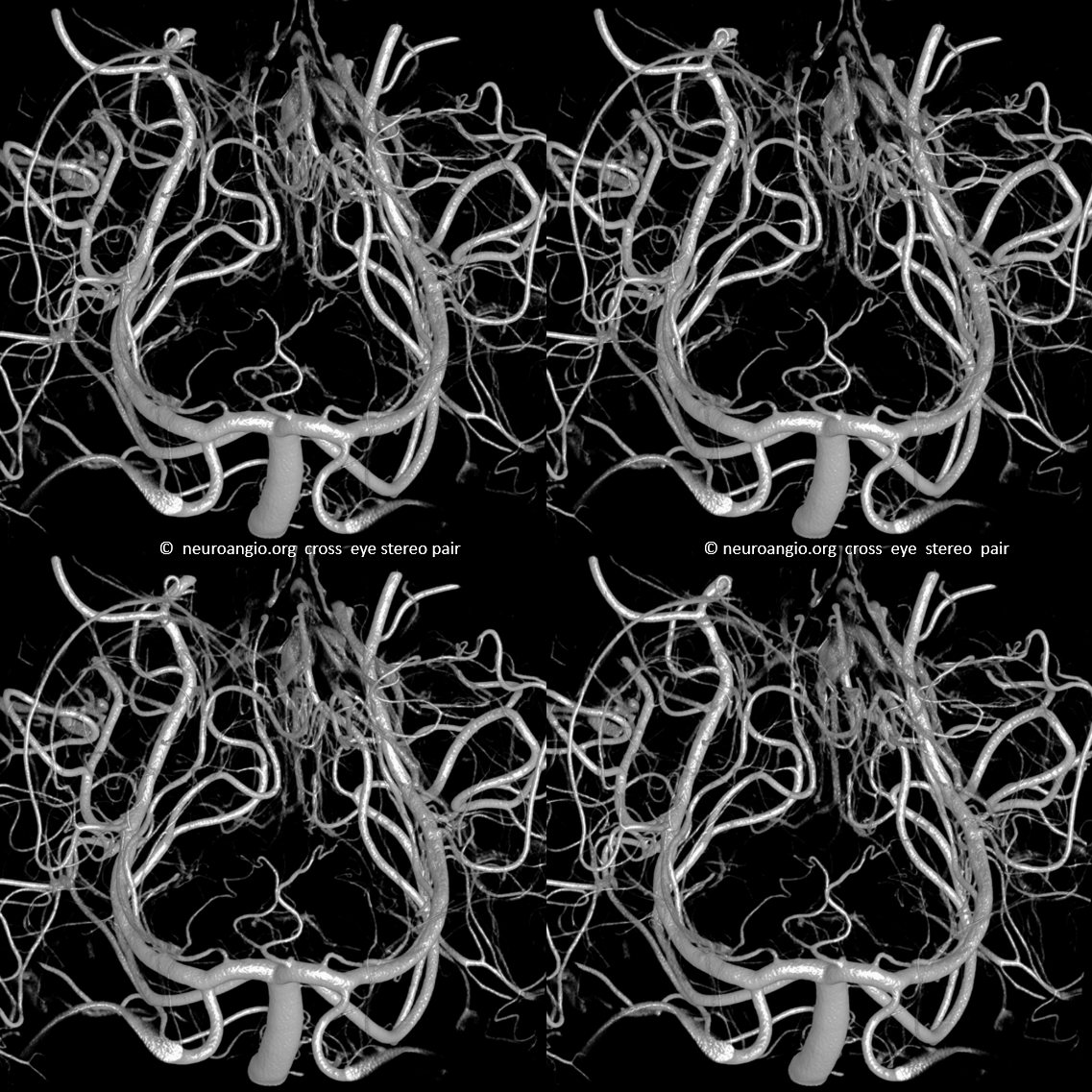
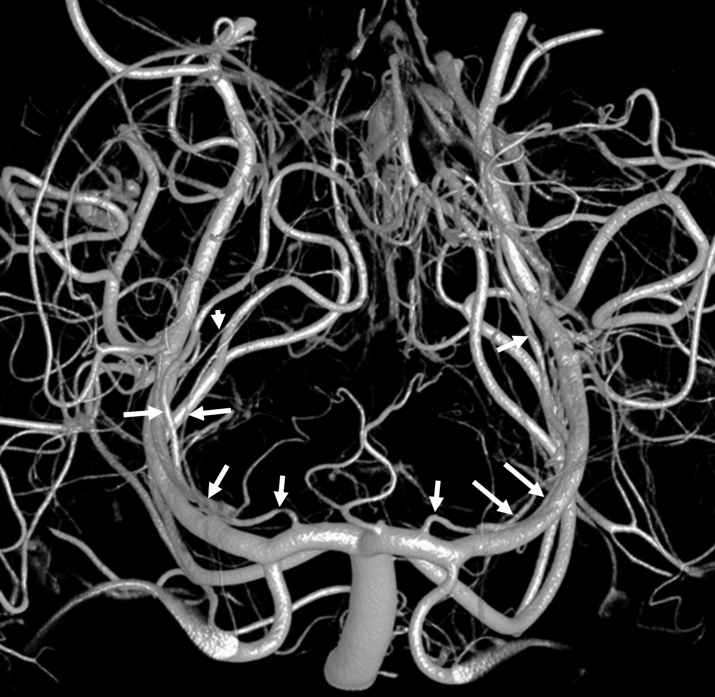
A specific and potentially very bad situation when catheterizing the PCA can arise if the microguidewire or microcatheter enter one of several small caliber vessels which course very much along the main PCA trajectory — but are definitely not the PCA. This can be particularly treacherous when both PCA and these vessels — namely the proximal posterior medial choroidal artery and the long circumflex (circumcollicular) arteries are both beyond the site of occlusion and thus both not opacified with contrast. Deploying a stenttriever in one of these pseudo-PCA impostors will likely lead to rupture of this vessel and potentially a large hemorrhage, in addition to likely infarct in the impostor vessel territory — which are usually eloquent, including lateral thalamus, cerebral peduncle, and quadrigeminal plate. How to avoid it? Look carefully at guidewire path. Do not force microcatheters into PCA territories they dont want to go into. Do a “test injection” before deploying the stent-triever. There is no good reason not to in this page’s opinion.
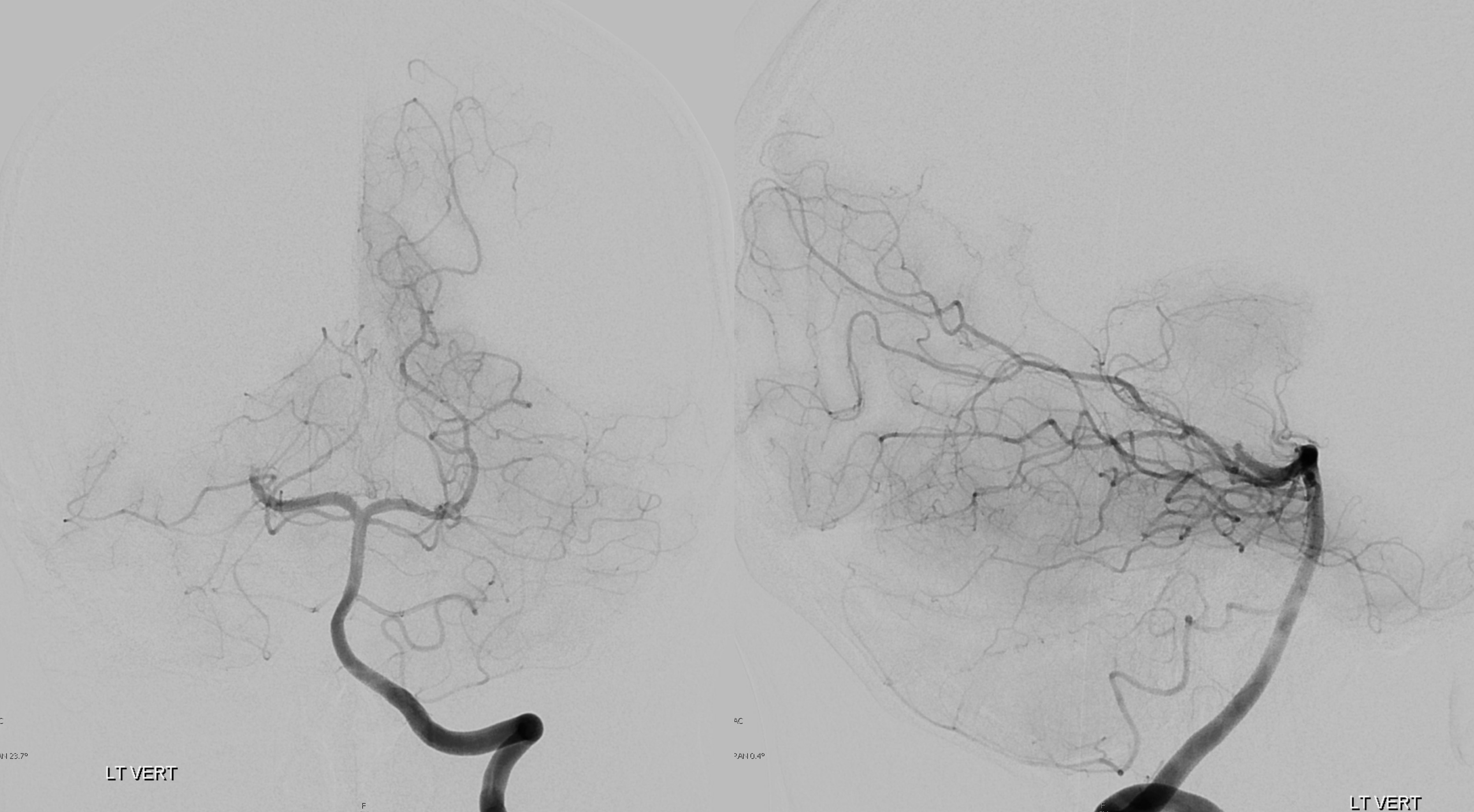
Arrows below point to the multiple PCA impostors running alongside the true PCA trunk in this normal example
Acute Stroke Case
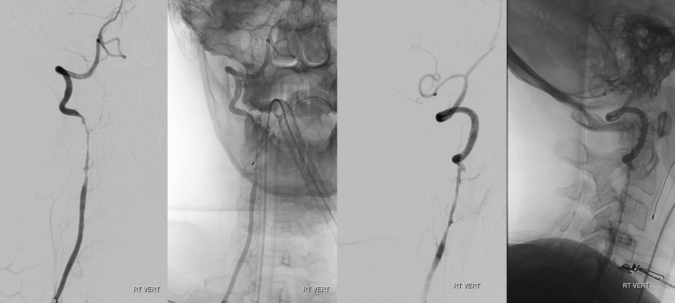
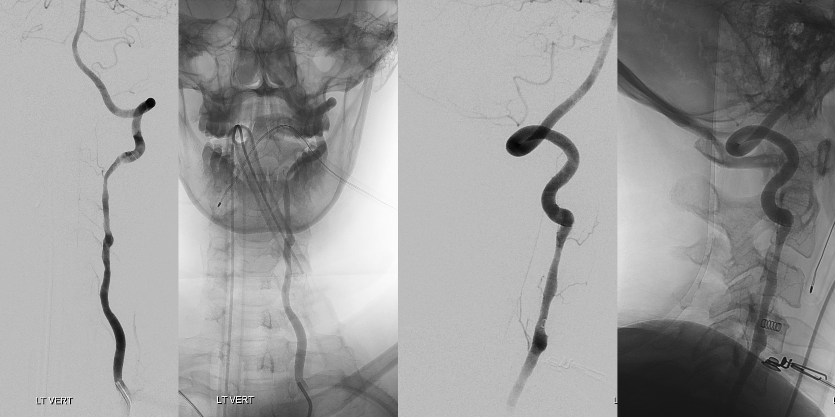
Below is example of this caution in a patient with bilateral vertebral artery dissections and left P2 occlusion
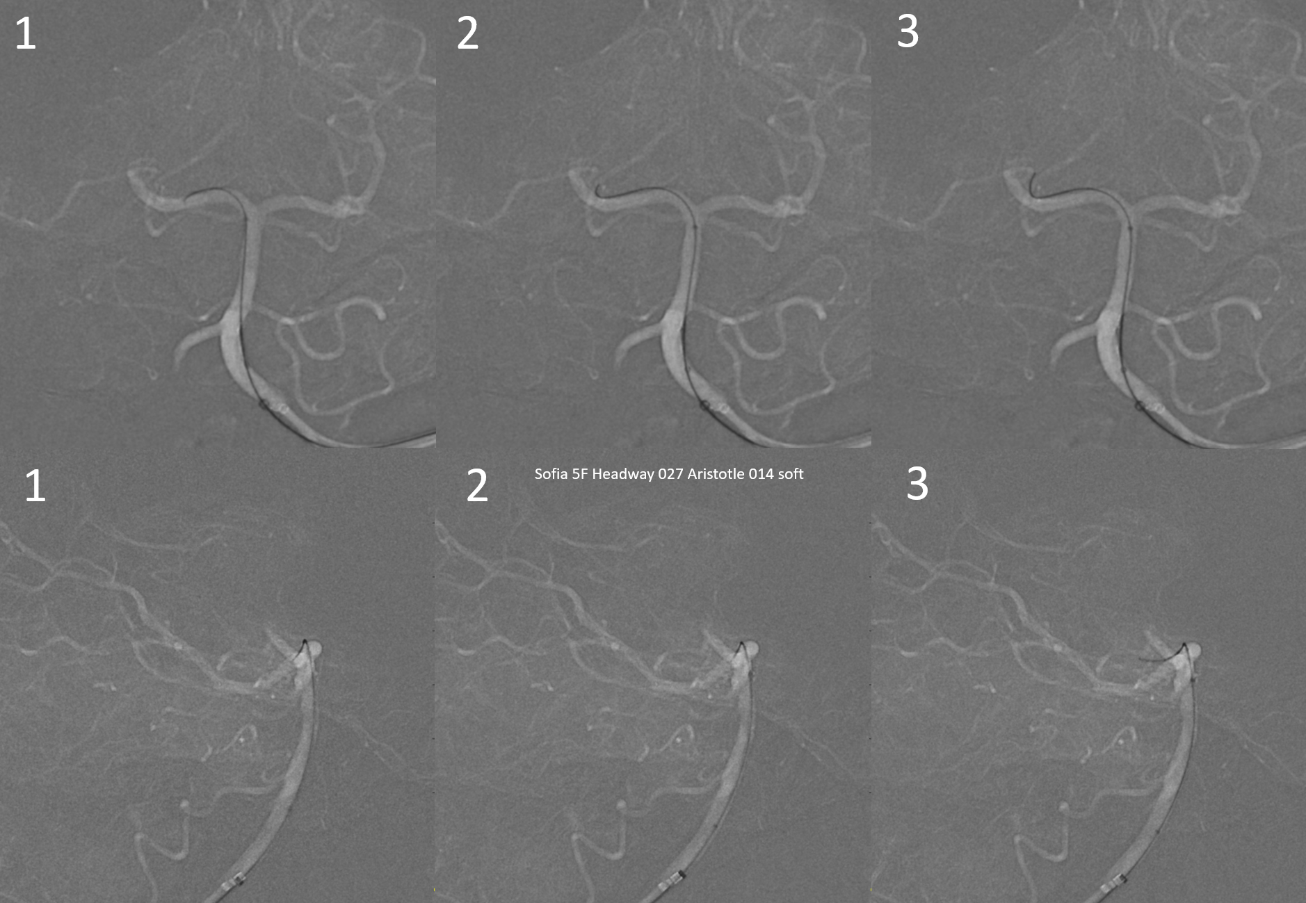
Wire enters the pseudo-PCA (1,2,3). Because the occlusion is more distal (P2), the false course of the wire becomes obvious in later frames (3) as it progresses distally. Had the occlusion been more proximal (P1), it would have been impossible to tell the difference by looking at the wire alone.
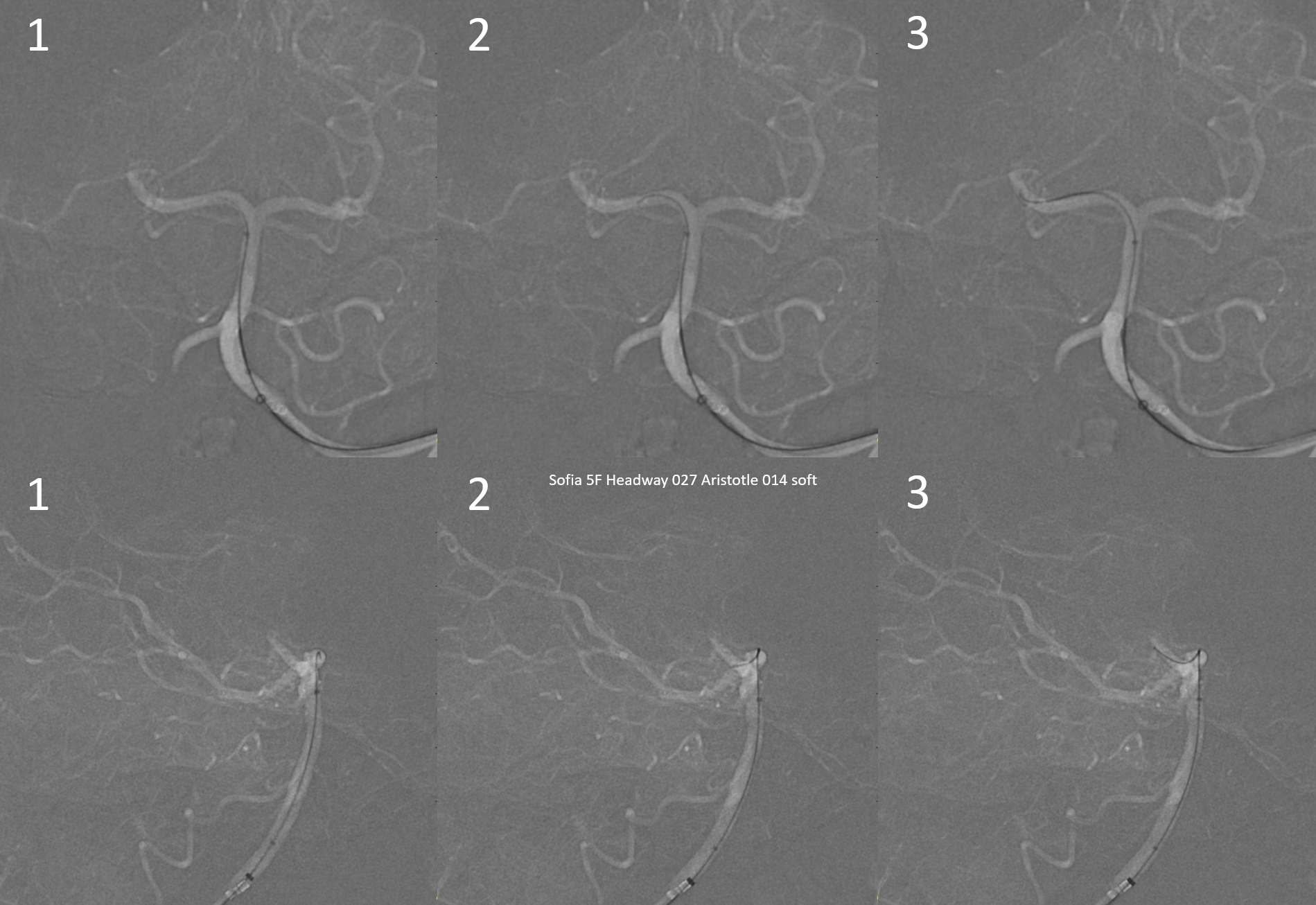
Selecting the right course
Movie
Both Headway 21 and Sofia 5 go in smoothly. Aspiration alone is another strategy to avoid disaster — as long as the catheters do not encounter undue resistance, chances are they are in the right places
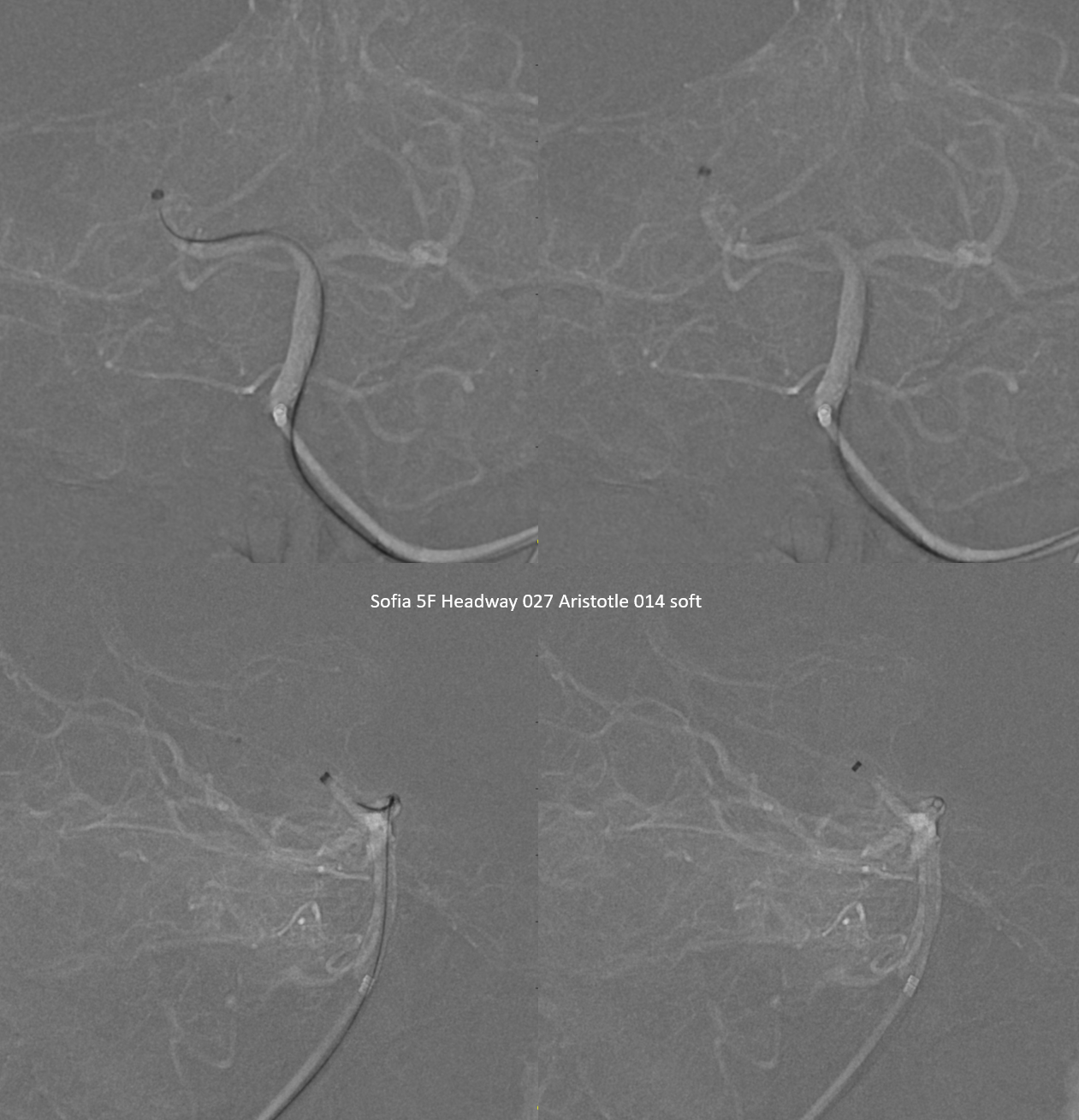
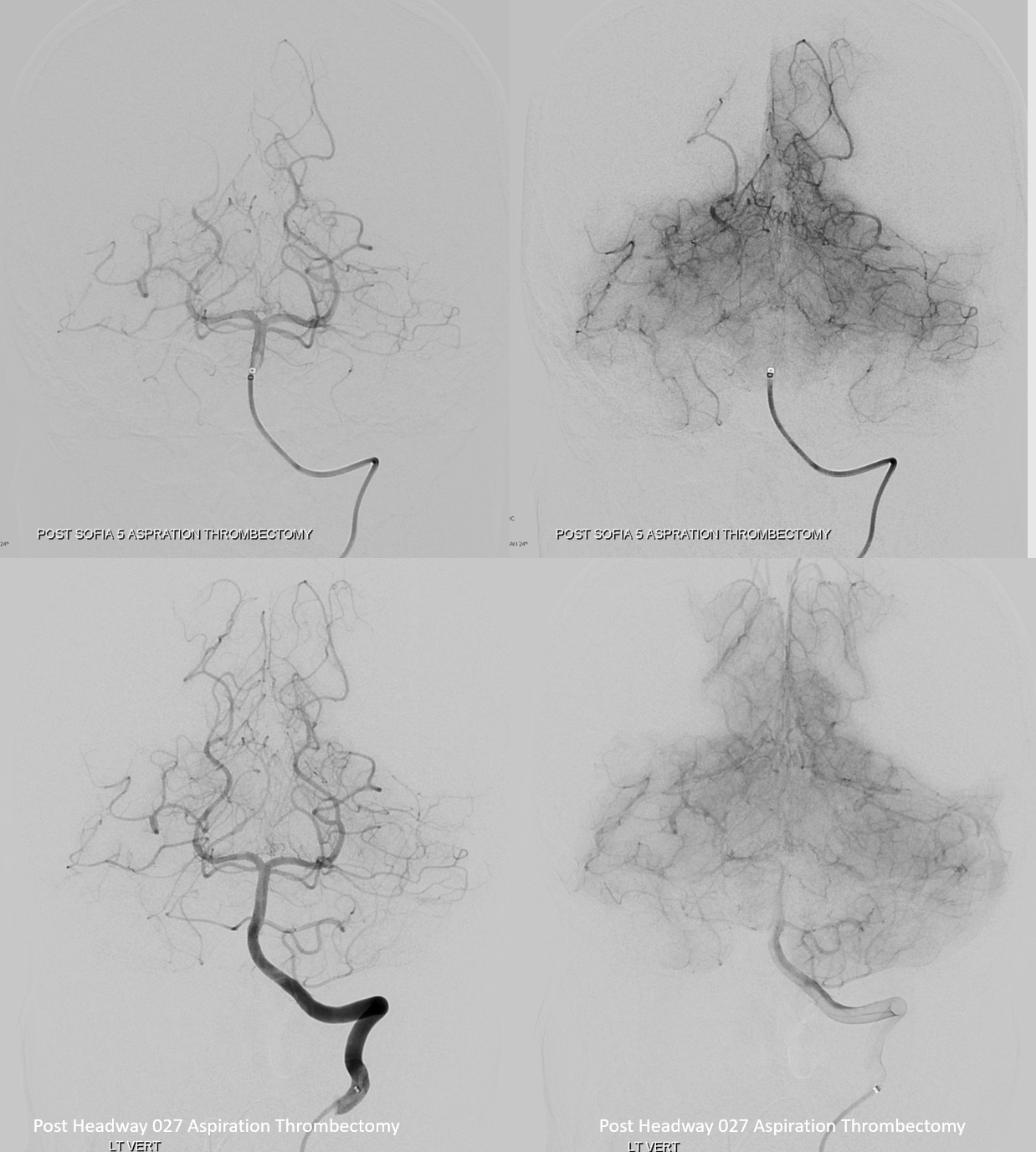
Post (top) has a small residual embolus in the parieto-occipital branch. This is successfully aspirated (bottom images) using a Headway 027 microcatheter (see other cases of 027 aspiration (1, 2, 3) in Case Archives page). No movie was saved
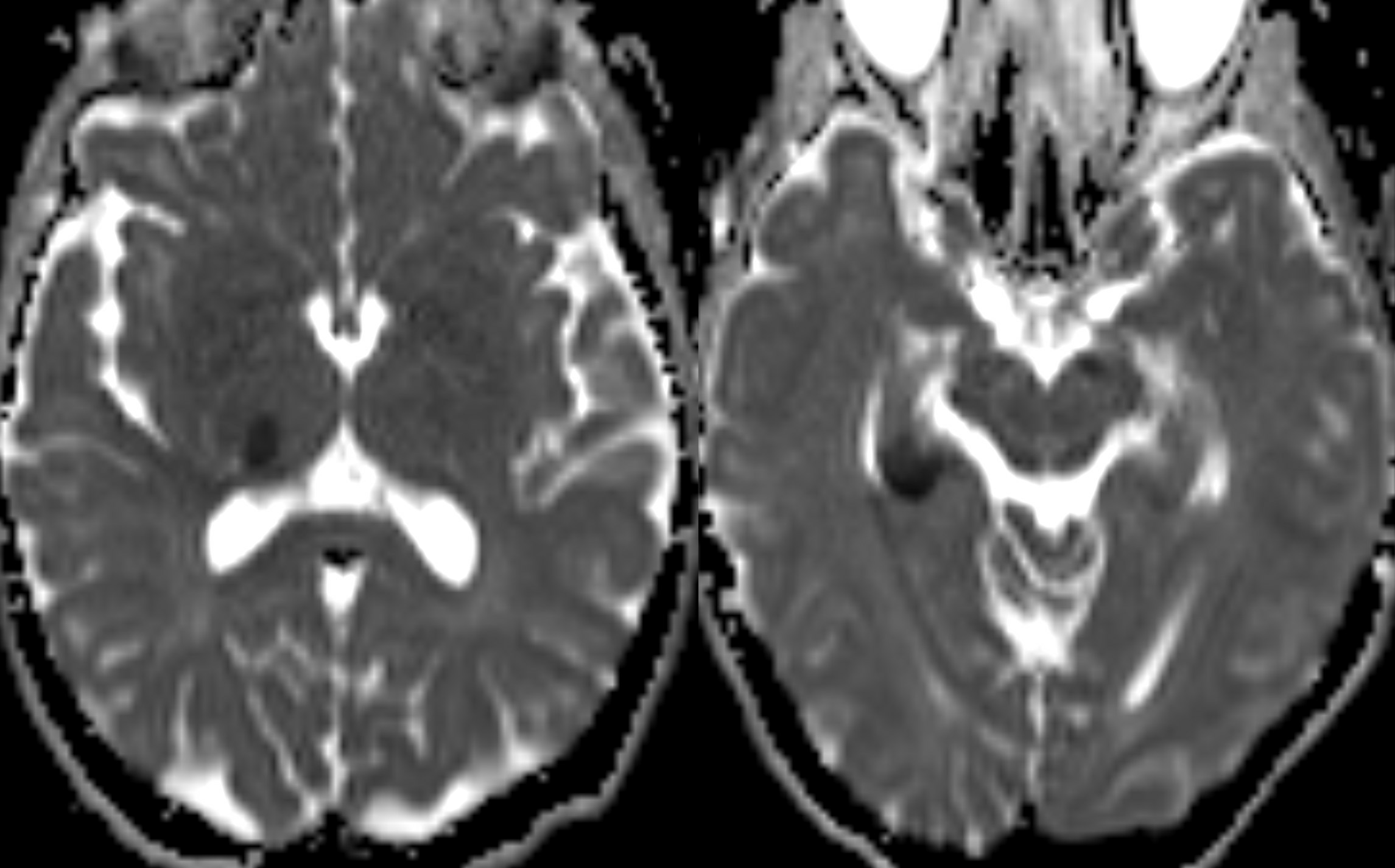
Post MRI — both infarcts are from other small emboli into well -known PCA territories (hippocampus — inferior tempora branches) and thalamus (posterior thalamic P1 perforators). Occipital lobe is clear
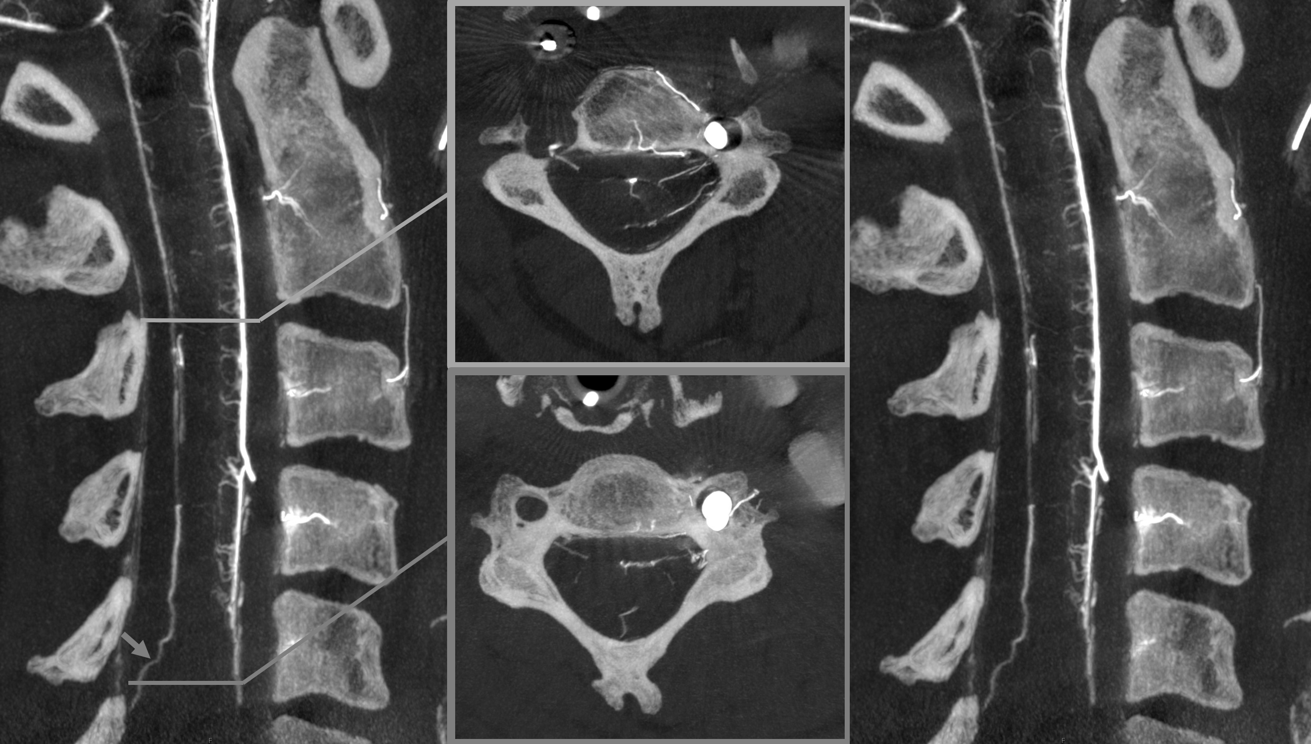
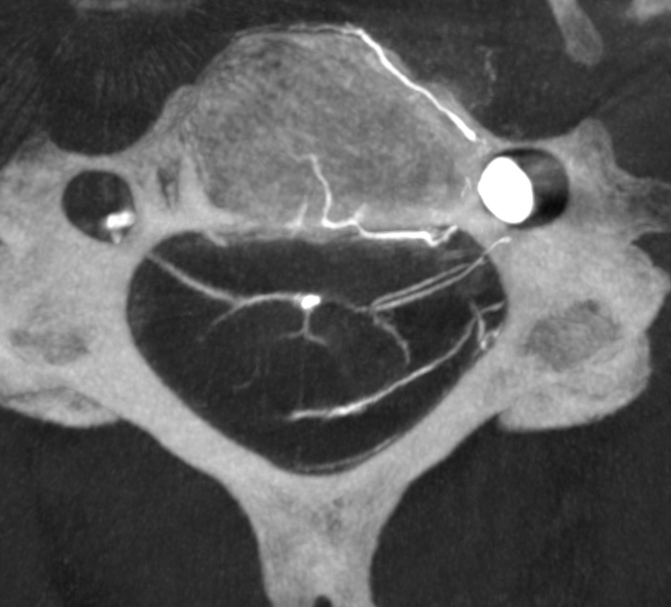
Beautiful DYNA of the dissected segment
Gorgeous venous cord images — see Spinal Venous Anatomy Page for more info
