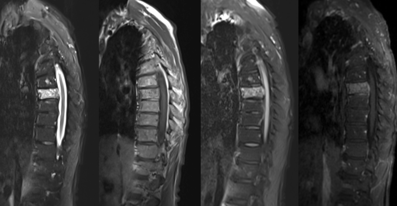
Preop embo of T7 hemangioma
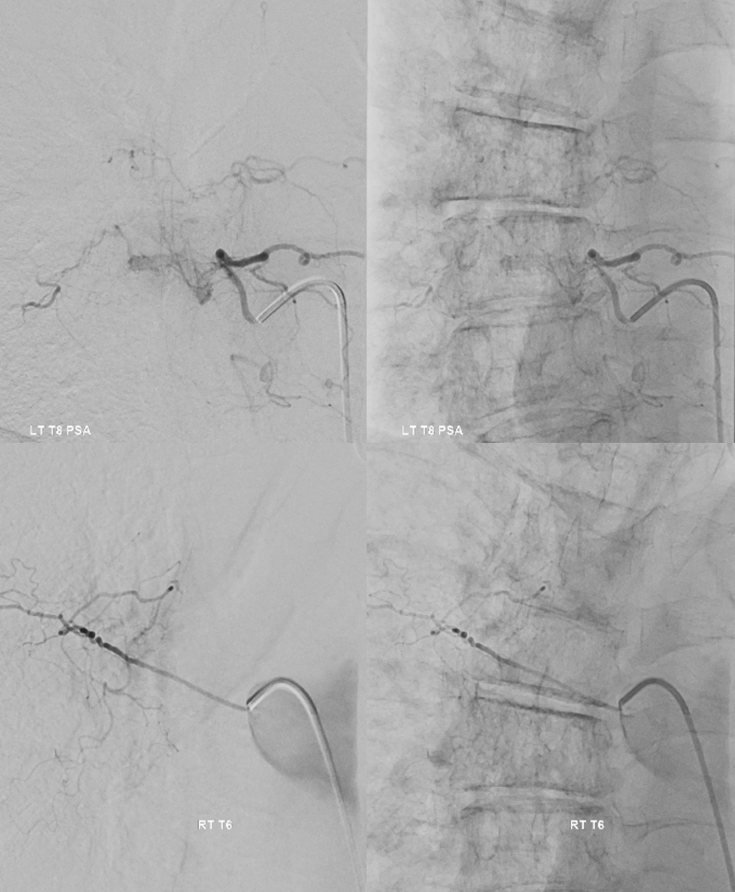
Importance of identifying spinal supply nearby
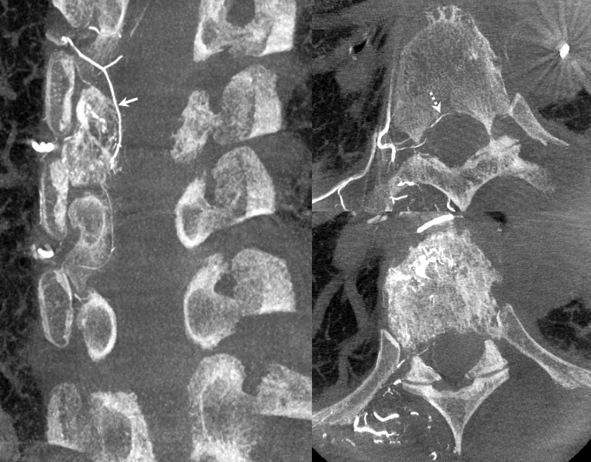
Right T7 Below
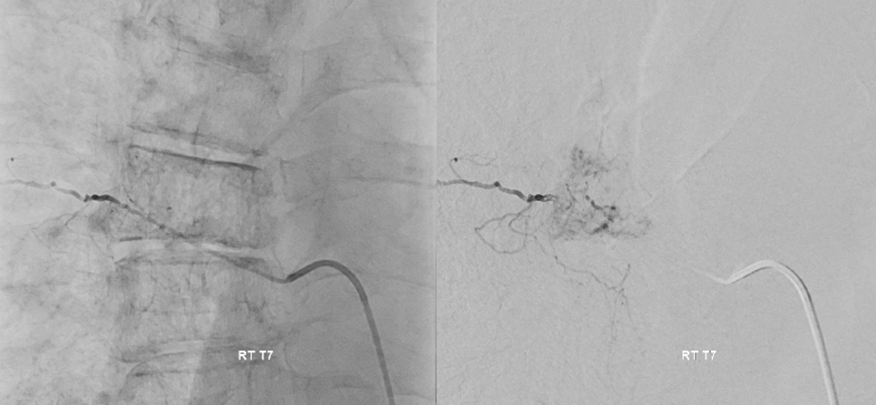
Right T7 DYNA CT — what is seen here that was not seen on 2D-DSA?
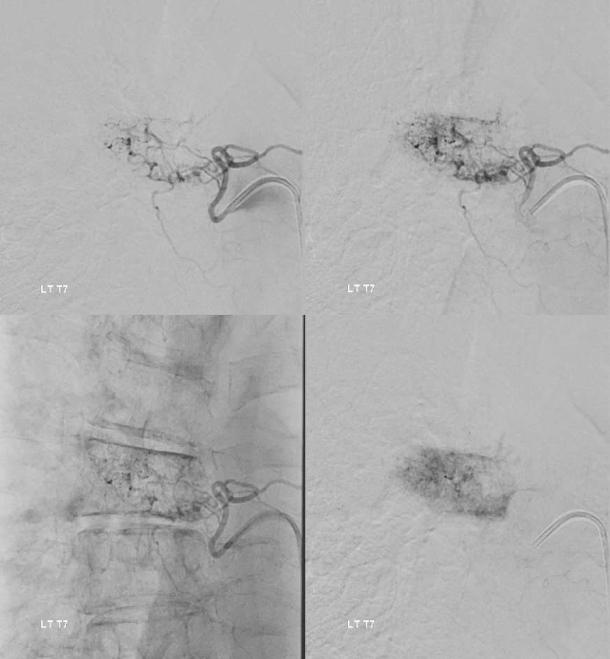
Left T7
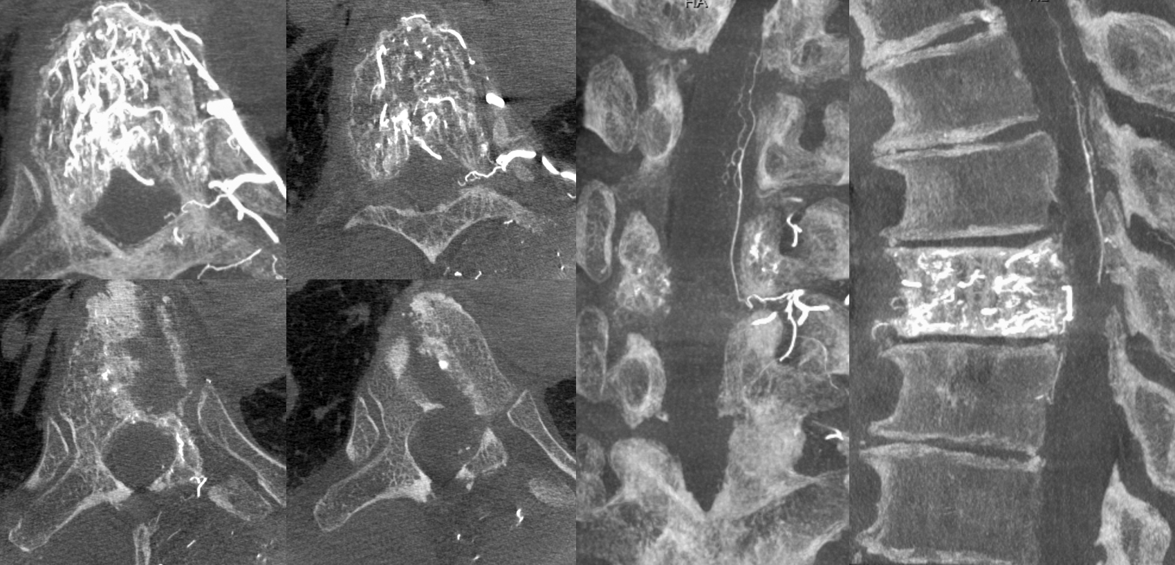
Left T7 DYNA — what is really seen here? Incredible…
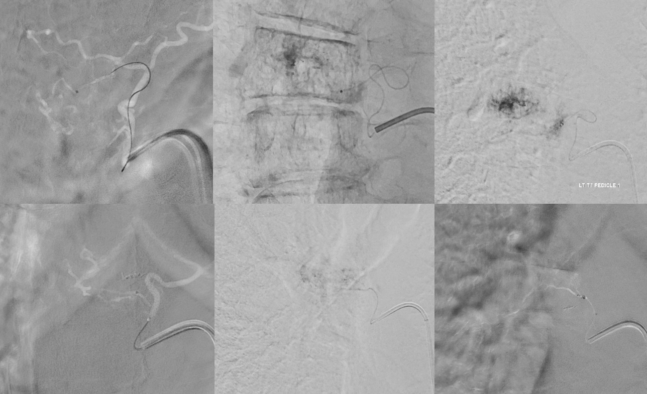
Can left T7 still be embolized? of course. Top row — posterior epidural / somatic branch subselective catheterization, followed by PVA embo — taking out the epidural component. Bottom row — anterior somatic branch — front of vertebral body subselective catheterization — more PVA embo from here
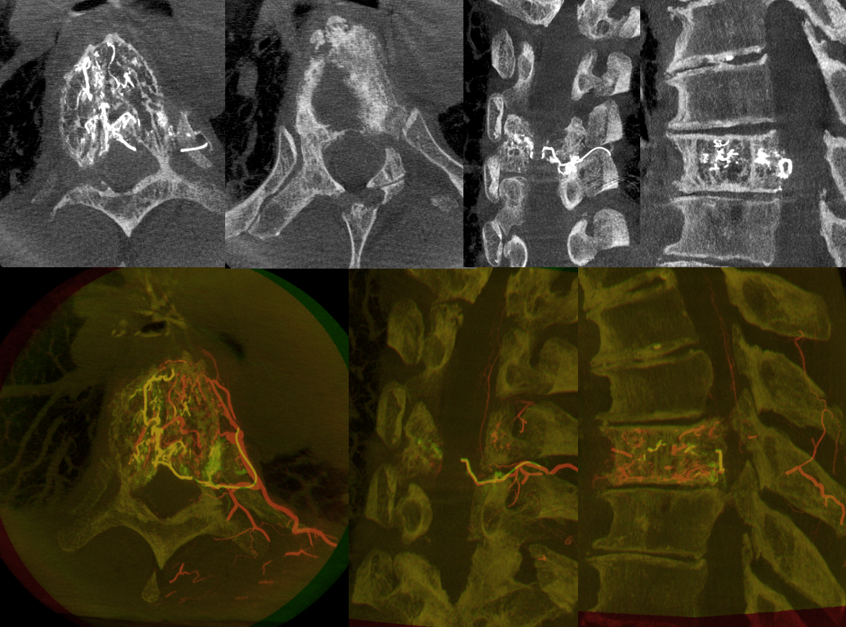
A micro DYNA was done from the posterior somatic / ventral epidural branch (top row above) — just to be sure. Top row below — MIPs. Bottom row below — “6D” MIPs showing co-registered images with the global left T7 DYNA

Post embo — partial embo but better. Now can you see the left PSA?
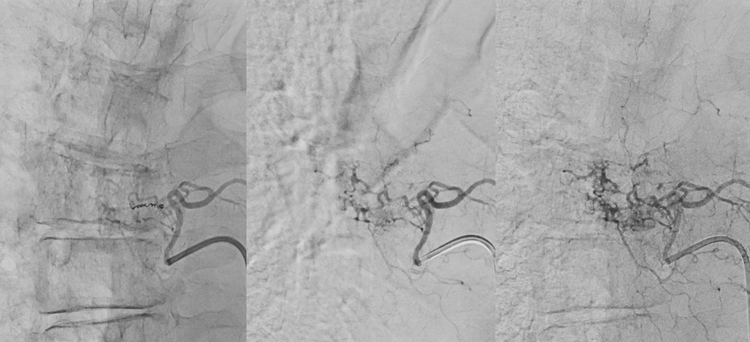
Standard right side embo — pushable coil closure of the intercostal artery, followed by particle embo and closure of the segmental artery proximally with more pushables
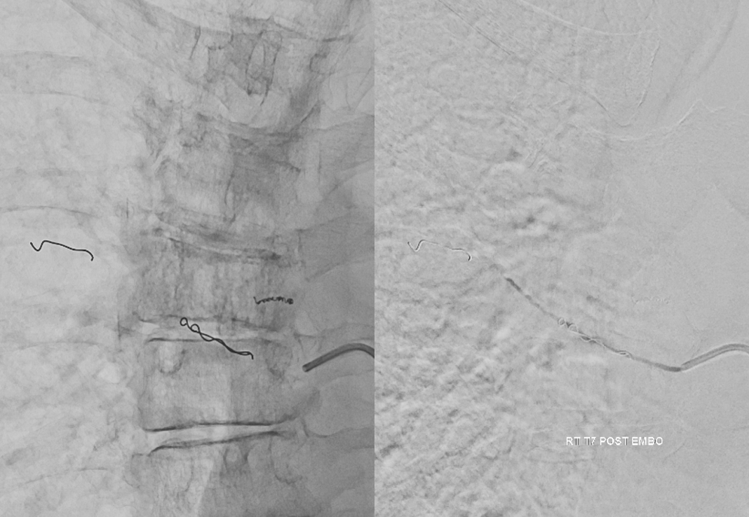
So, how about that for value of cone beam CT? See more cases in archives for pre-embo identification of cord supply…
