With Eytan Raz, MD PhD. Presentation is with several days of headache and SAH on CT
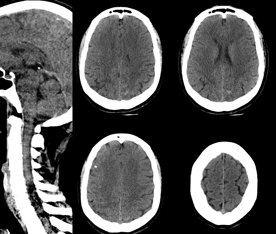
See it below?
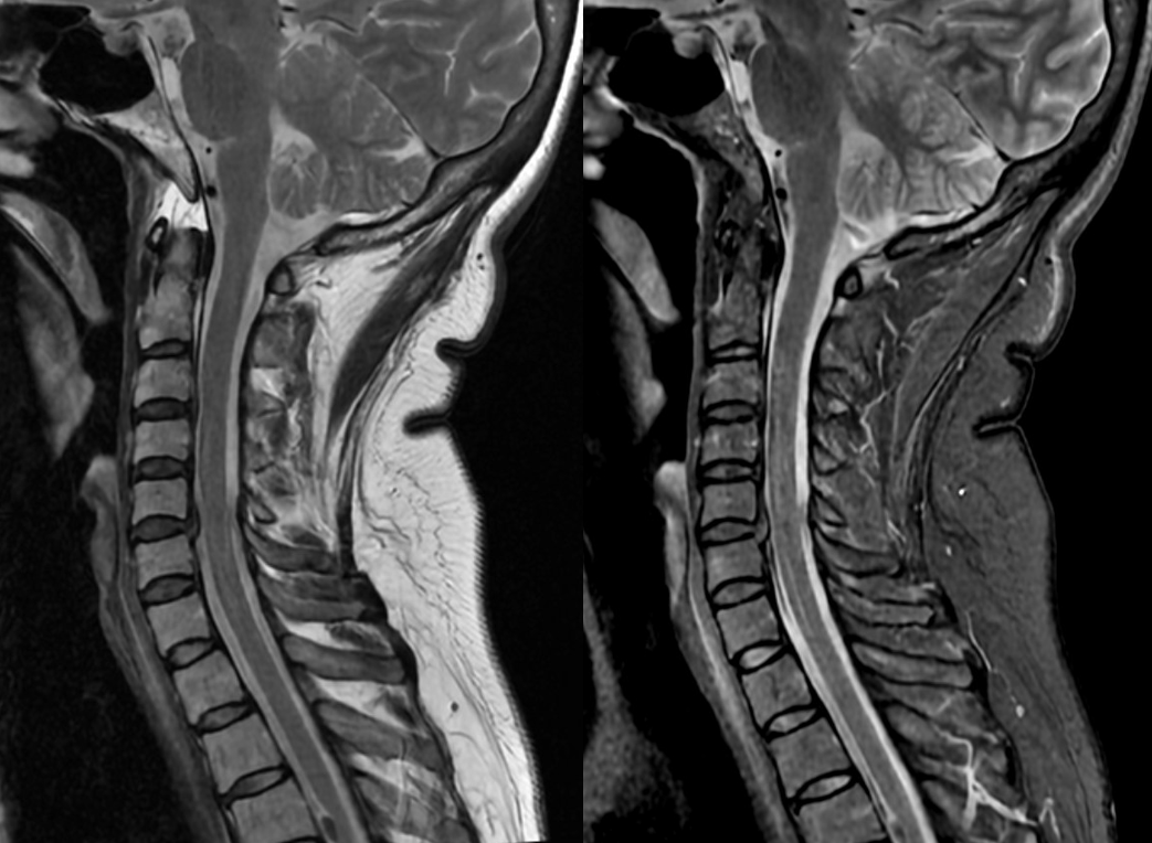
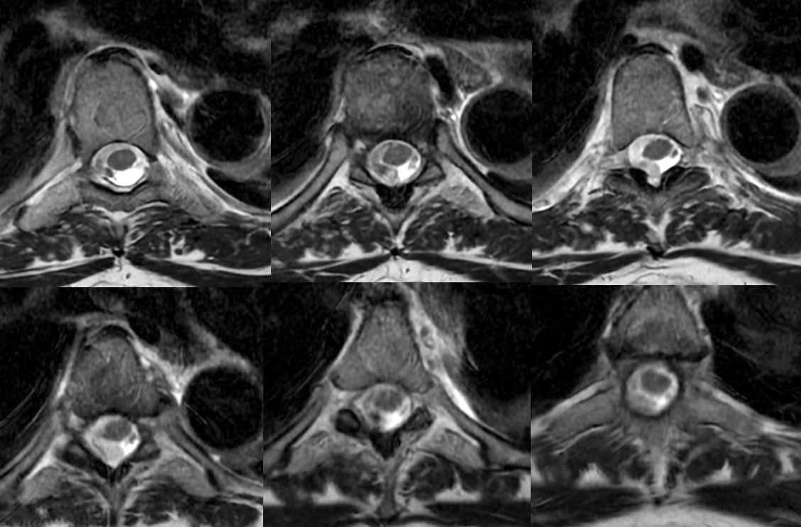
Not so good below
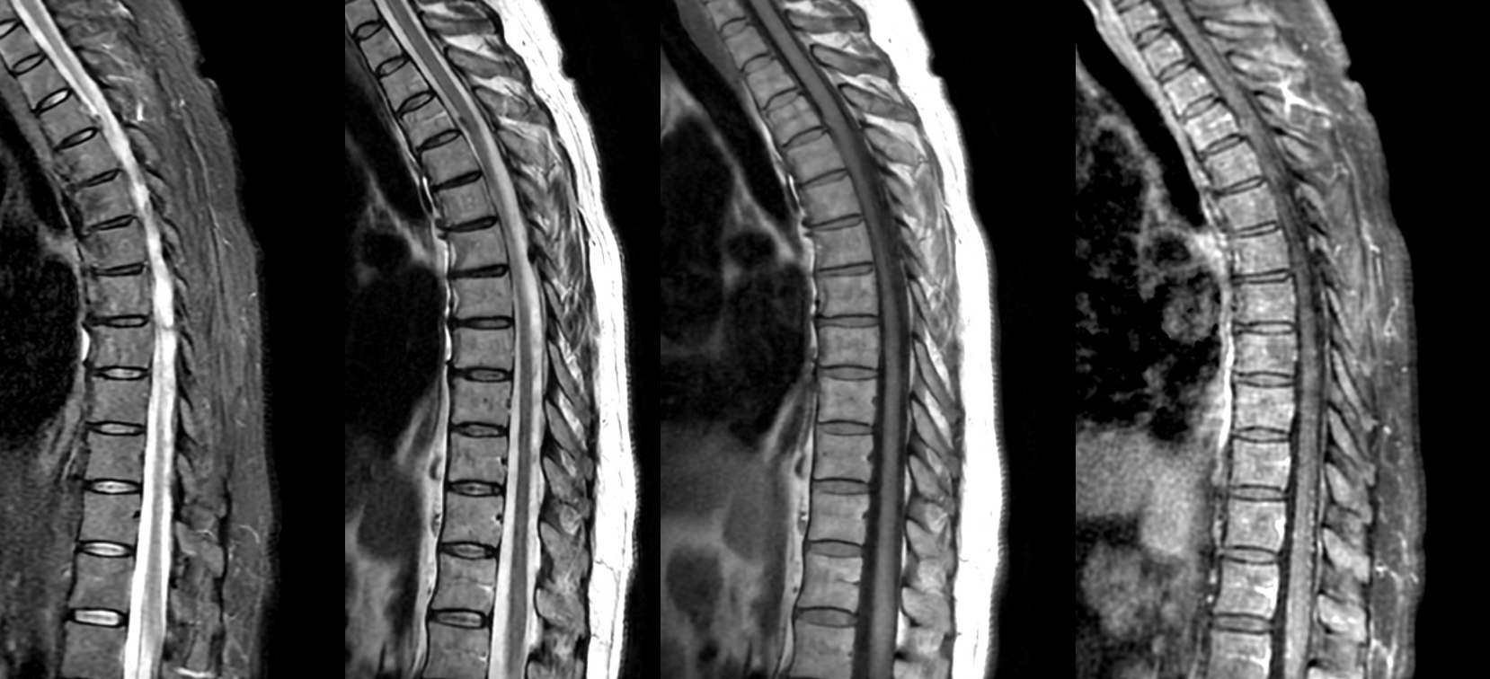
How about now?
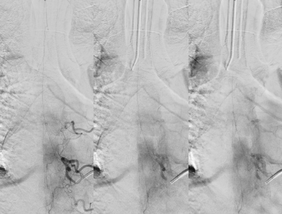
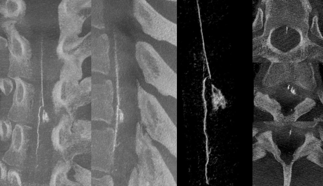
Angio
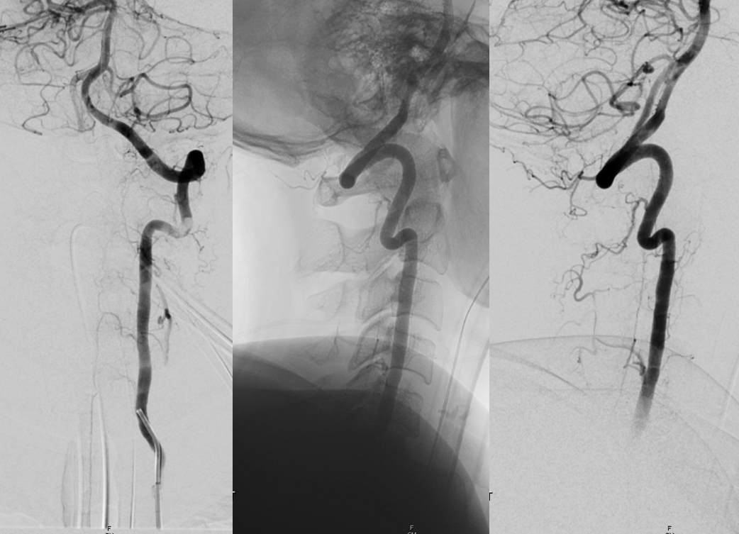
Classic look — partially thrombosed aneurysm of the radiculomedullary artery is retrogradely opacified from another radiculomedullary contributor to the anterior spinal axis

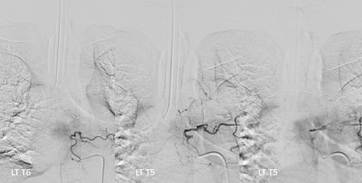
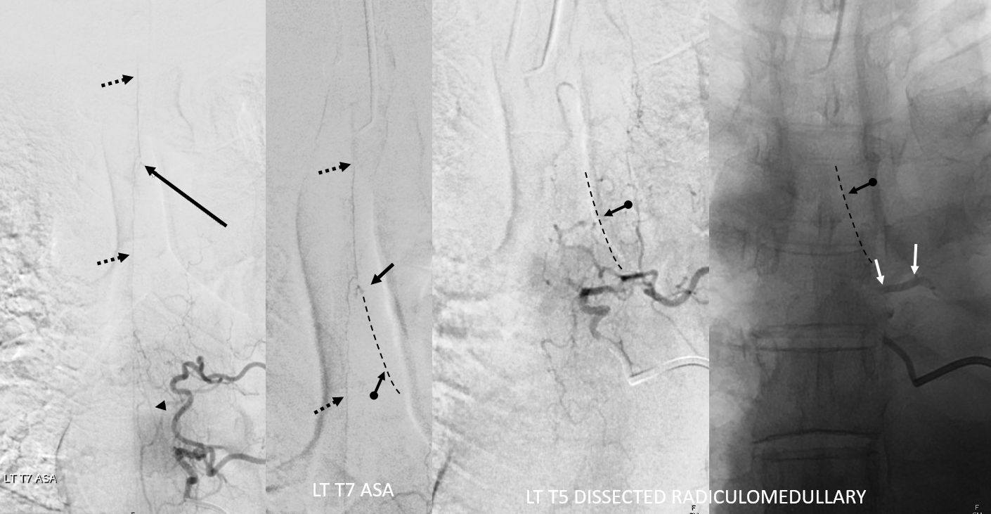
The dissection involves the segmental artery (white arrows)? or is it contrast stasis from wedge position?
Anterior spinal axis — dashed black arrows; pseudoaneurysm — black arrows; the dissected and not visualized radiculomedullary artery — dashed lines and ball arrows;
FOLLOW UP — 2 weeks later — still there but a bit smaller. Flow reversal is all it takes usually.