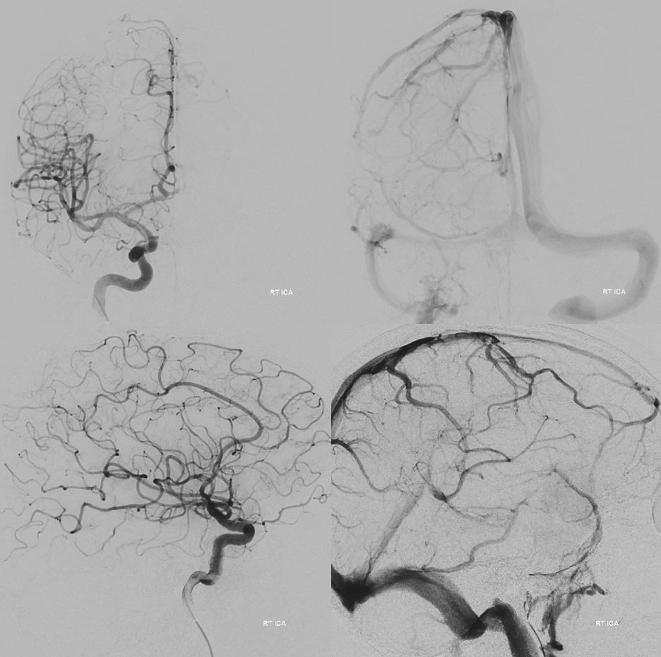
The power of HR-CBCT Imaging

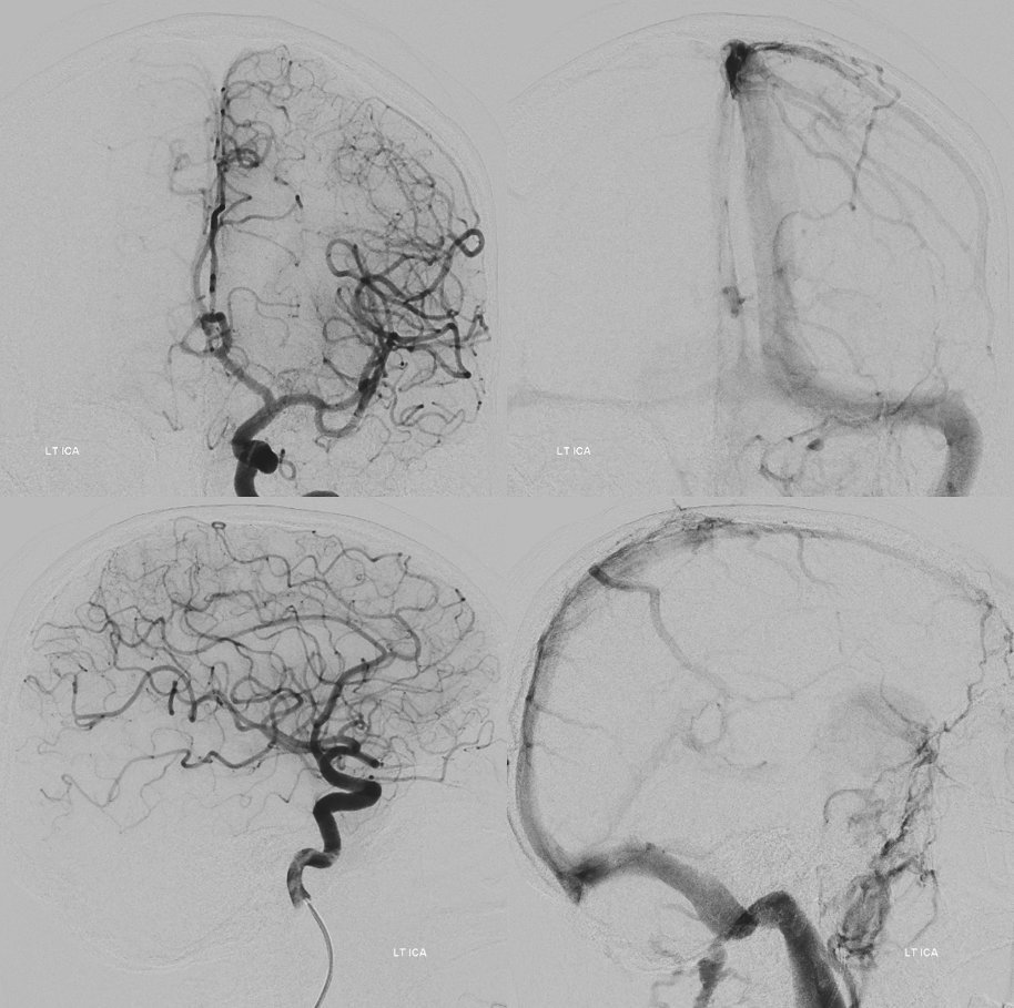
Pial supply best seen from subselective injection
MHT

Now for the real deal. HR-CBCT…
MHT Stereo — fusion of ICA and MHT micro HR-CBCTs
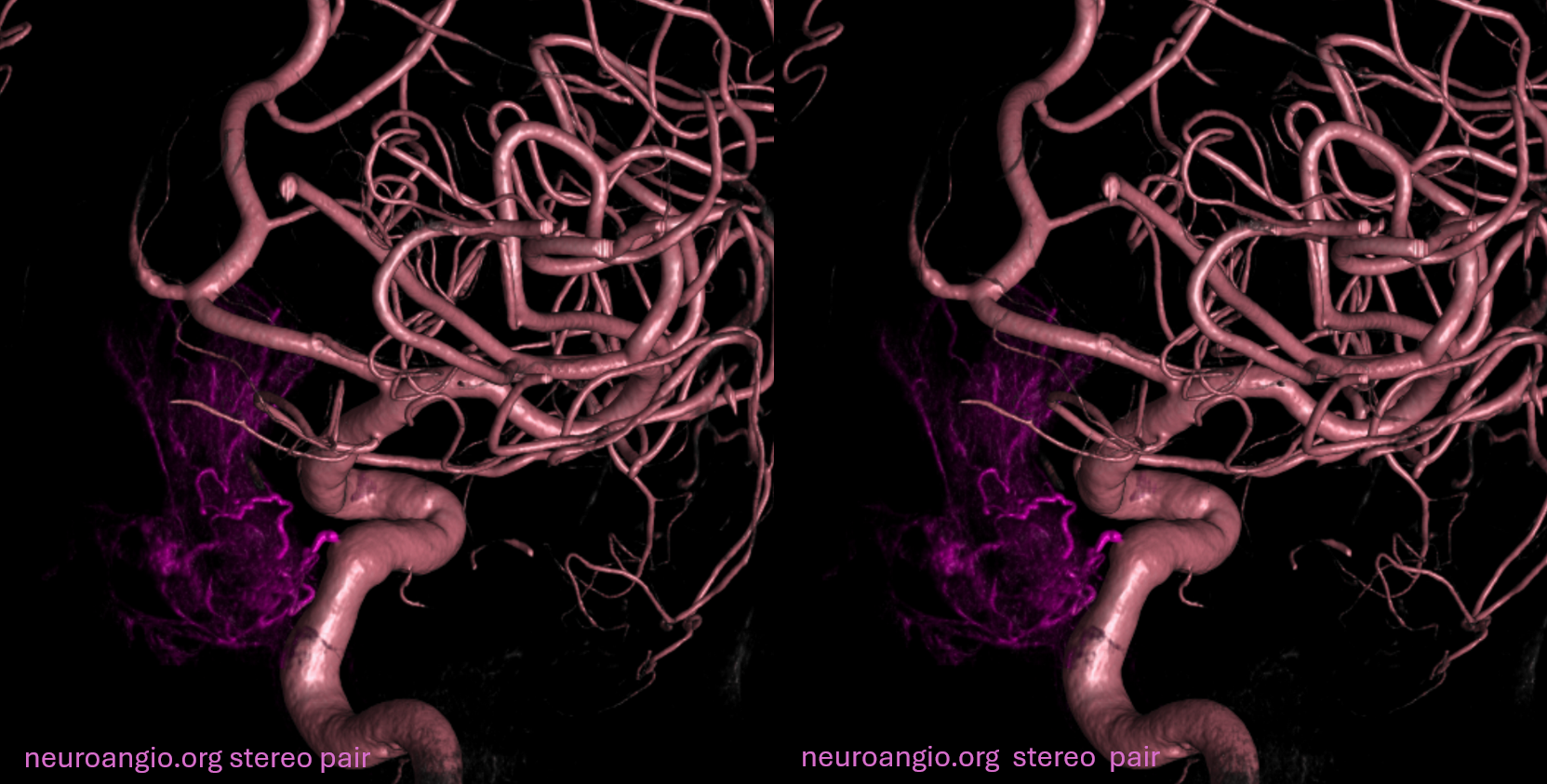
Fusion of R and L ICAs. The ACOM is wide. Therefore, a lot of the left ICA injection crosses to the right. This allows to differentiate between right ICA supply (red, MHT mostly), right ACA pial supply (yellow), and left ICA (mix of pial and dural, mostly pial) supply — green. The top row are 0.5 mm thick, bottom 3 mm. Note bilateral supply of the stalk on the upper left image.
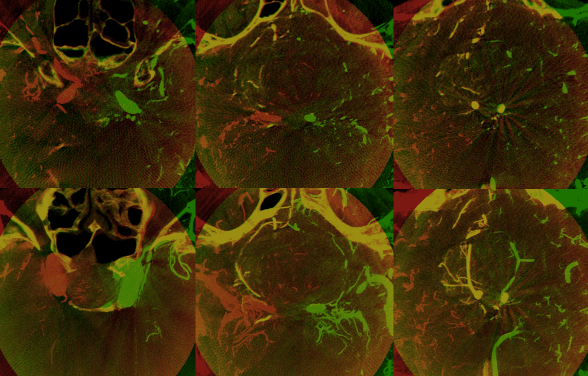
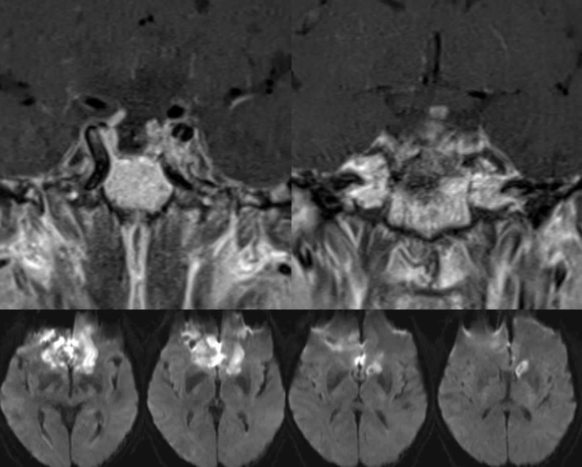
Coronal view — compare to the appearance of coronal MRI

Video — which has a lot more info. Including superior hypophyseal supply

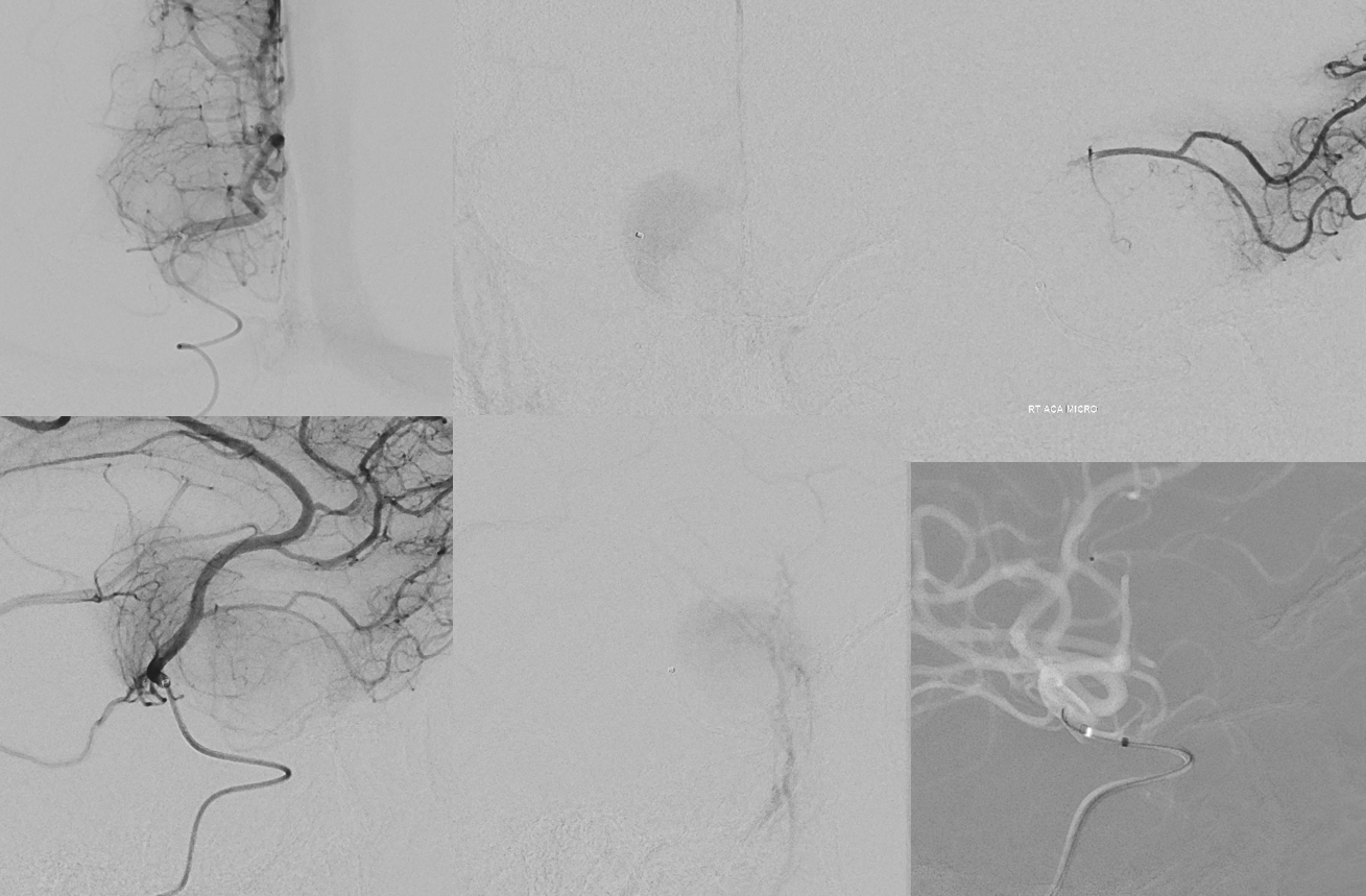
Reconstruction of A1 injection. Note various sources of pial supply (arrows), including from highly unusual proximal A2 location. The ones notoriously missing are from A1 and ACOM, projecting inferiorly. Not there (oval)

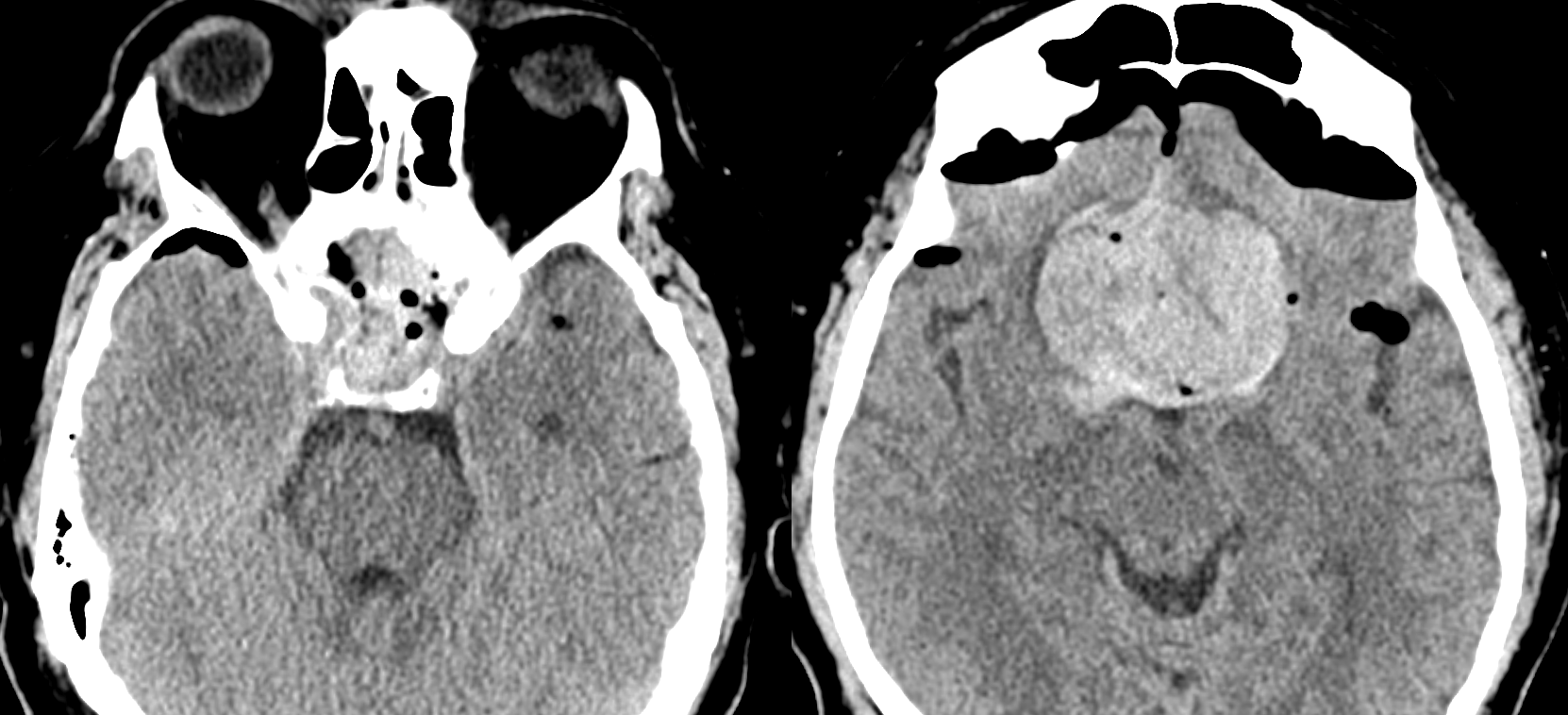
But wait. there is more. So, after transnasal resection there was an apoplexic hemorrhage (7 o’clock, right image)
Post emergent reop
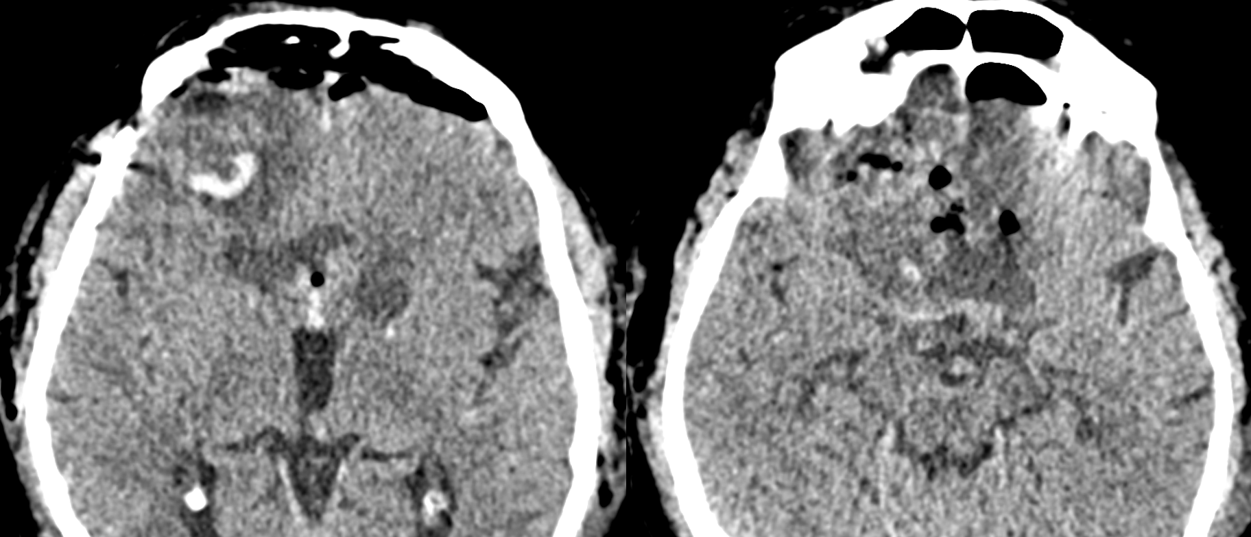
Vision improved
MRI