A key vessel in human embryology, the stapedial artery is the link between the embryonic precursors of the adult internal carotid, internal maxillary, and middle meningeal arteries.
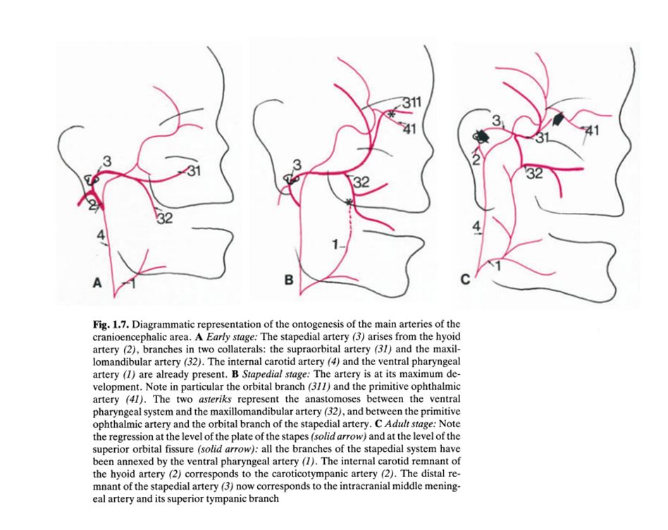
Briefly, in the developing human the territories of the middle meningeal and internal maxillary arteries are supplied by the internal carotid artery via the stapedial artery. Subsequently, probably due to increasing demand on the internal carotid system by the developing telencephalic vesicles, supply of the IMAX and middle meningeal is transferred to the ventral pharyngeal artery — a forerunner of the proximal external carotid artery. Thus, both IMAX and MMA are embryologically distinct from the remainder of the external carotid system.
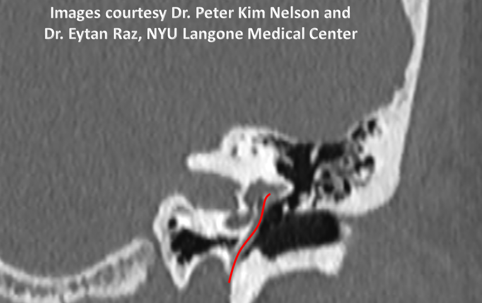
The link between the embryonic ICA and the homologs of the IMAX and MMA is the stapedial artery. In the adult, this vessel arises from the distal cervical or proximal vetical petrous ICA, ascending vertically posterior and lateral to the ICA and entering the middle ear cavity. It continues to ascend along the cochlear promontory, passing between the crura o the stapes (amazing!) and turning anteromedially to exit the middle ear cavity into the middle cranial fossa with the greater petrosal nerve to become the middle meningeal artery. Strictly speaking, the proximal portion of the vessel ouside of the middle ear cavity is referred to as the hyoid artery.
The following diagram, from an excellent and easily accessible article by Richard Silbergleit, Douglas J. Quint, Bharat A. Mehta, Suresh C. Patel, Joseph J. Metes, and Samir E. Noujaim: The Persistent Stapedial Artery. AJNR 200: 21:572–577 illustrates these stages:
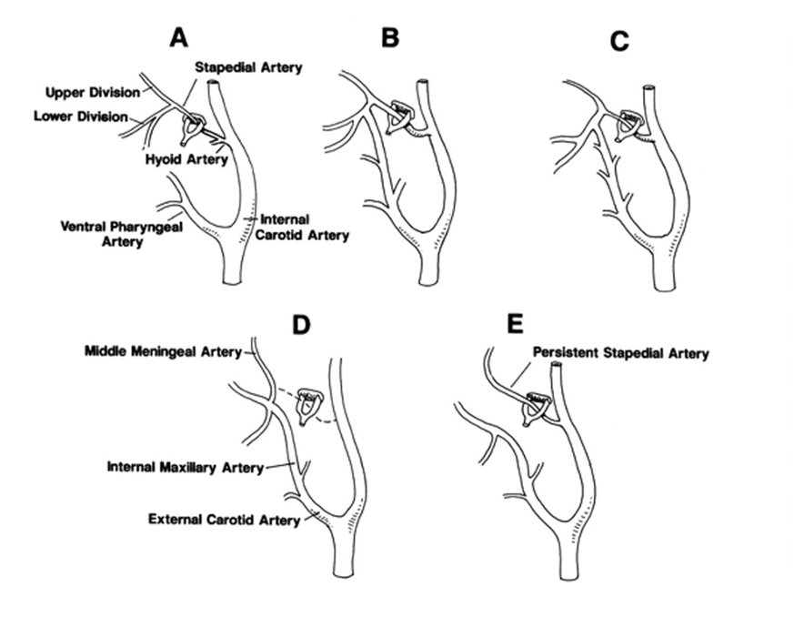
A: Early embyronic arrangement. The upper division of the Stapedial artery remains intracranial and is the forerunner of the MMA. The lower division exists the cranium via foramen spinosum to supply tissues of the face, and is the forerunner of the Internal Maxillary Artery (IMAX)
B: Establishment of anastomosis between the lower division of the stapedial artery (future IMAX) and the ventral pharyngeal artery.
C: Transfer of the IMAX and MMA territory to the Ventral Pharyngeal Artery. This is accompanied by reversal of flow within the foramen spinosum portion of the MMA from craniocaudal to caudocranial.
D: Typical adult arrangement. The hyoid and stapedial arteries persist, below angiographic resolution. The hyoid and proximal stapedial arteries are now named the “caroticotympanic” artery. Although everpresent, it is too small to be angiographically resolved. The distal part, related to the petrosal nerve is the superior tympanic branch of the middle meningeal artery.
E: Persistent stapedial artery: The Upper Division remains attached to the stapedial artery, while the lower division (IMAX) remains within the territory of the external carotid artery. There is no foramen spinosum in this case — no structure, no hole
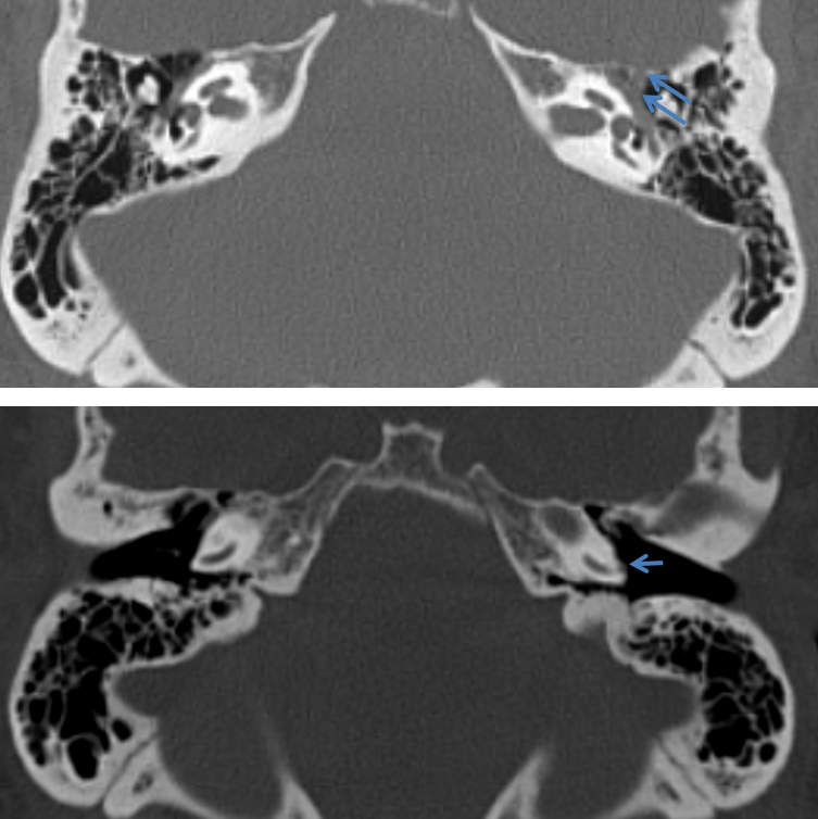
Here is an incidental persistent stapedial artery. On all images, the following arrow designations apply:
Black arrow = middle meningeal
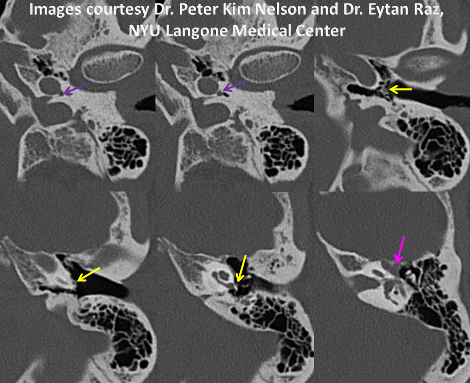
Pink arrow = geniculate / greater petrosal segment
Yellow arrow = stapedial segment (inside middle ear cavity, along the cochlear promontory, inbetween stapes crura)
Purple arrow = distal cervical and vertical petrous segments (remnant of the hyoid artery)
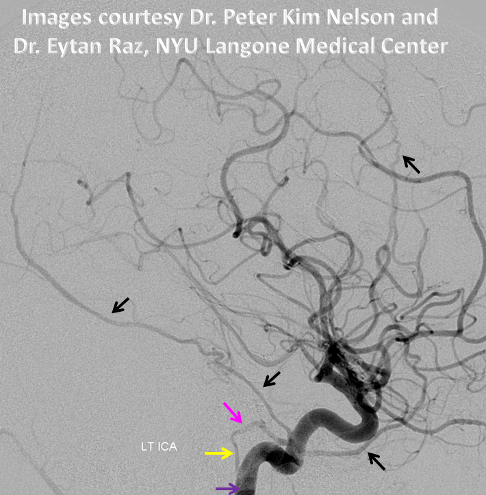
Stereo images below, frontal projection; notice characteristic thinning of the stapedial artery inside the middle ear cavity, compared with the more proximal cervical/petrous segment
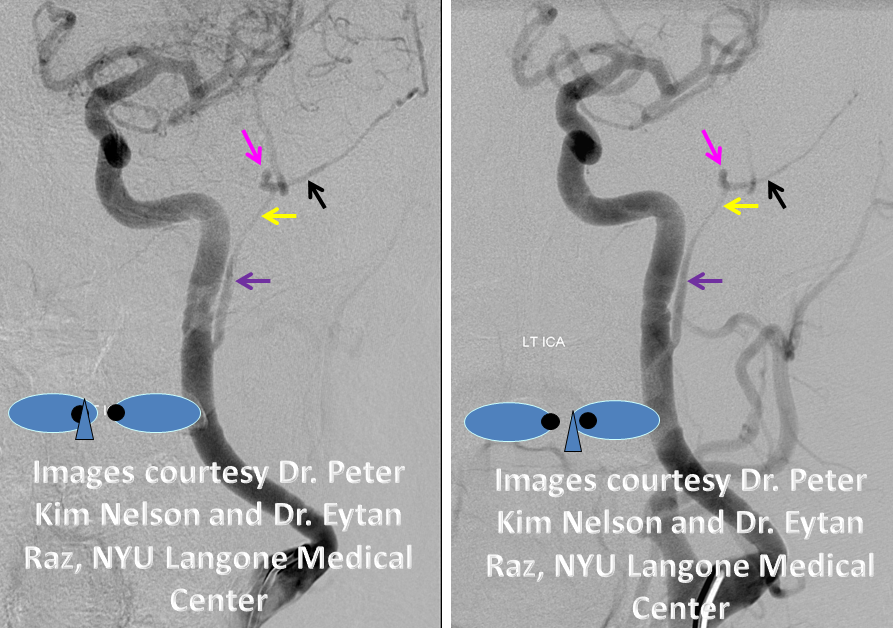
Stereo in lateral projection, where the narrowing of the middle ear cavity segment is even better seen
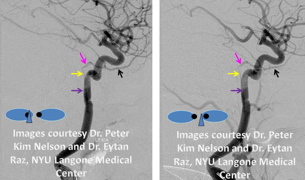
Usubtracted view:
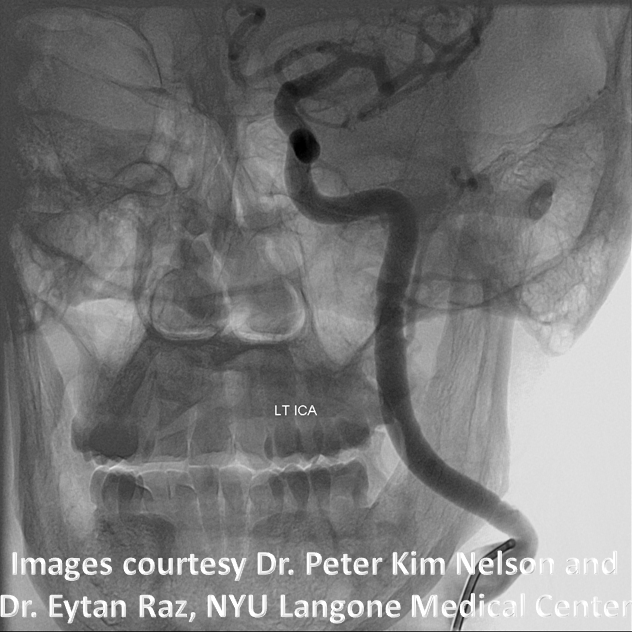
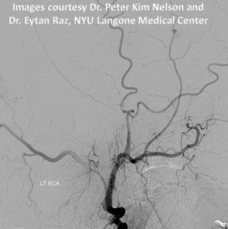
Ipsilateral External Carotid injection, demonstrating near-complete absense of middle meningeal branches
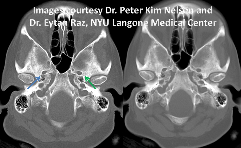
CT demonstrating lack of ipsilateral foramen spinosum (green, present is blue), by itself not specific as the MMA can come from other places also, most commonly the Recurrent Meningeal variant of the Ophthalmic Artery
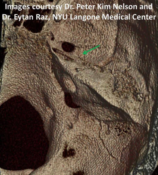
Same difference
Artery in the geniculate/petrosal segments (top) and along the cochlear promontory (bottom)
Course of the artery on serial axial CT slices, with the same color code as above:
A movie version of the same:
Curved reconstructions delineate vessel course through the temporal bone
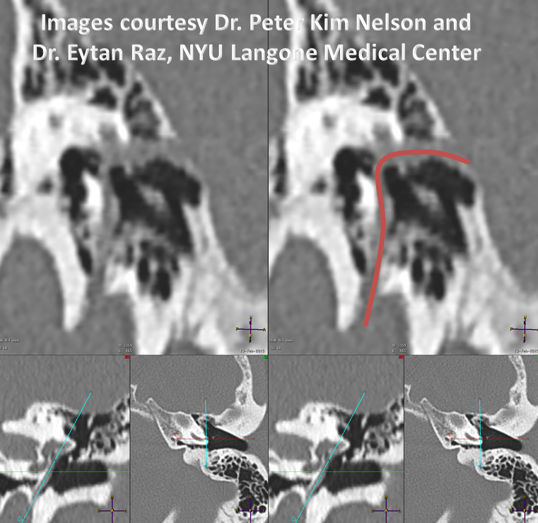
Beautiful oblique MIP in plane of the stapes crura
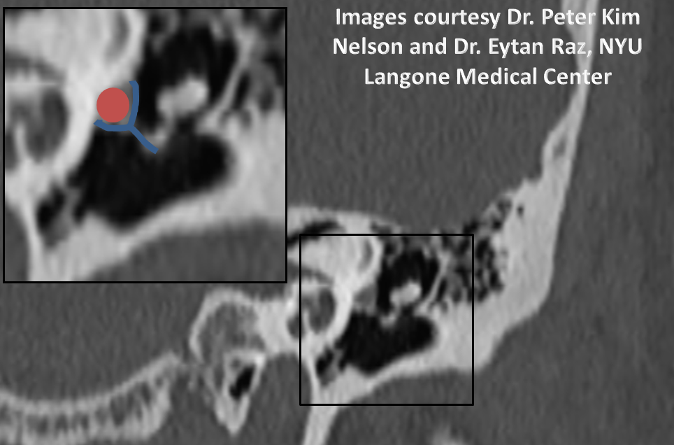
Another curved MIP
Finally, a figure from Surgical Neuroangiography from Lasjaunias, Berenstein, and Ter Brugge
Also see Middle Meningeal Artery page
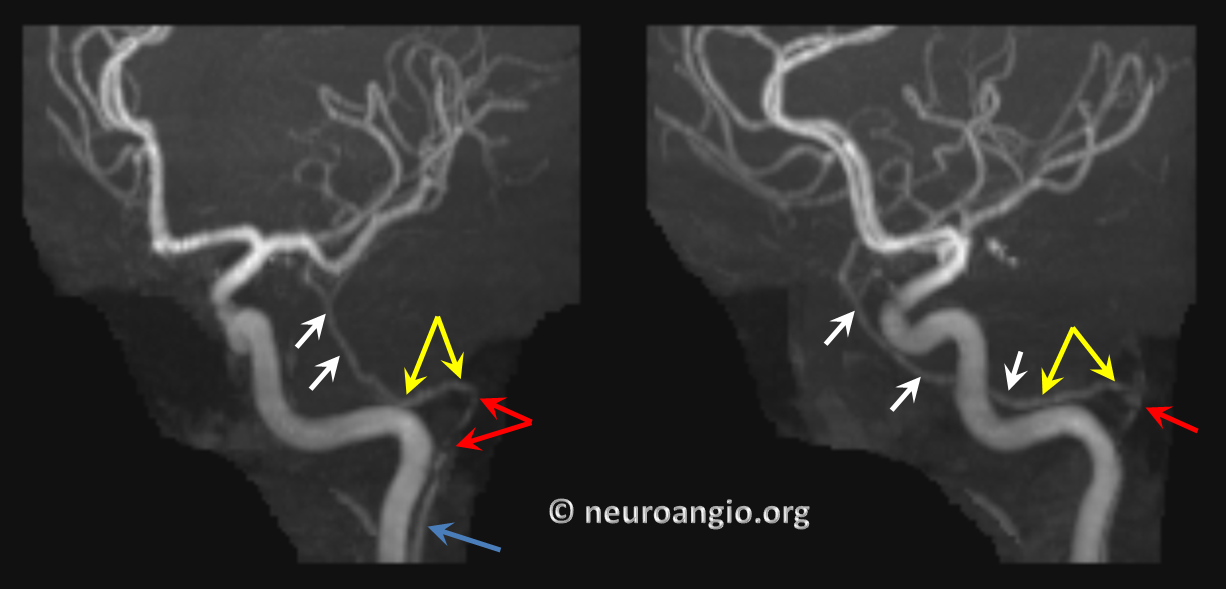
Persistent Stapedial Artery. In the same way the inferior tympanic-carotidotympanic connection results in ascending pharyngeal reconstitution of the petrous carotid artery, a persistent inferior tympanic can maintain an embryonic connection to the middle meningeal artery via the petrous branch of the MMA that makes us the facial nerve arcade. Embryonically, the middle meningeal artery does not belong to the IMAX — it instead arises from the petrous ICA via the primitive hyoid artery. The hyoid connects to the inferior tympanic, which goes thru the stapes crura (called stapedial artery at this point) and then via the facial arcade supplies what is ultimately the MMA. This connection is rudimentary in adulthood for most of us. However, in some cases the ascending pharyngeal-inferior tympanic-petrous-MMA connection persists. Simply put, the MMA is a branch of the ascending pharyngeal. Here is an example of this, on MRA; images courtesy of Dr. Eytan Raz. Blue – inferior tympanic branch; red = stapedial; yellow – petrous/horizontal facial nerve canal branch; white = MMA
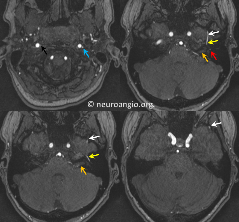
Axial views, with right AP in clandestine black; the IAC is orange
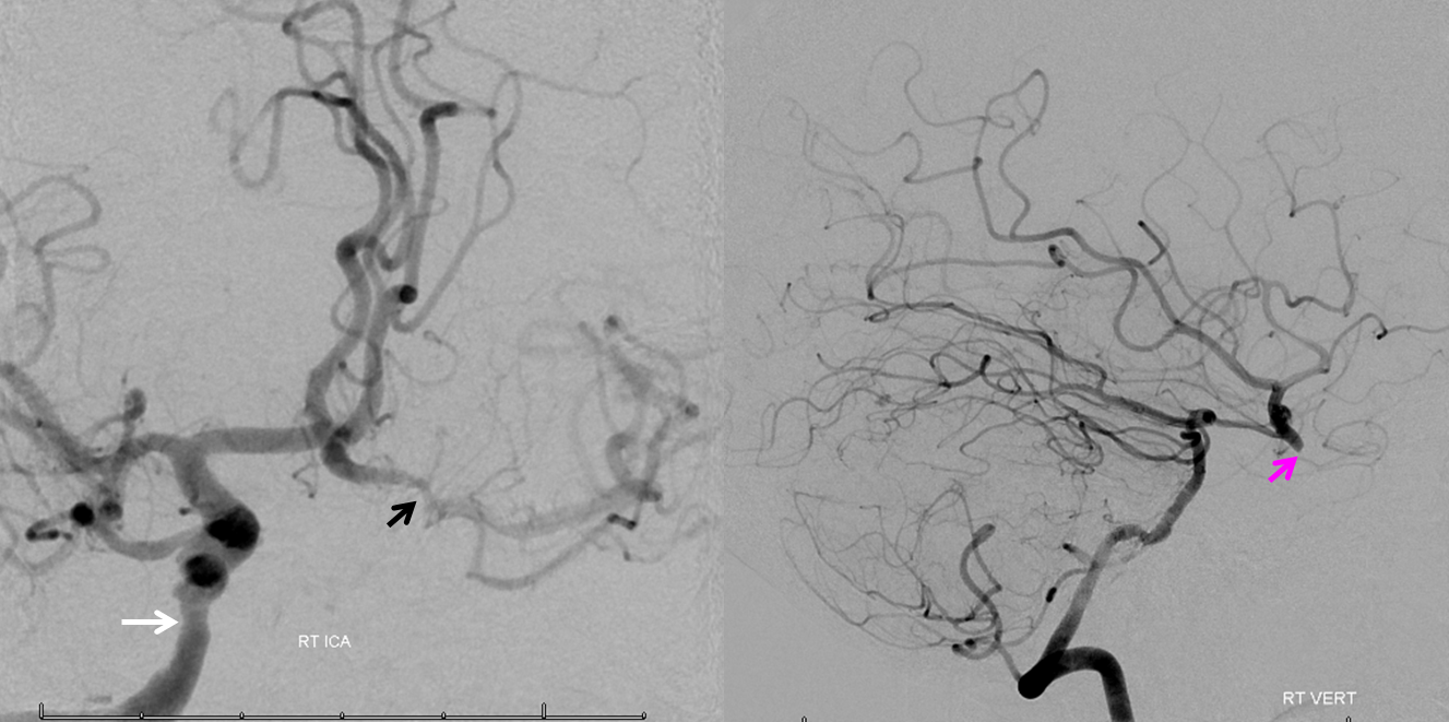
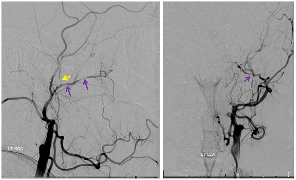
Embryonic Memory of the Stapedial System? A Case of Atherosclerosis Isolated to the Middle Meningeal and Internal Maxillary Branches of the ECA
A patient with multiple atheromatous disease risk factors presents for evaluation of cerebrovascular insufficiency. Right ICA and vertebral angiography demonstrates occlusion of the supraclinoid left ICA (pink arrow) with collateral reconstitution via the anterior and posterior communicating arteries. Notice stenosis of the proximal left A1 segment (black arrow) and atheromatous disease of the vertical cavernous segment (white arrow)
Injection of the left CCA demonstrates complete occlusion of the ICA. Notice irregular caliber of the left internal maxillary artery (purple) and proximal middle meningeal artery (yellow). The remainder of the ECA, including the coveted superficial temporal artery, are free of atherosclerotic burden. Inflammatory workup (ESR, CRP) were normal. Could it be that embryologic origins of the IMAX and MMA are the reason only those two vessels among the entire ECA are subjected to atheromatous disease? Personally, I believe this is the explanation
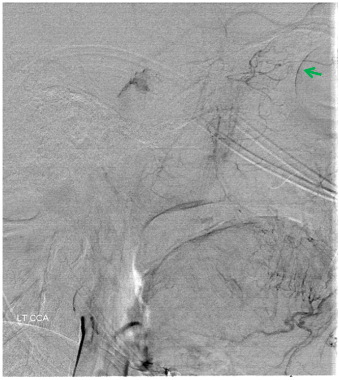
Just for your info, see the choroid blush of the eye (green) in late phase of injection, due to collateral reconstitution via the anterior deep temporal artery. For more on collateral ophthalmic patterns, see dedicated Ophthalmic Artery page
Clinical Application? See this awesome case of Persistent Stapedial Artery ICA reconstitution in acute ischemic stroke
