
Here we show how to do microcatheter DYNAs to understand compartments of brain AVM and plan embolizations
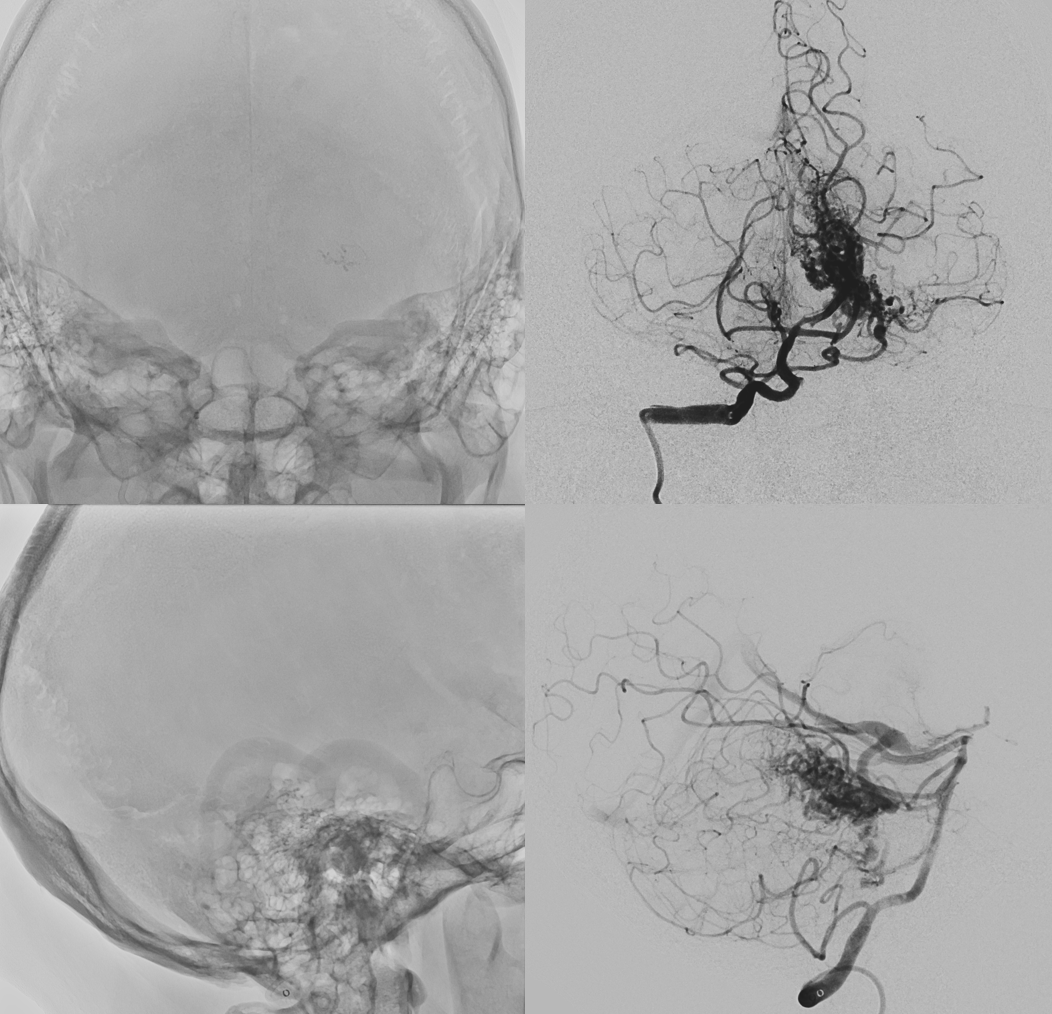
An incidental AVM was managed conservatively for 3 years, until it ruptured. Suspicion of perinidal aneurysm at rupture location (oval)
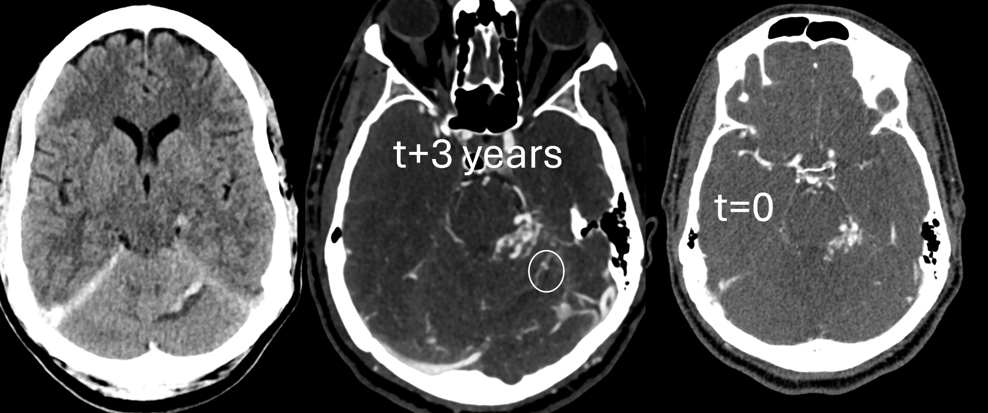
Angios — there are two enlarging proximal AICA aneurysms, however these are not where blood is. The culprit aneurysm is hidden in the AVM

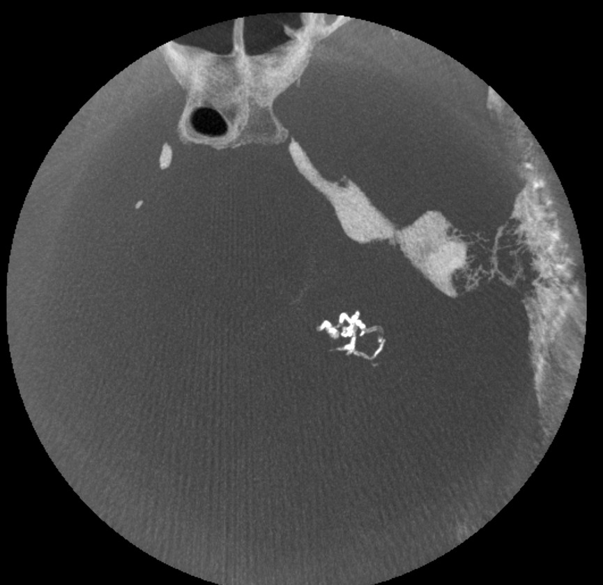
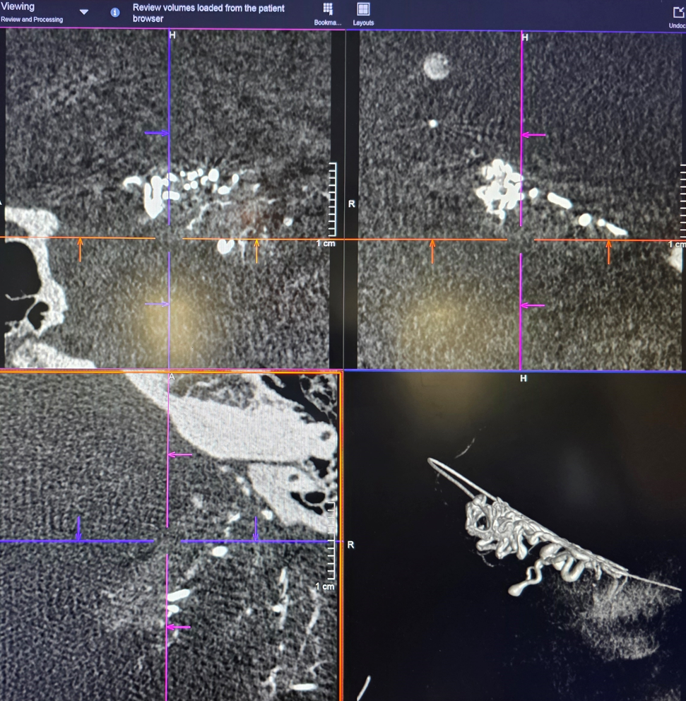
DYNA identifies the suspected rupture site. Axial = top; sagittal = bottom left; coronal = bottom right

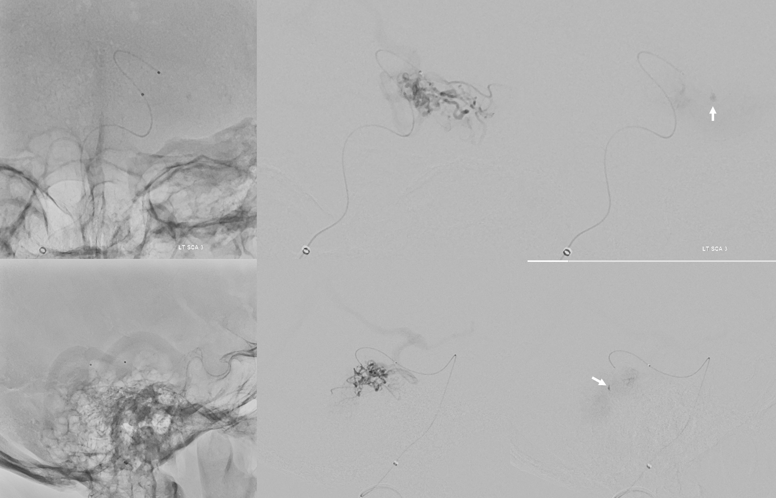
Microcatheter Injection of SCA — contrast stasis in the pseudoaneurysm
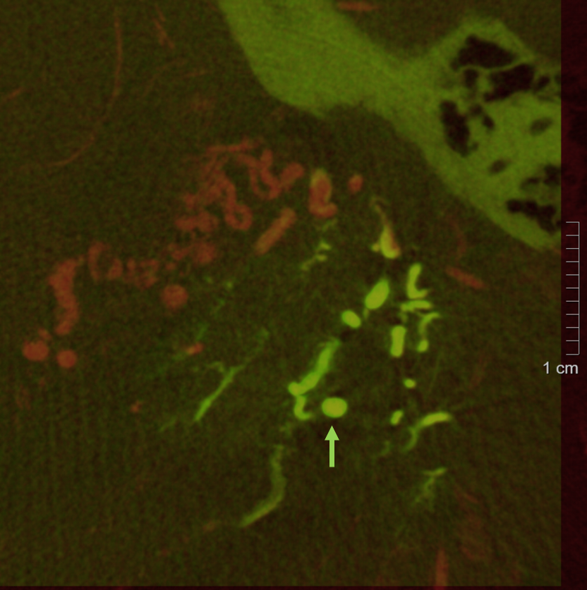
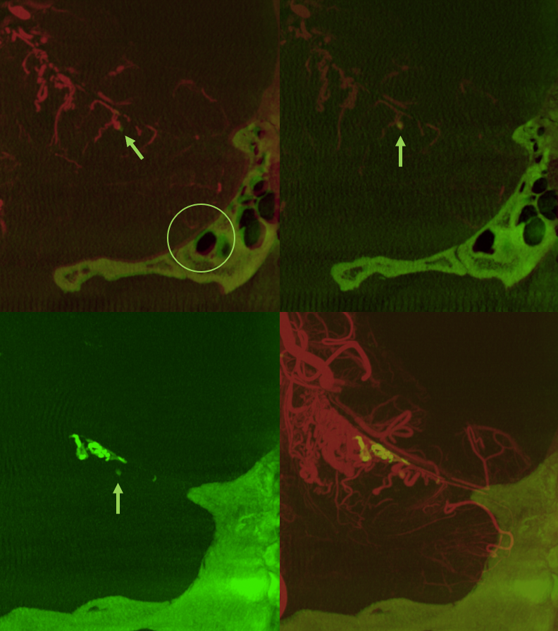
From this position, microdyna is obtained (green) and fused with the right vert global dyna (red). MIP images show the target, as well as some cerebellum
A more selective position is obtained based on the microdyna
Accelerated images of nBCA injection
Post nBCA:lipidol 1:2 injection
DYNA of nBCA cast
A post-injection DYNA is obtained. This needs to be a dual volume injection (native/mask + injection) to see the nBCA better. Protocol as before: Global right vert DYNA — 7 second dual volume (DSA DCT) FOV 22 micro dyna no binning protocol. Injection — 3.5 cc/sec for 35 cc, 3 sec delay. Undiluted contrast. Below is a secondary reconstruction:
Fusion of cast and initial global DYNA. Arrow points to aneurysm with some glue in it. The top right image is misregistered (see halo effect on bone = oval) therefore the glue drop is lateral to the aneurysm. On the upper right image, the misregistration is corrected and glue is where it should be.
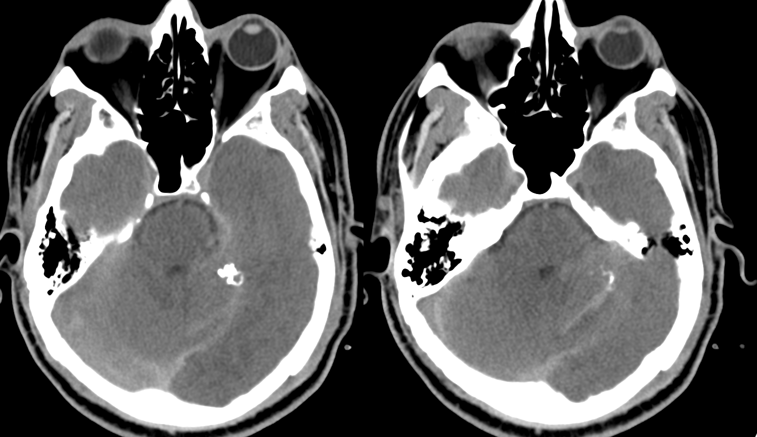
Post CT
How to do this?
Global right vert DYNA — 7 second dual volume (DSA DCT) FOV 22 micro dyna no binning protocol. Injection — 3.5 cc/sec for 35 cc, 3 sec delay. Undiluted contrast
Here is the original VR reconstruction
We need to do better. Creating secondary reconstruction
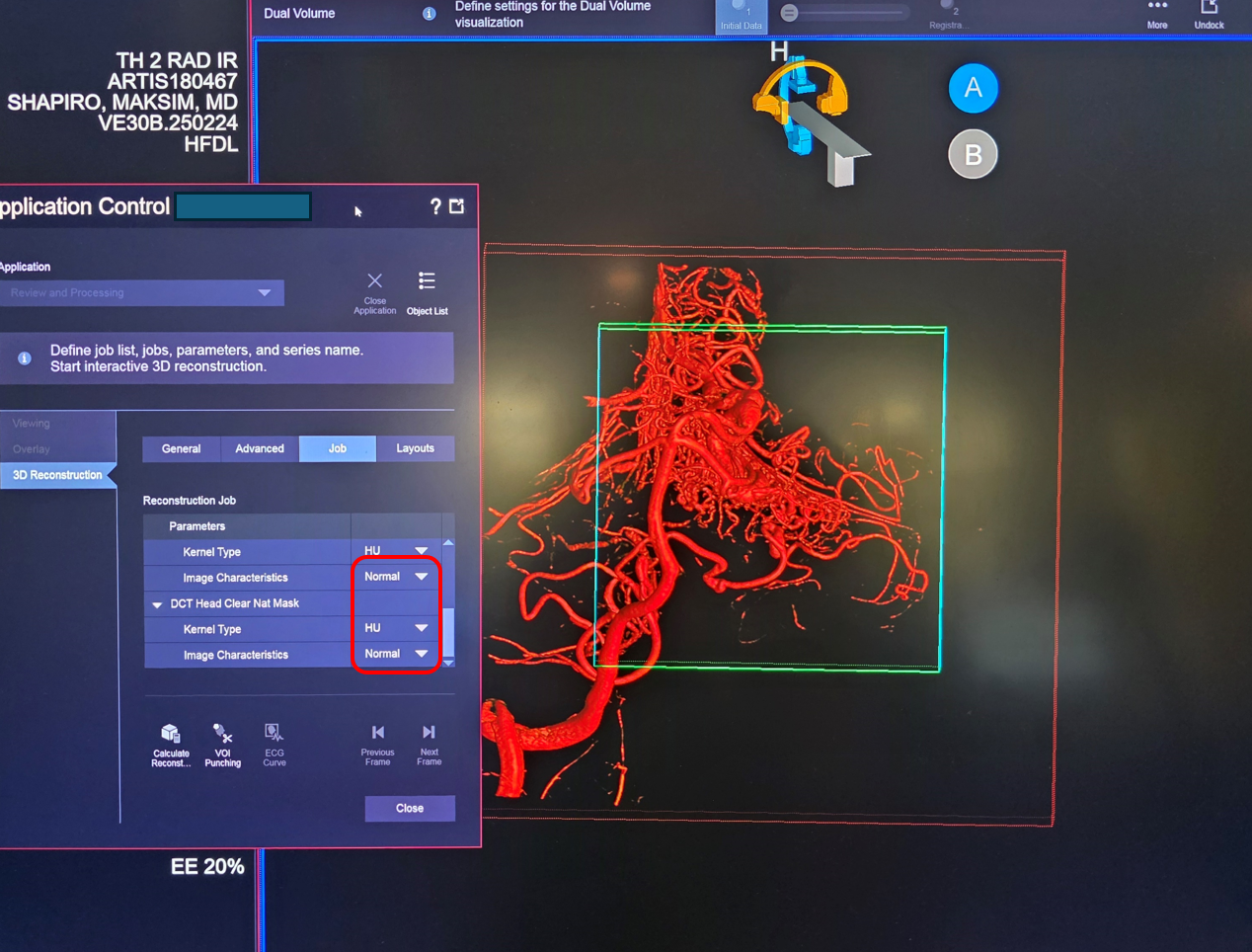
Secondary volume rendered — Subtracted dataset (SUB). A lot of small vessel info is lost in subs. For small vessels, “Nat fill” or natural fill are better.
Nat fill. See those AICA aneurysms? And our target aneurysm too. But how to find the road to it in all this mess?
We do microcatheterizations to find the target. We expect the target to be off the paravermian / medial branch off the left SCA. Here is the microcatheter injection. Contrast stasis inside aneurysm
Now we do DYNA from this position. Microcatheter = Headway duo 156 cm. Hand injection with 3 cc polycarbonate syringe. Protocol = 7 sec single phase (DCT) 22 cm FOV micro DYNA no binning. Inject for 3 seconds before imaging
This is the original reconstruction
Again, we do a secondary reconstruction, as we did with the global right vert one before
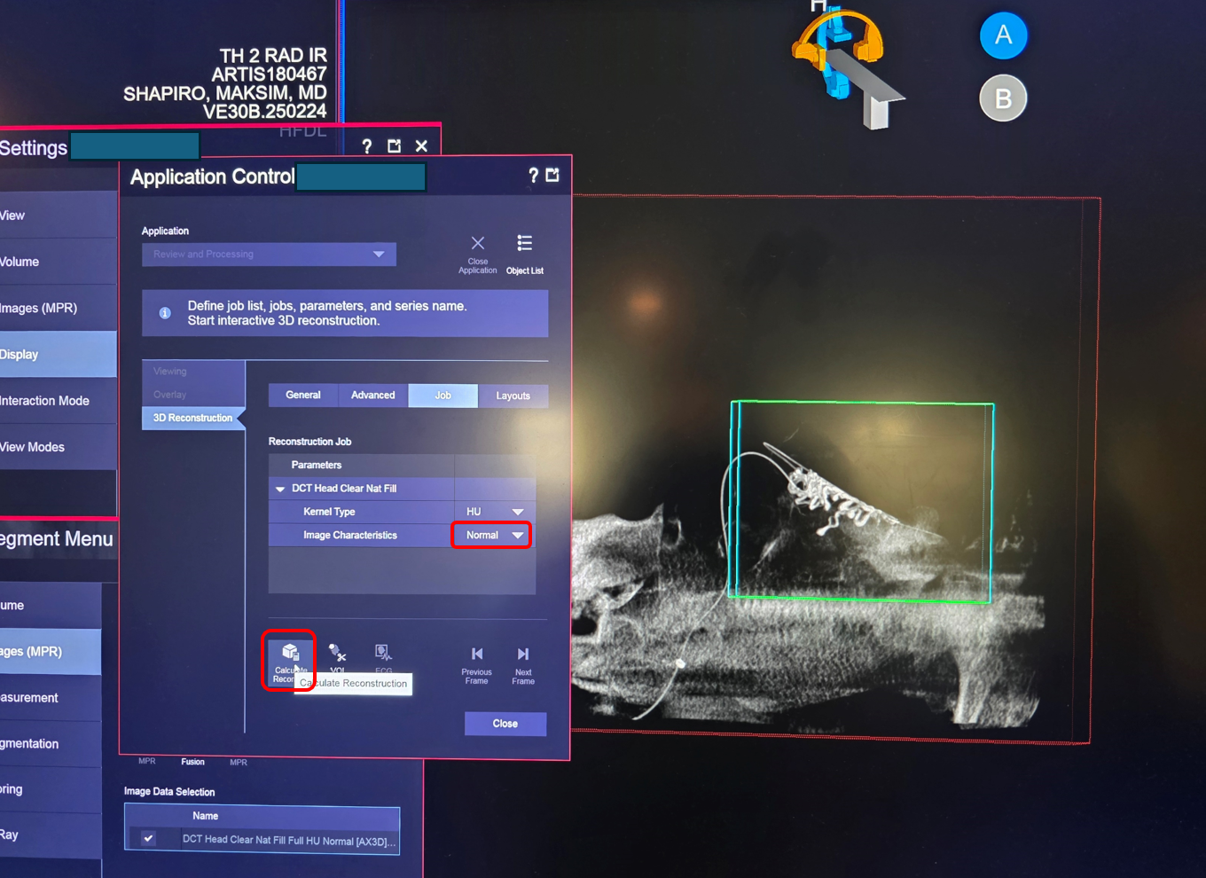
Result
Now we fuse this microDYNA with the Natural Fill secondary recon of the global right vert injection. With the above series open, choose the Nat Fill of the gobal vert DYNA
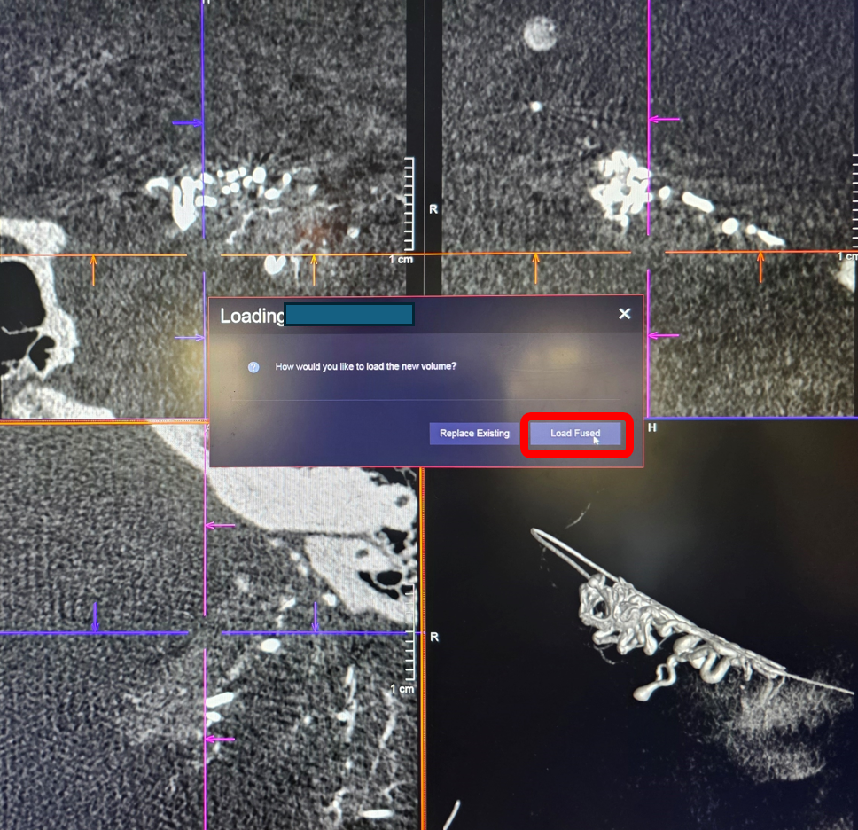
Load fused
A 3D/3D Fusion window should pop up. sometimes it does not — open it from “images” menu

Orange / white — choose “automatic registration” — it might work sometimes
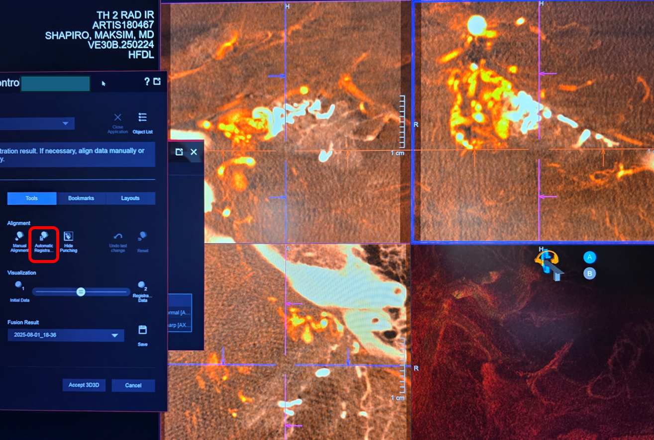
Worked — now we should assign separate colors to each injection
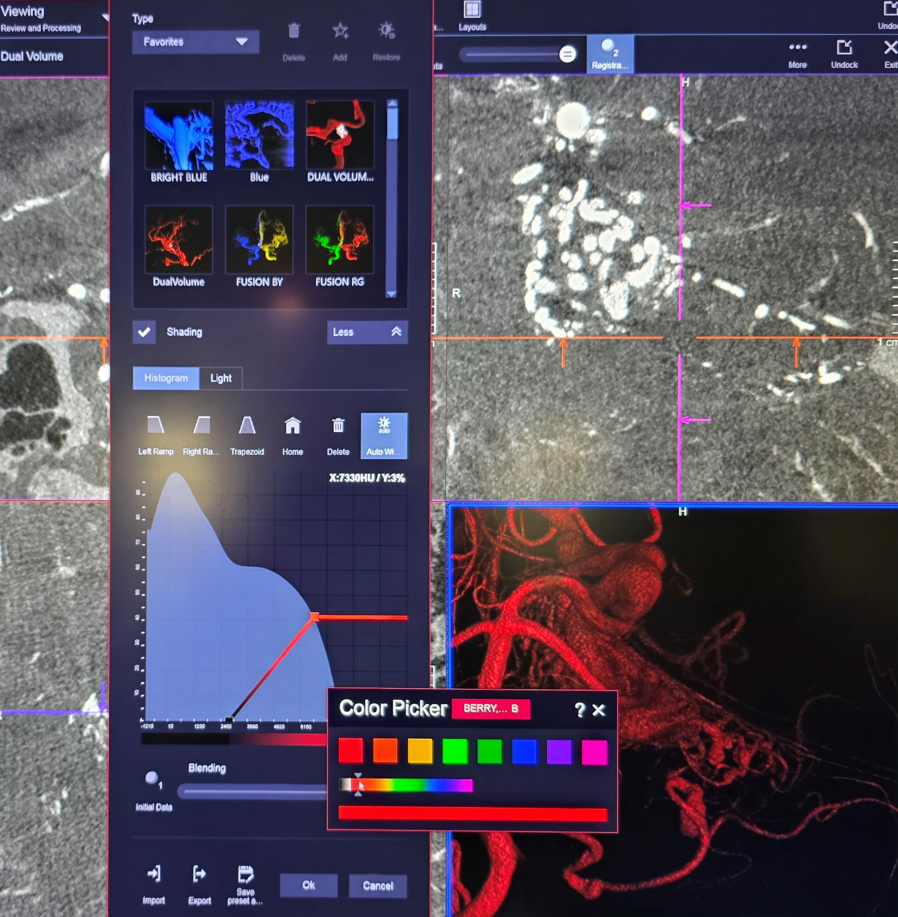
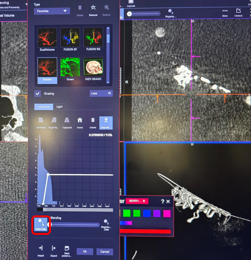
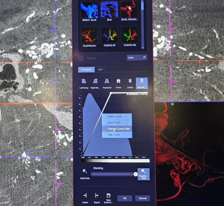
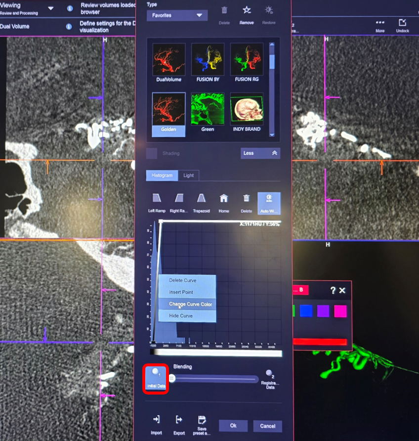
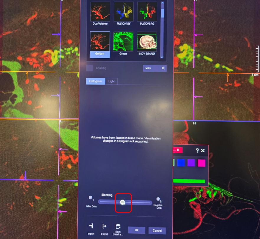
Open “Preset Gallery” . As long as both volumes are displayed you cant manipulate them individually fully. Choose either dataset 1 or 2 (registered or initial data, either one, red outline below)
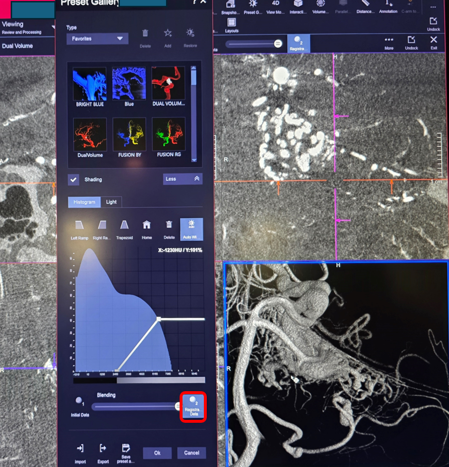
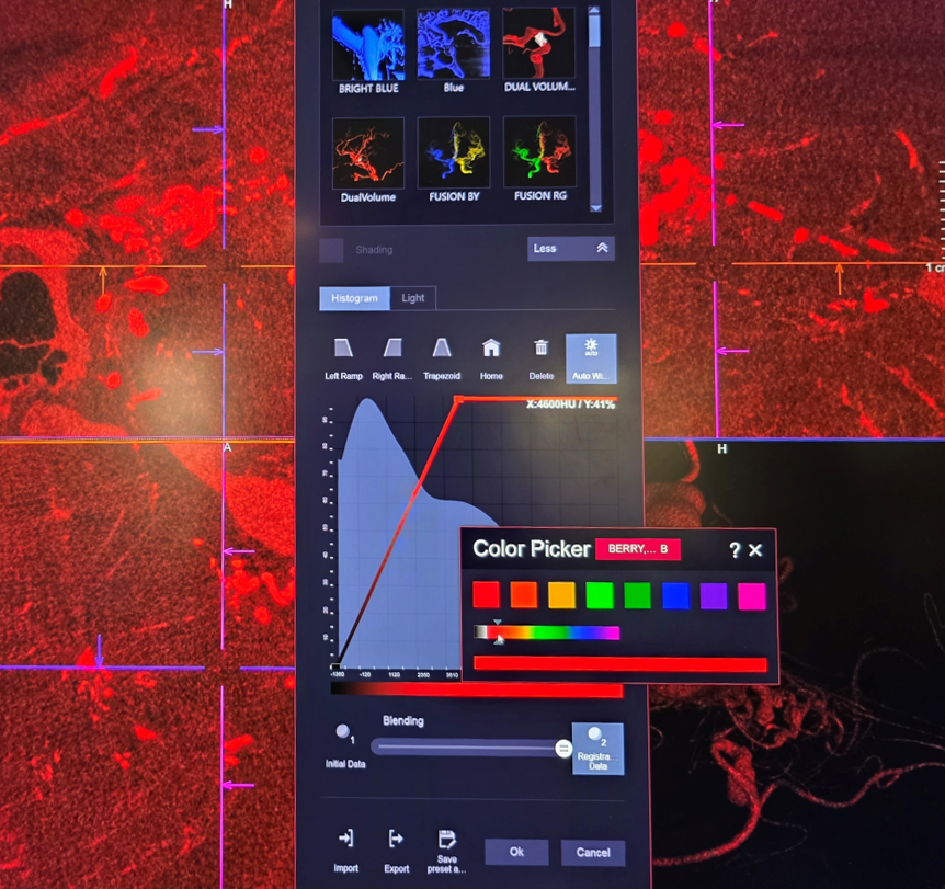
Now you can select color
Now go to the other dataset and choose another color
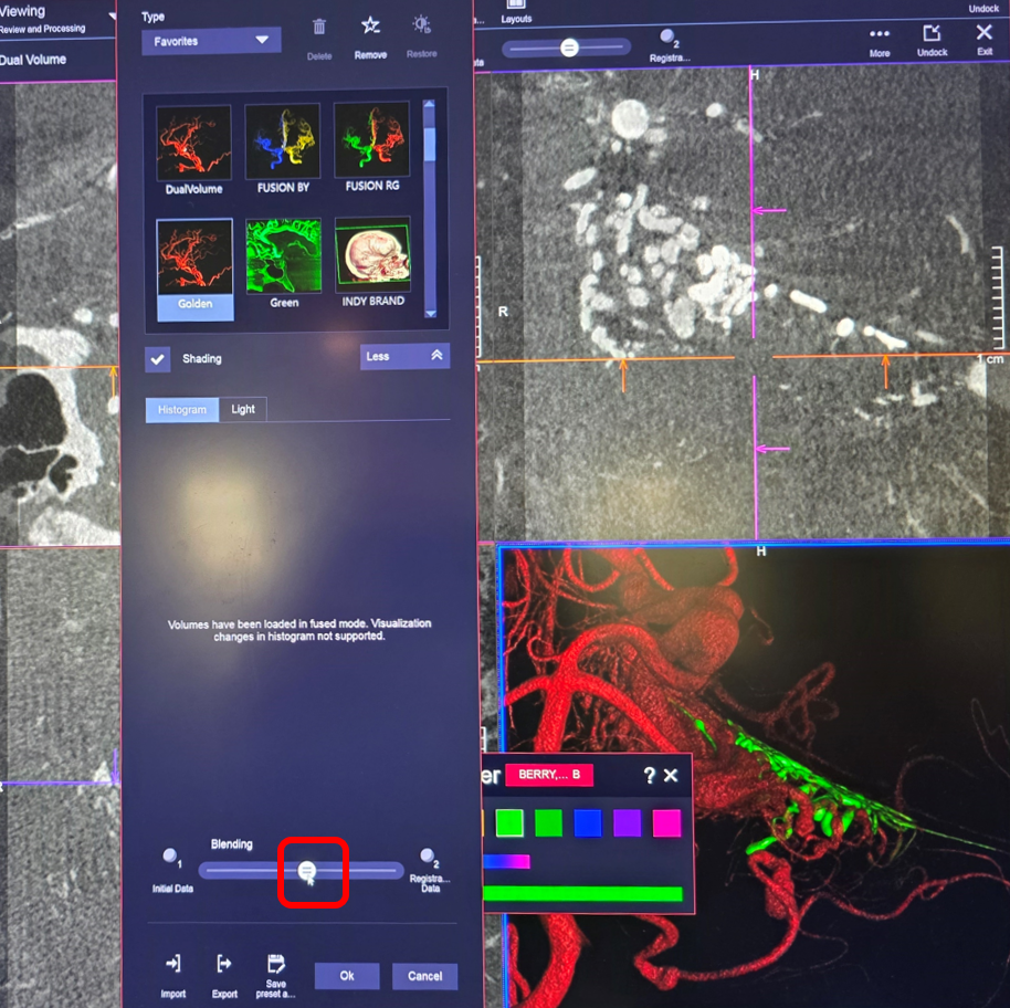
Now move the ball to blend the two datasets. You have two color set in Volume Rendered window
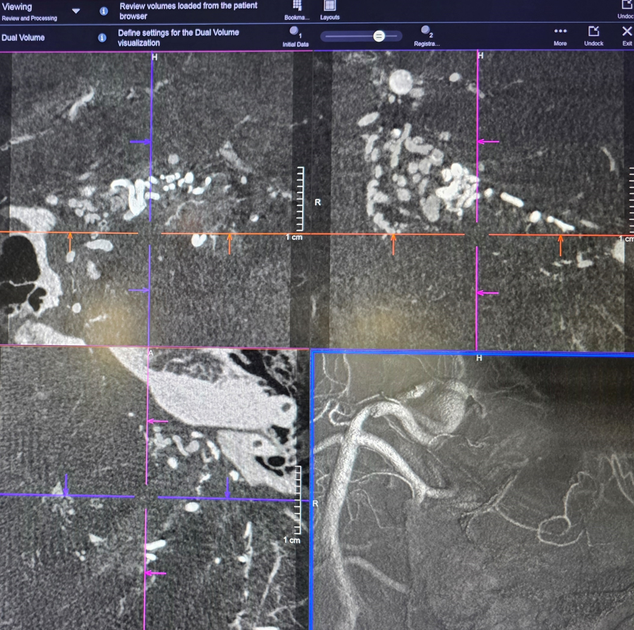
This is what it looks like. Pretty good.
Now we need to do the same for the cross-sectional MIP datasets
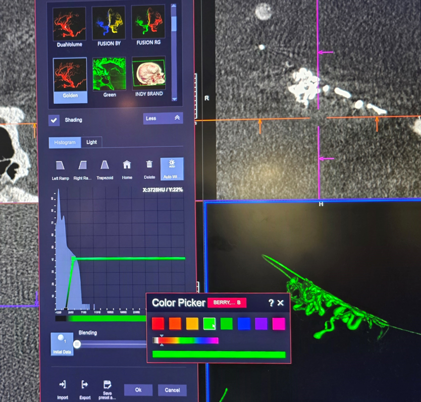
Click on any MIP window. Do the same thing — go to dataset 1 or 2, select change curve color on the histogram…
Helps to have same colors
Now we choose “Initial Data” 1 set
Green again
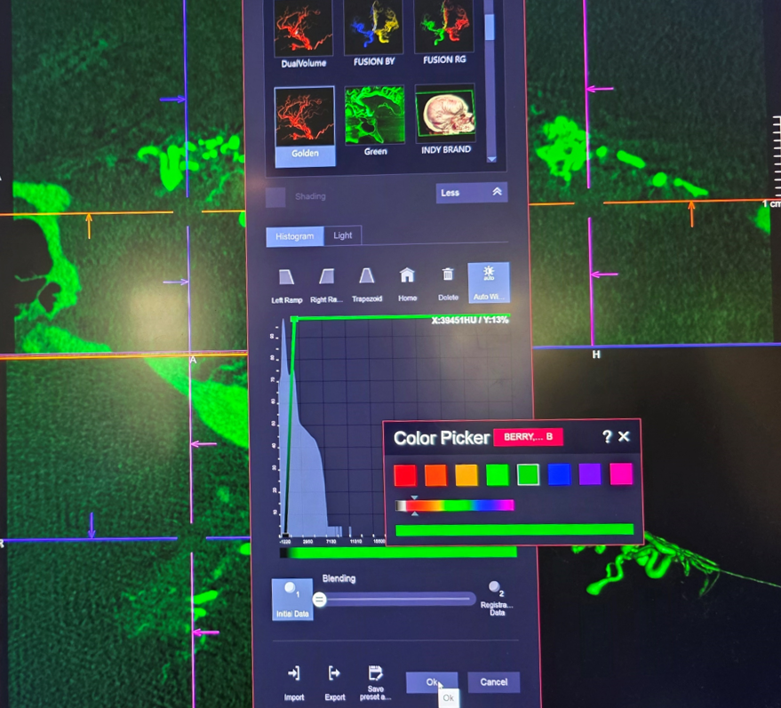
Now blending
Play with Image Slice Thickness for different effects
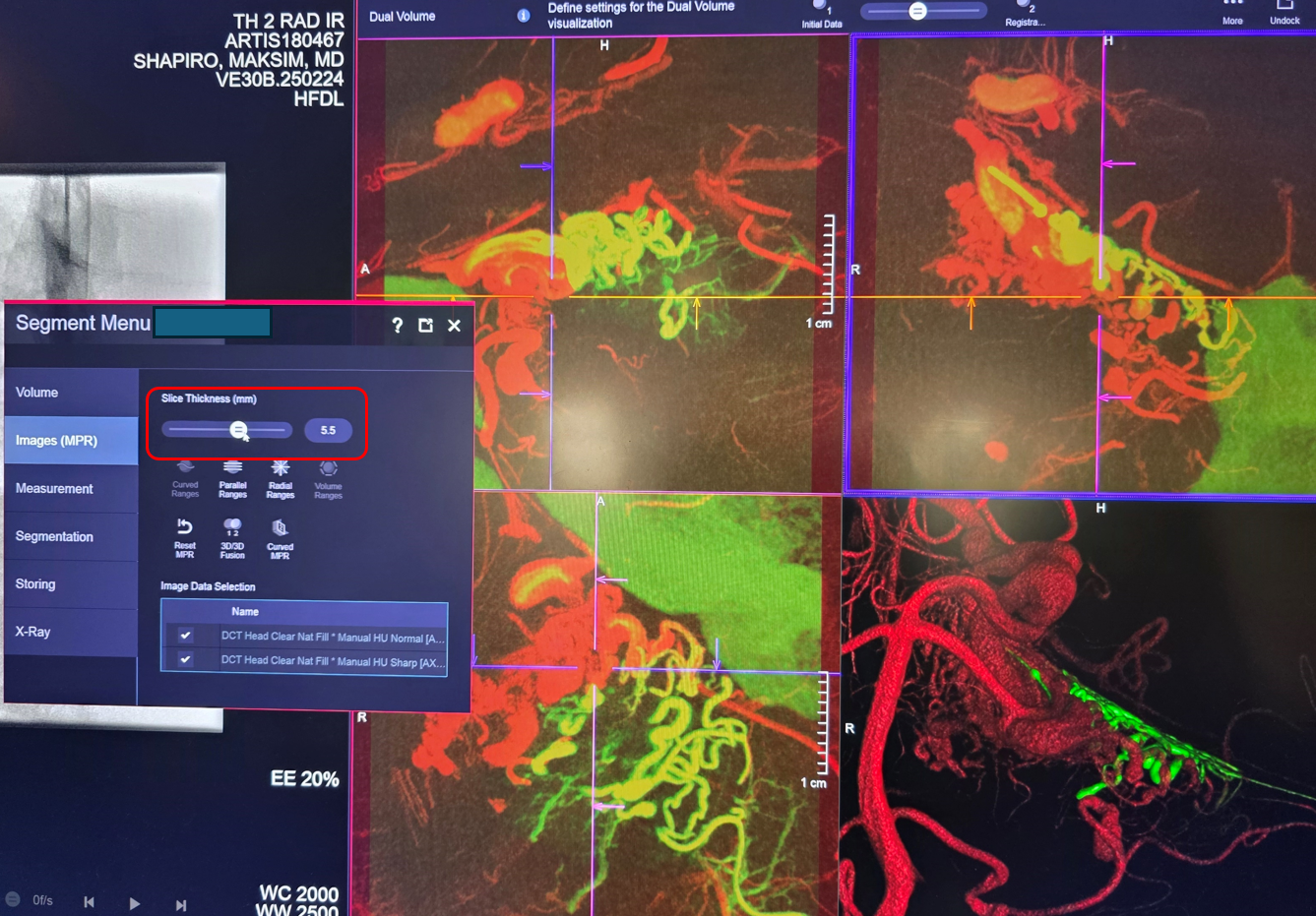
Parallel ranges of the axial fused MIP set
Coronal
There is target and cerebellum there. Based on the above, a more subselective position was reached (see beginning of the case) and target embolized with nBCA
A post-injection DYNA is obtained. This needs to be a dual volume injection (native/mask + injection) to see the nBCA better. Protocol as before: Global right vert DYNA — 7 second dual volume (DSA DCT) FOV 22 micro dyna no binning protocol. Injection — 3.5 cc/sec for 35 cc, 3 sec delay. Undiluted contrast. Below is a secondary reconstruction:
Enough? Well, we did more
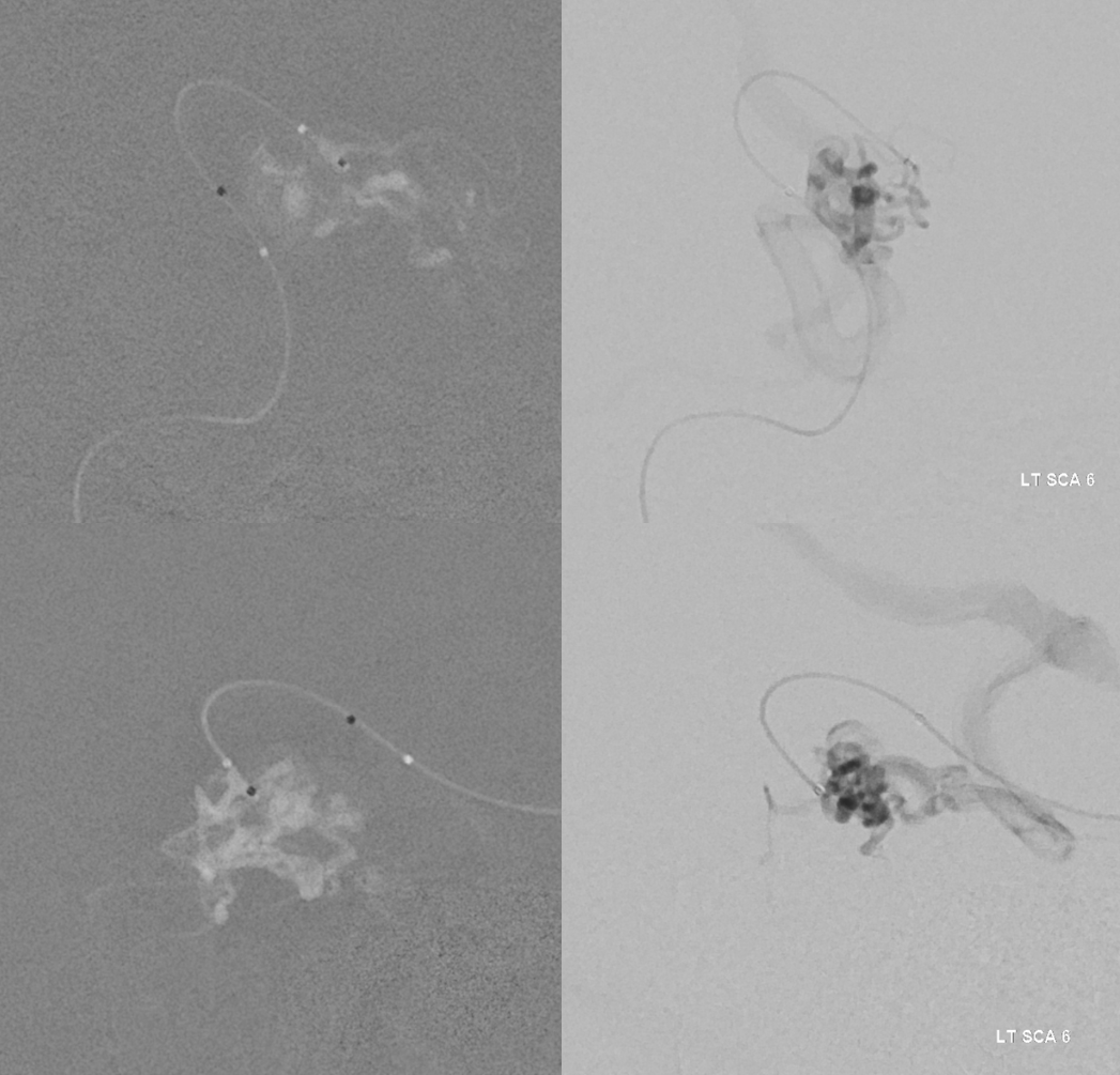
Also did MICRO DYNA of the LATERAL branch of the left SCA
Microcatheter = Headway duo 156 cm. Hand injection with 3 cc polycarbonate syringe. Protocol = 7 sec single phase (DCT) 22 cm FOV micro DYNA no binning. Inject for 3 seconds before imaging
Fuse secondary reconstruction the “natural fill” of the global right vert DYNA with the secondary reconstruction of the microDYNA. We know the aneurysm is NOT in this distribution, but want to do this to plan future embo
Axial MIPS
Finally, we did the same for the AICA… Shown here without secondary reconstructions… so quality is not so good, but enough to see two perinidal AICA aneurysms