
Another great vasa vasorum case from Guglielmo Pero, MD
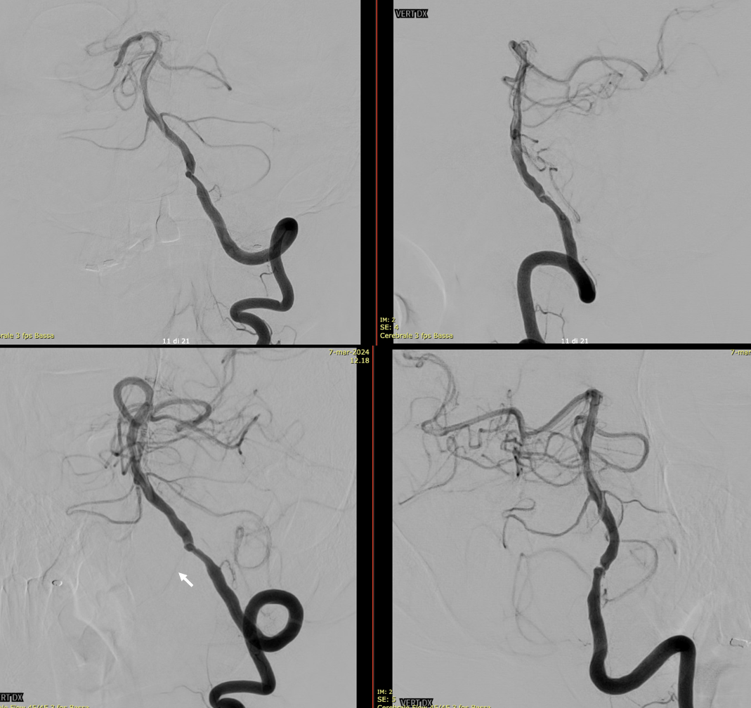
Diagnostic DSA with left vert injection. We can see an irregular stenosis of the pre-junctional vert, AICA-PICA and a small lateral spinal artery (arrow) originating below the stenosis.
So much more to see on advanced imaging
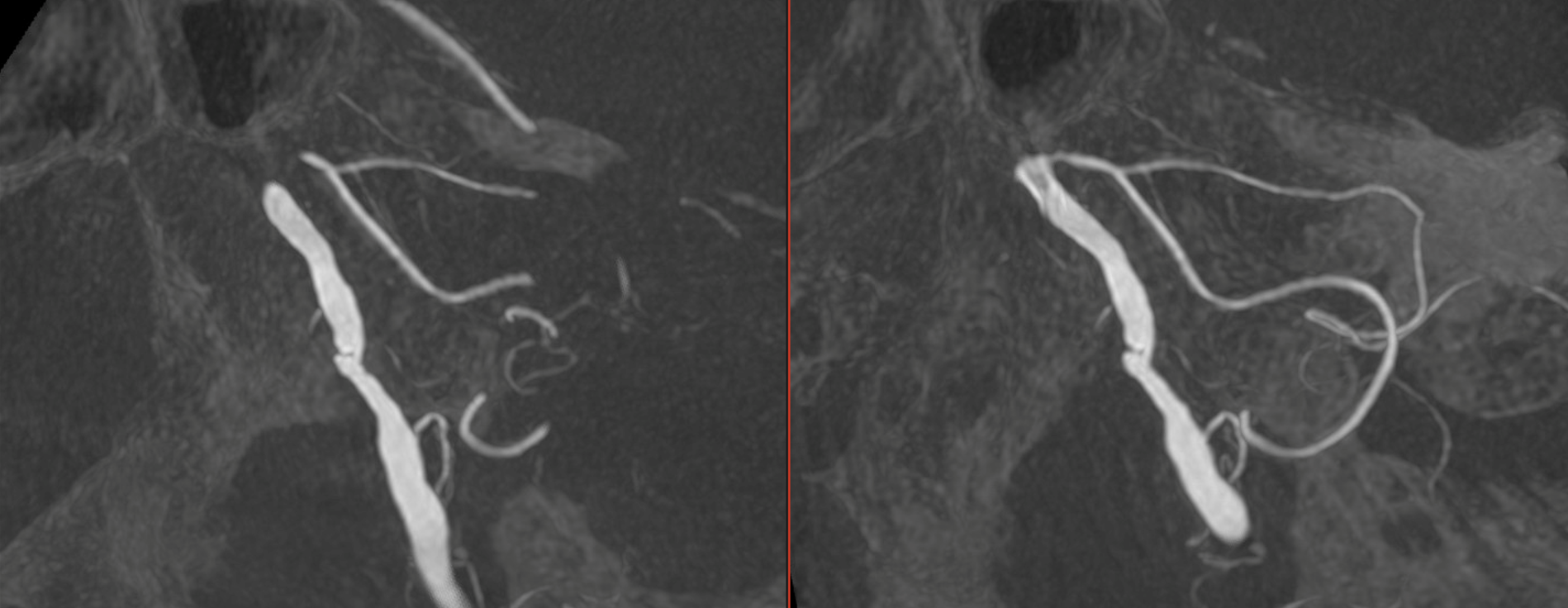
Thin MIP reconstructions of a 3D acquisition (5 seconds, 22 FOV, 20 ml pure contrast – 300 mg/ml – manual injection), confirming the stenosis, the AICA-PICA and the lateral spinal artery. (awake patient).
How much even more with more…
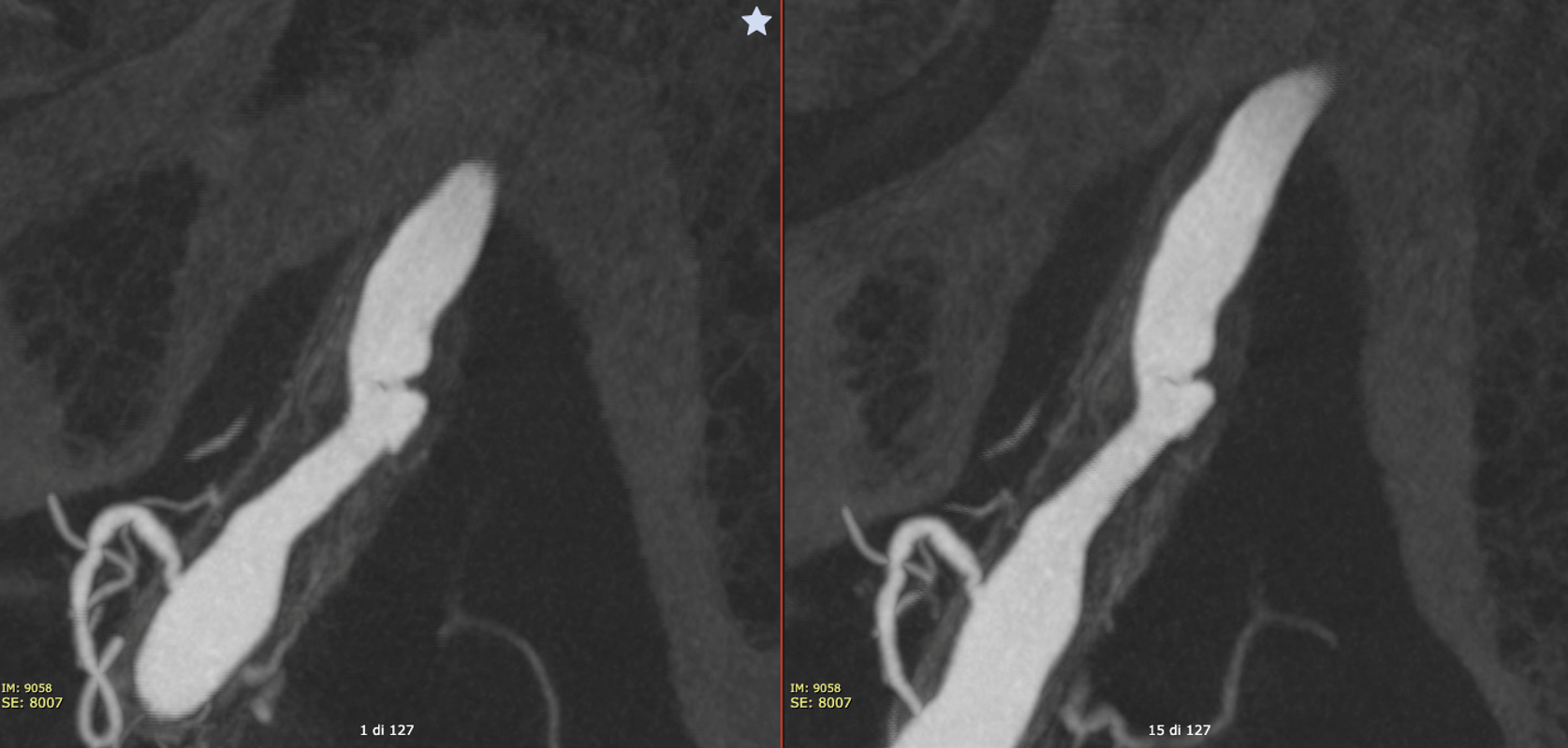
Vaso-CT images are much more detailed. (Vaso-CT protocol 22 cm FOV, 20 seconds acquisition, 20 ml pure contrast (300 mg/ml) manual injection, reconstructed with 50% FOV and 5123 matrix).
Here we can clearly see the vessel wall. The irregular shape of the plaque with ulceration is clearly visible. How much worse does the plaque look?
What are the arrows pointing to?
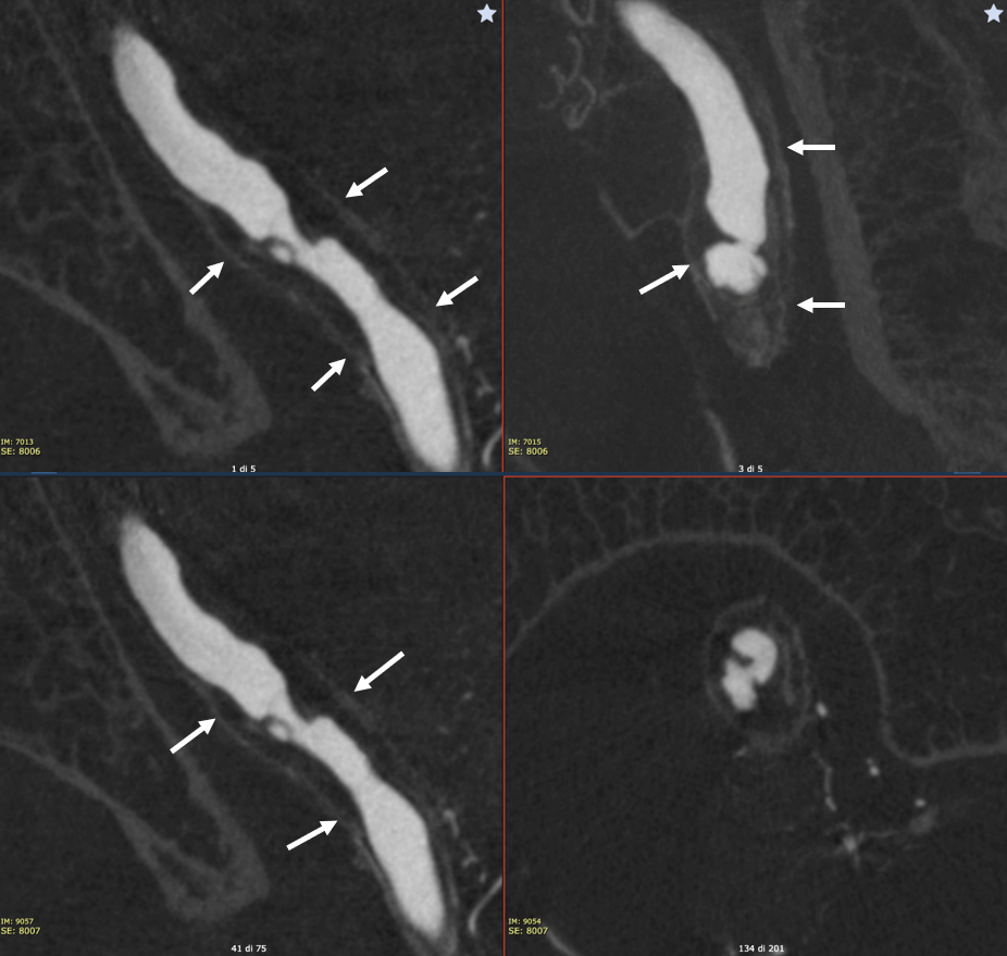
More! The thickened wall and relationship with several small branches suggests mix of calcification and vasa vasorum
Here we can see how the pial connections with the lateral spinal artery (arrow) recanalize the branch that lost its origin from the vertebral artery because of the plaque (dashed arrow)

Stenting
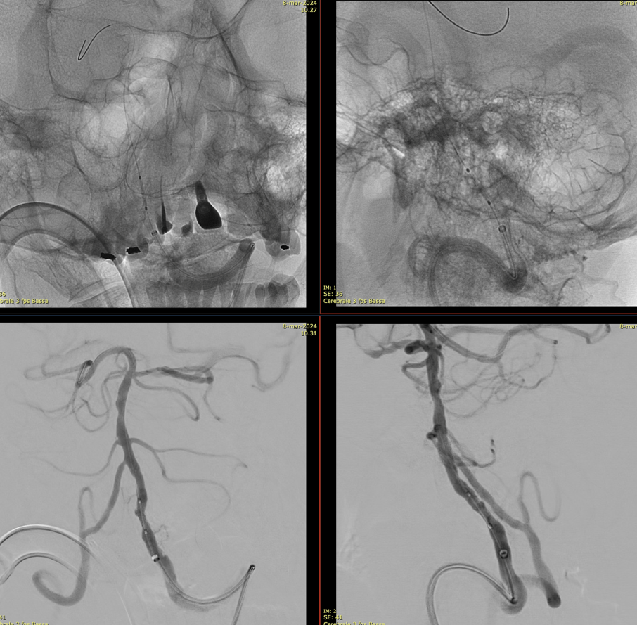
After stenting (Vaso-CT protocol 22 cm FOV, 20 seconds acquisition, 20 ml 50% contrast (300 mg/ml) manual injection, reconstructed with 50% FOV and 5123 matrix), we can still see the ulceration of the plaque, that is filled by the contrast, and the the distal end of the stent not completely apposed to the vessel wall. In these cases, we prefer avoiding overinflation of the balloon because of the risk of rupture of the artery.
