Fusions are not limited to same day procedures. One can fuse remote CT studies etc. The process is the same as for same day procedures. DICOM data must be available on the workstation, that’s all.
Here we show how this works for an AVM with post-resection residual
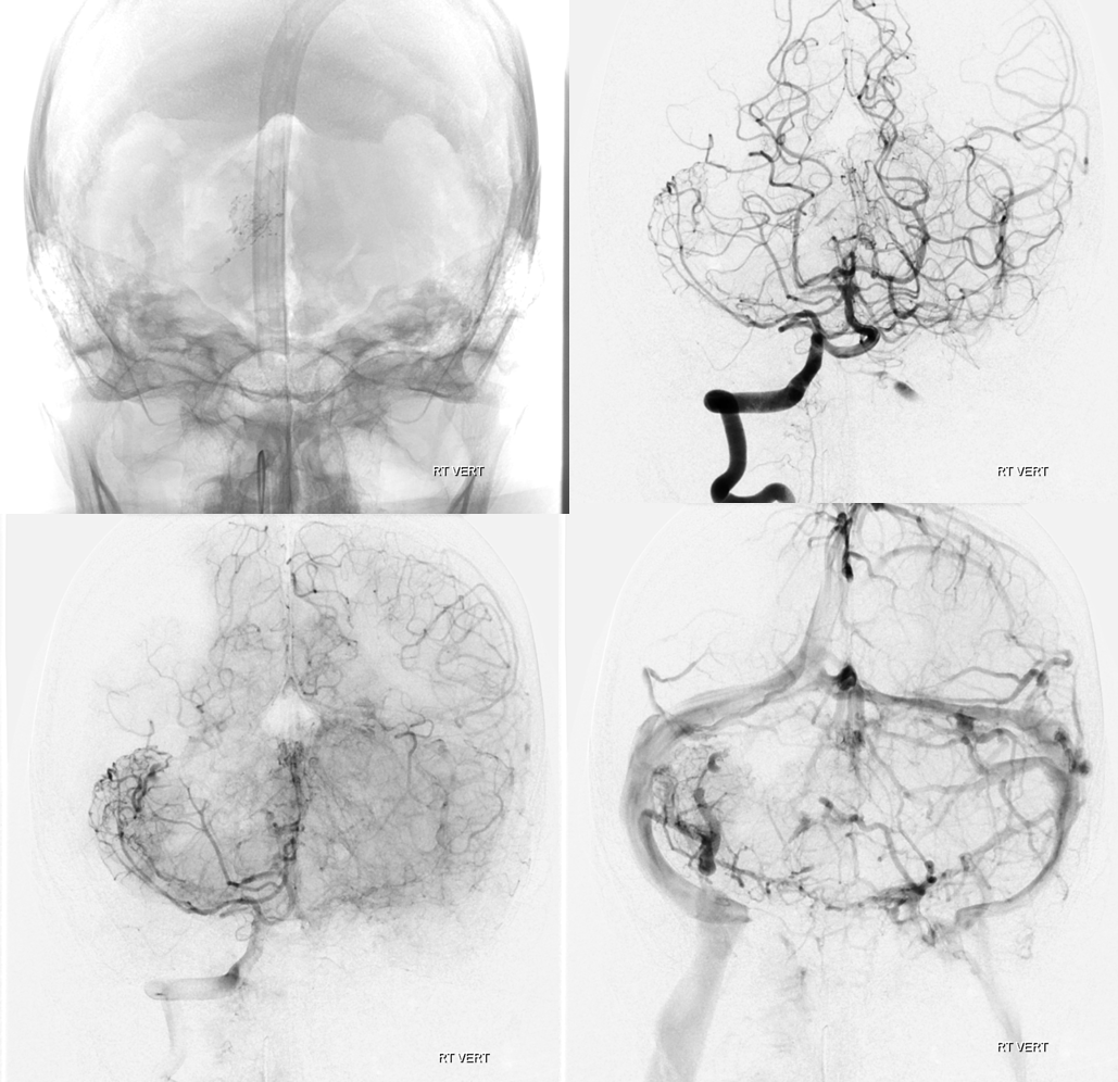
Presentation is hemorrhage
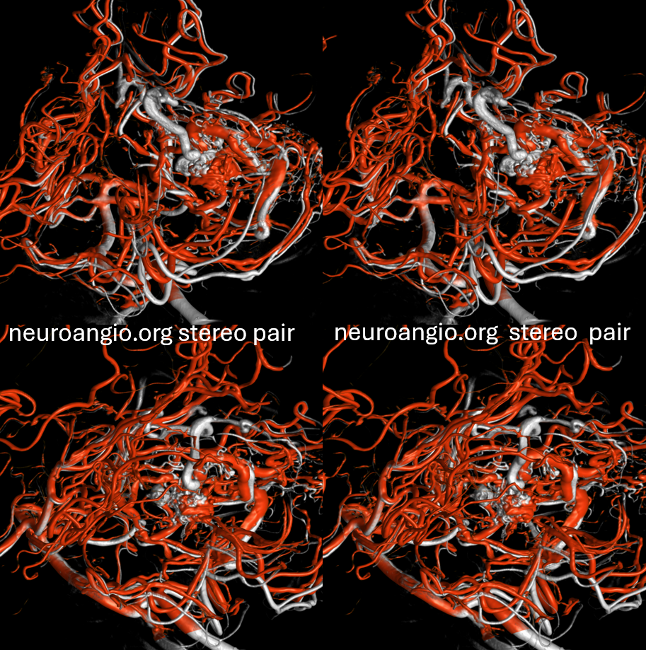
Bottom row are stereo pairs
Post preop embo
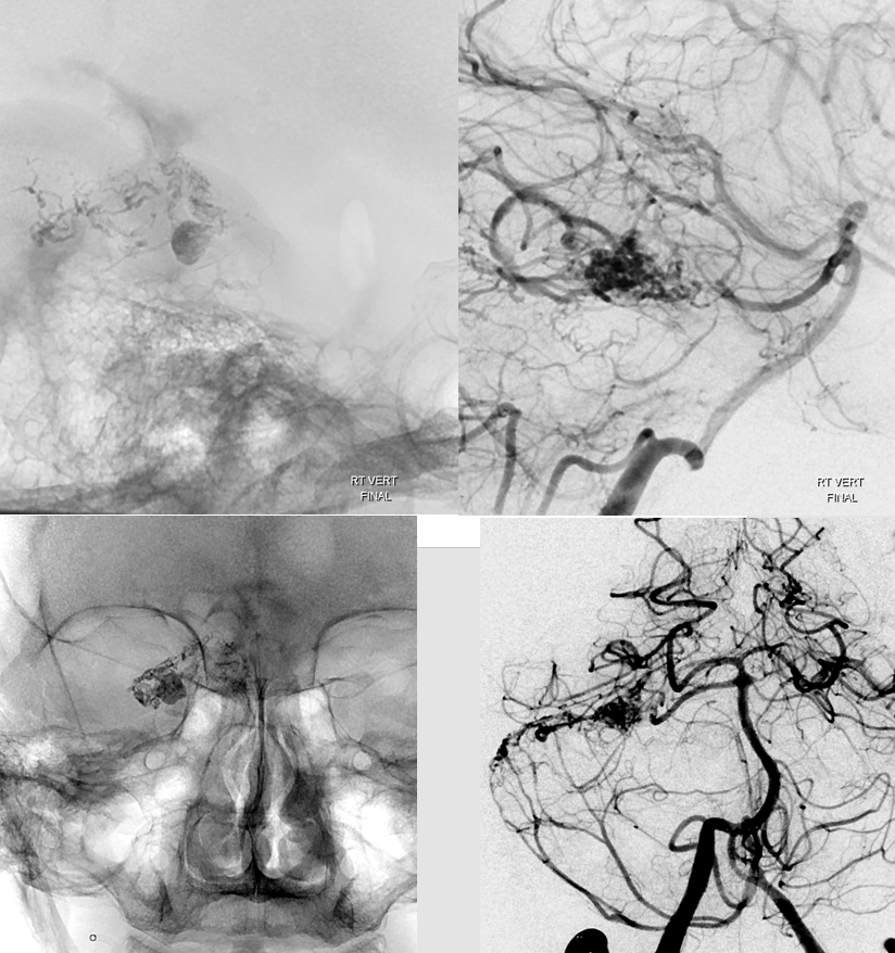
Post-resection there is a large residual
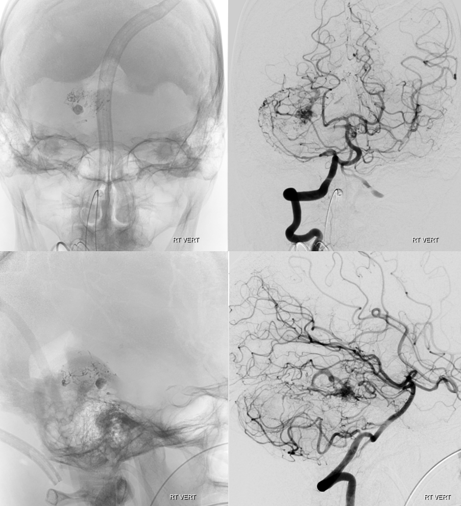
Ok. This happens once in a blue moon. Gotta go back in and take the rest out. Does one need navigation for that? Not really. But in smaller residuals it can help. Here we have a big one to show the idea of how to do it.
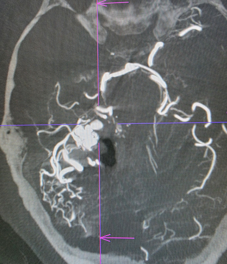
Make a DYNA of the residual. Siemens Q, 10 second DSA/DCT dual volume acquisition. FOV = 42. Large FOV to allow for navigation co-registration if needed. Injection 3 cc/sec for 36 cc, 2 sec delay, 100% contrast.
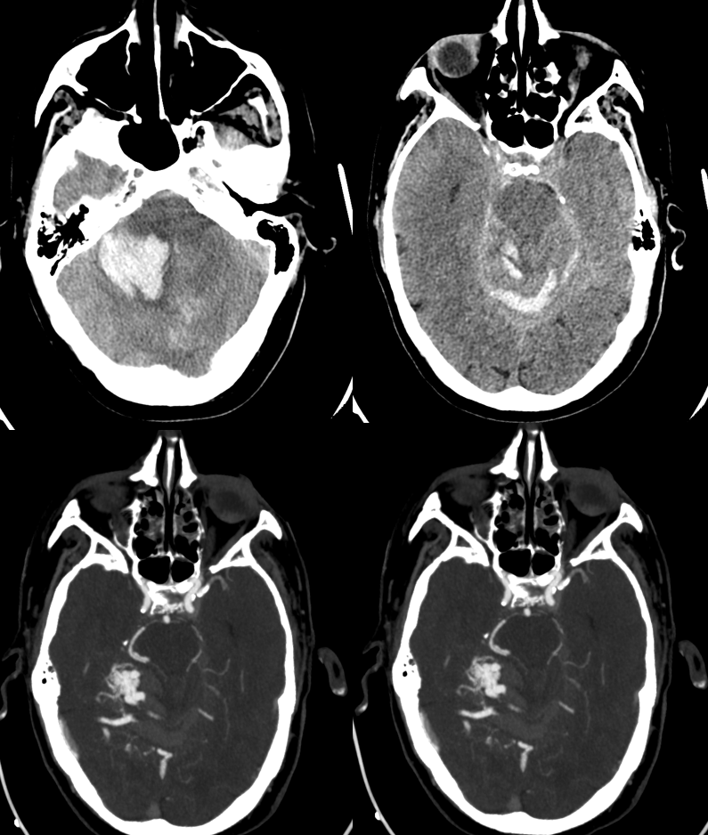
Here is the residual with the surgical cavity medially
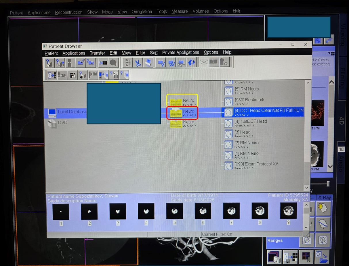
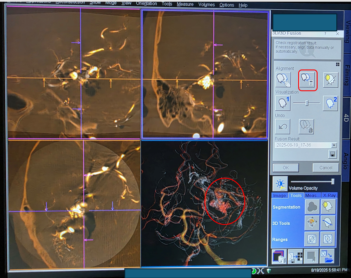
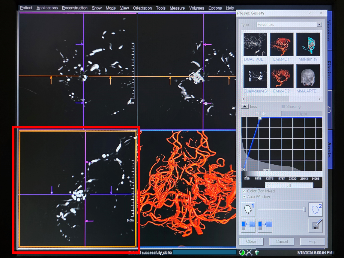
Load the post-resection “SUB” dataset (can use nat fill also) (most recent dataset is in the folder outlined in yellow. Now add the “Nat Fill” pre-embolization DYNA dataset (outlined in red)
This is the fusion. Almost perfect alignment. Hit “automatic” alignment to see what happens.
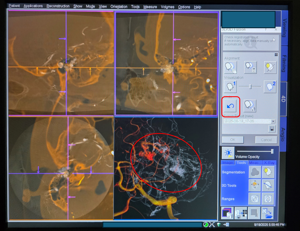
Opps. looks much worse (red oval). Click “undo”
align manually and painfully
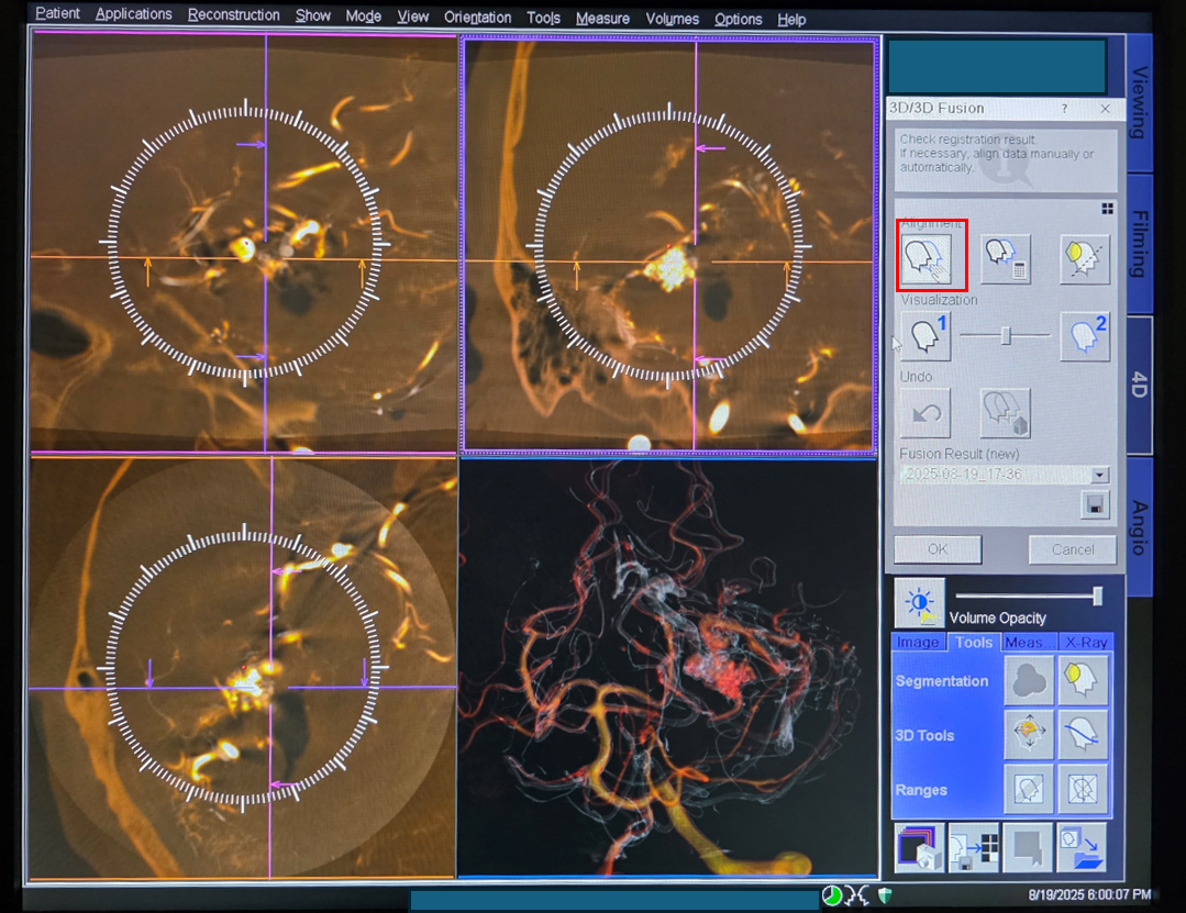
Not bad
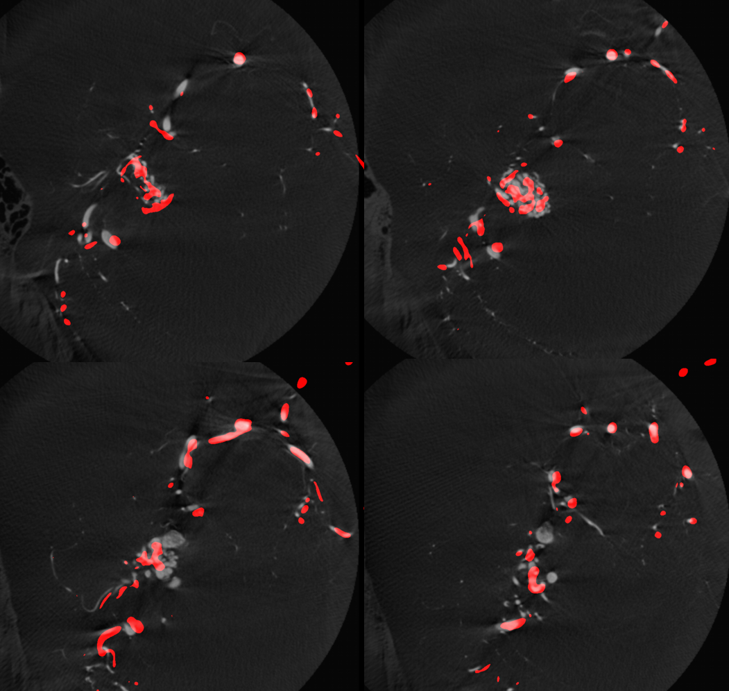
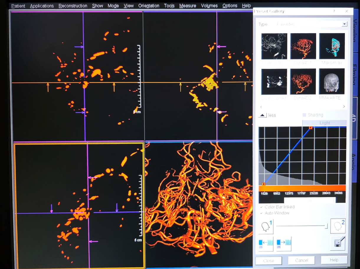
Now select any MIP window (red square). Change color to red (the preop dataset is in white).
Siemens Q does not do well with MIP color changes. The resulting dataset is binary, without hues. But its good enough.
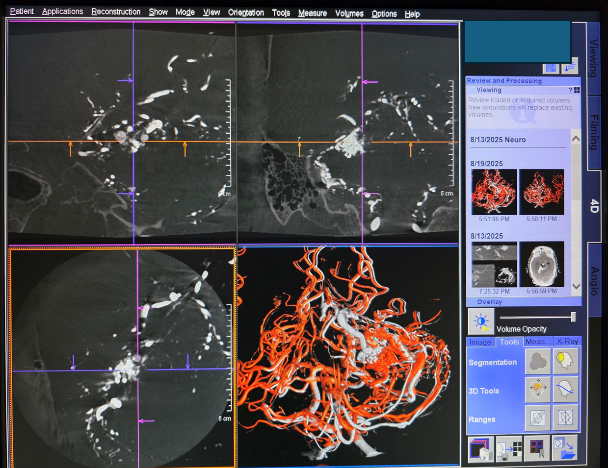
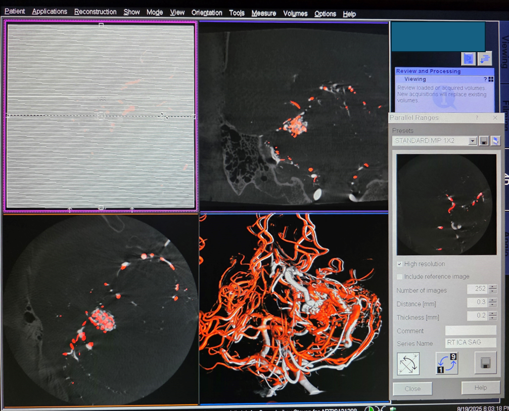
Now blend in the pre-embo, pre-resection dataset. Make rotational volume ranges (see other pages for examples). Select “Parallel Ranges” in the sagittal window to make axial or coronal parallel ranges set. Vary Distance between slices and slice thickness as needed. Make them high resolution.
Here you go
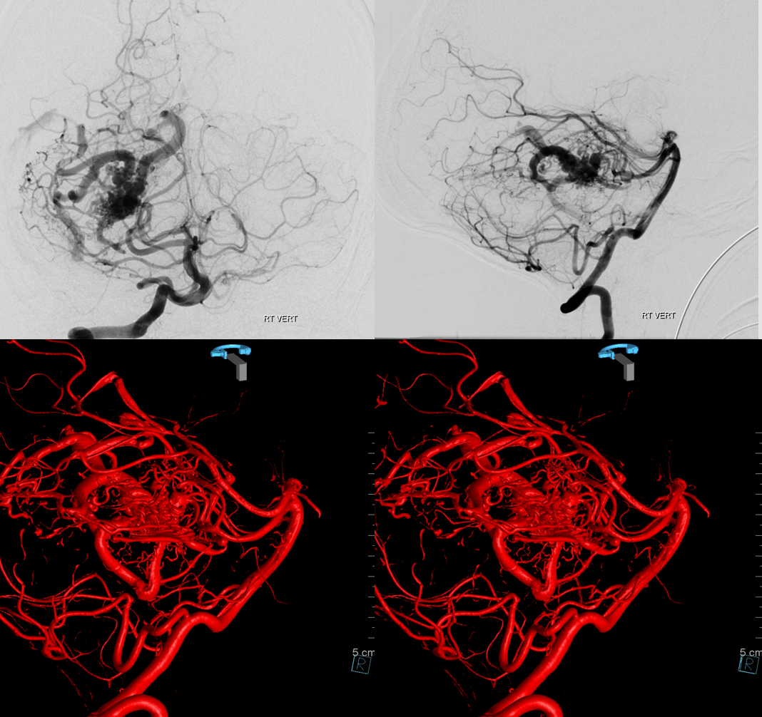
Post-re-resection — all is well now