There is indeed literature on this — largest series from Boston Childrens here

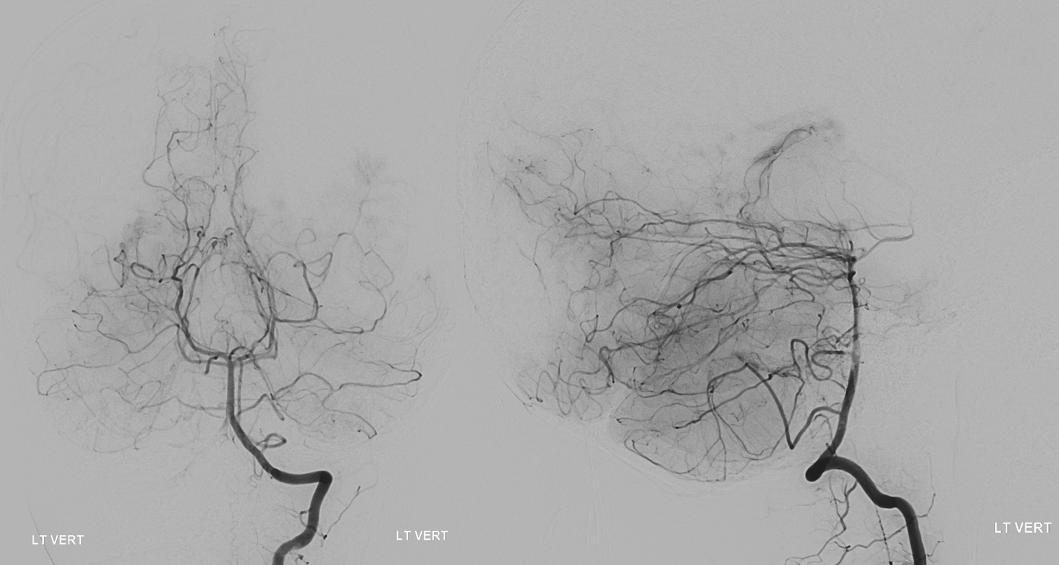
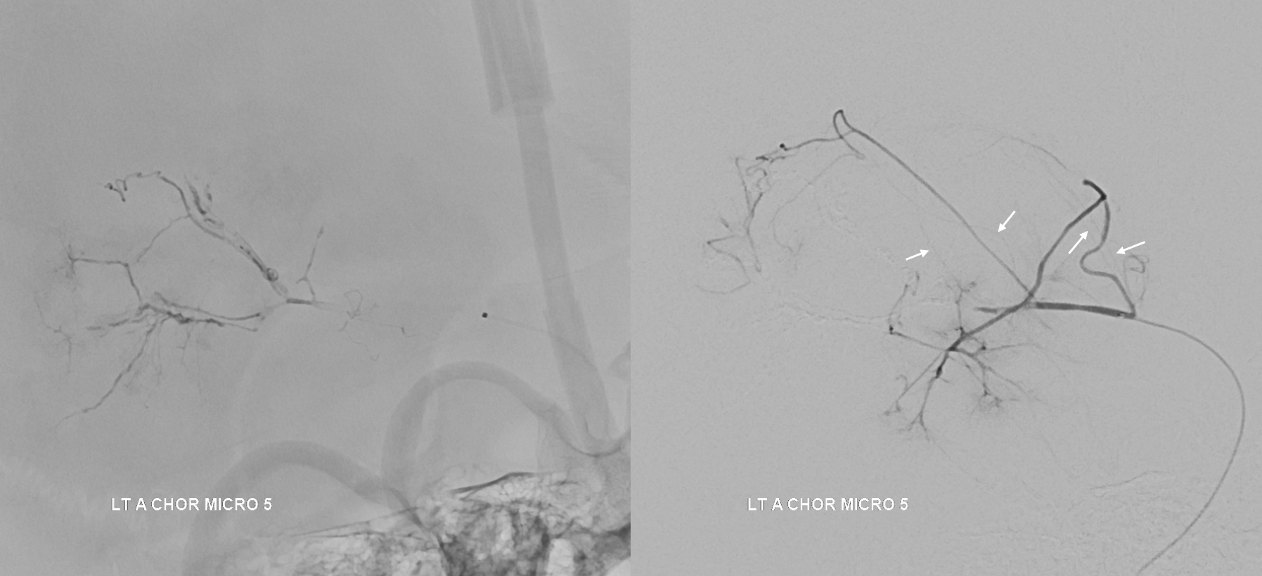
Majority of supply is from anterior choroidal. What is the mistake in the acquisition of following images?


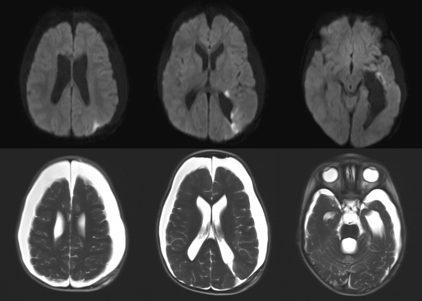
Can you see the normal parenchymal territory below?
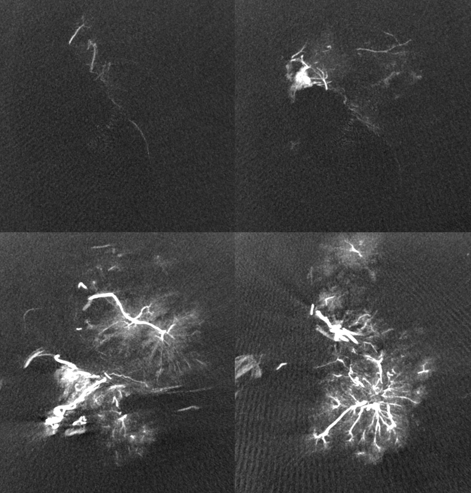
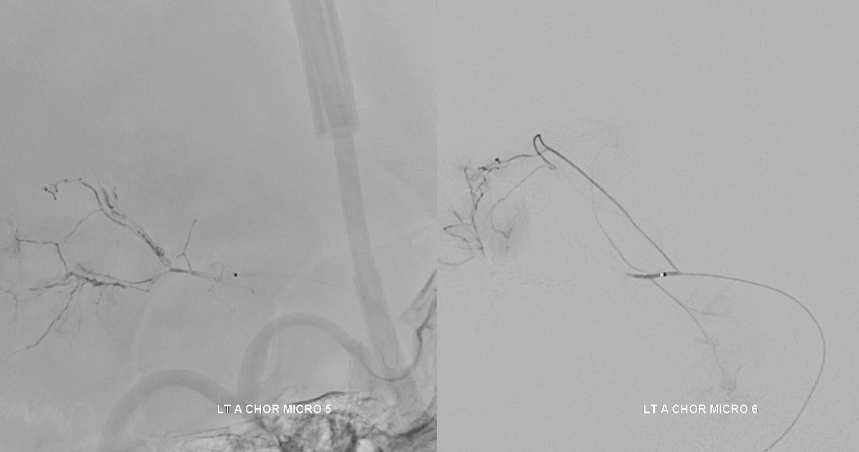
Multiple sequential catheterizations of intraventricular pedicles supplying the tumor, followed by dilute nBCA injection (nBCA:Lipidol 1:3)

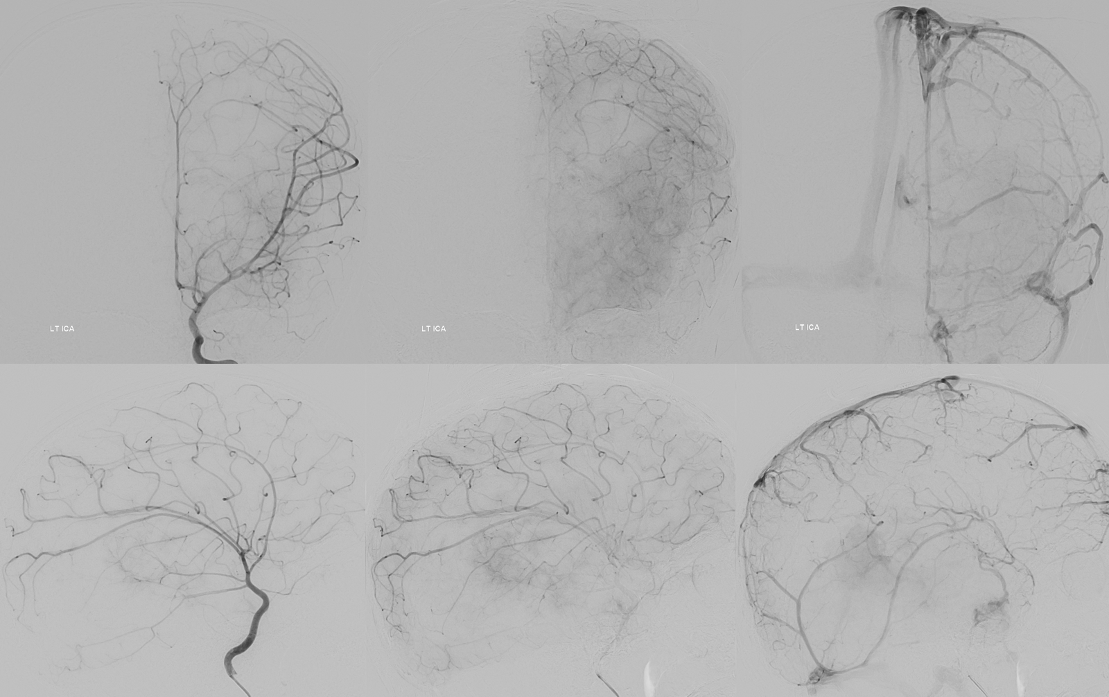
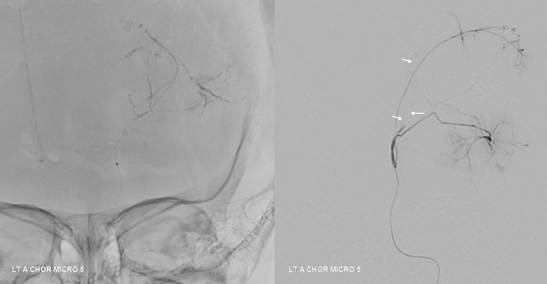
As the tumor is embolized, extremely small (but eloquent, and small because this child is very small) are better seen (arrow). What is the hallmark of residual tumor supply in regard to the classic choroidal anatomy? What is the equivalent of “pial supply” here?
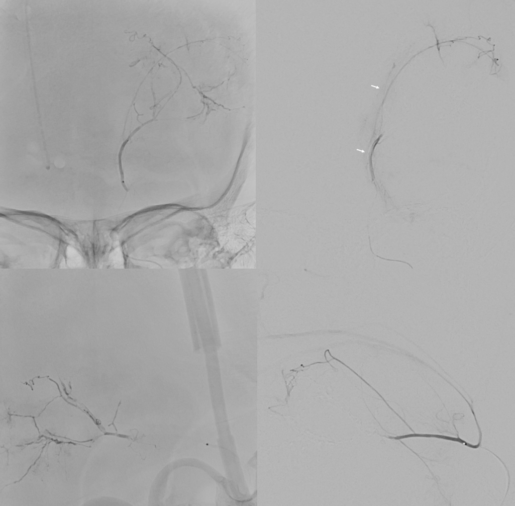
The most proximal pedicle was too small for catheterization, and developed severe spasm in the attempt. This allowed an even better view of the normal choroidal territory previously not so well opacified. The residual tumor supply is also well seen. Again, what is so interesting here?
Final hint