Increasingly, it is recognized that for many, perhaps most large/giant partially thrombosed aneurysms, vasa vasorum are the principal factor keeping the aneurysm alive. They are blood vessels which feed the thickened, pathologic aneurysm wall — recruited from either pial or dural sources. In other words, the wall has a life / blood supply of its own, independent of whatever is going on inside the aneurysm. This is why stuffing these types of aneurysms with coils or anything else is useless. Flow diversion is also frequently not successful — either because of insufficient device coverage — the misguided one-and-done strategy — or vasa vasorum which are not touched by flow diverters.
With Cone Beam CT, vasa vasorum are seen in pretty much all large/giant partially thrombosed aneurysms. Below are some examples
A — vision loss
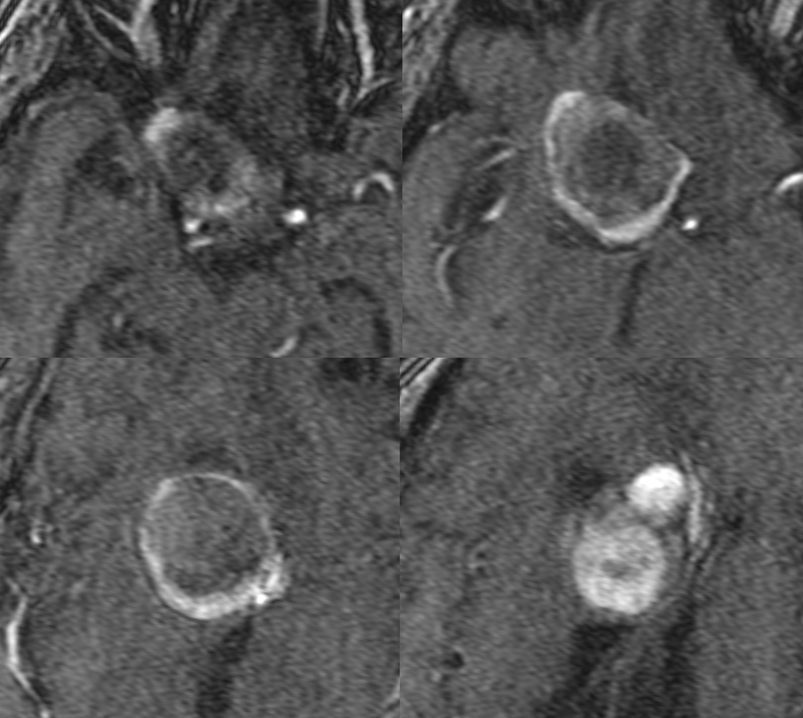
MRI – pre-T1 (top), post (middle), FLAIR, T2, Diffusion (bottom)

MRA
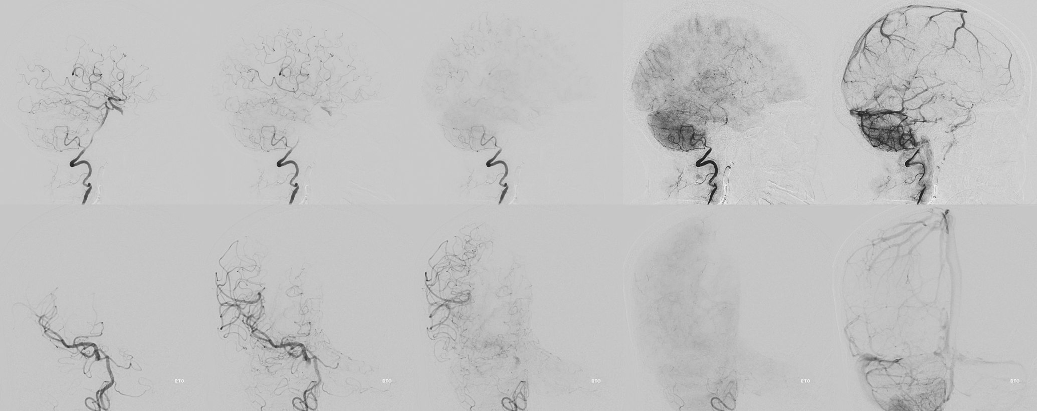
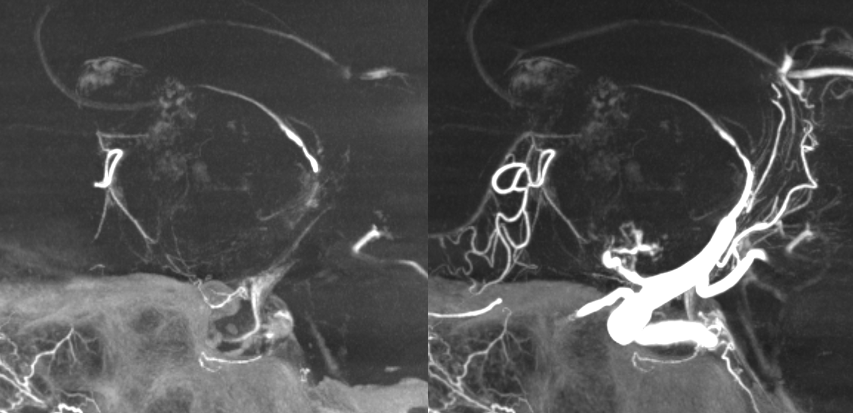
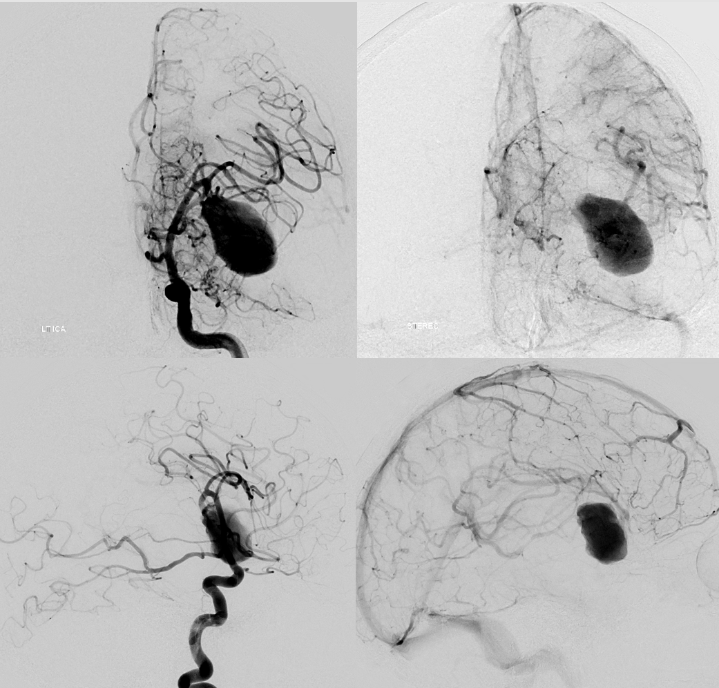
Angio — barely any inflow at the base. Vasa vasorum are basically impossible to see on 2D-DSA. Cone Beam CT is needed

BTO. The A1 is hypoplastic
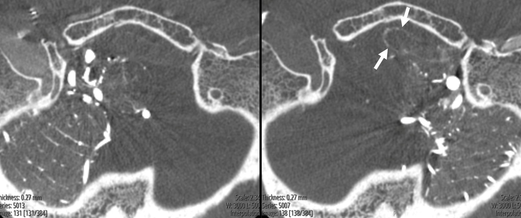
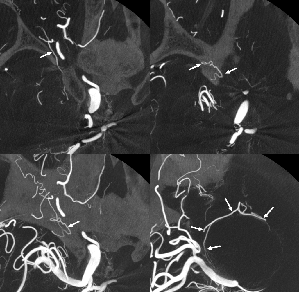
Cone Beam CT

With arrows — vasa vasorum are curiously dural in origin here — via deep recurrent meningeal (dashed arrows). Solid arrows are surface vasa vasorum
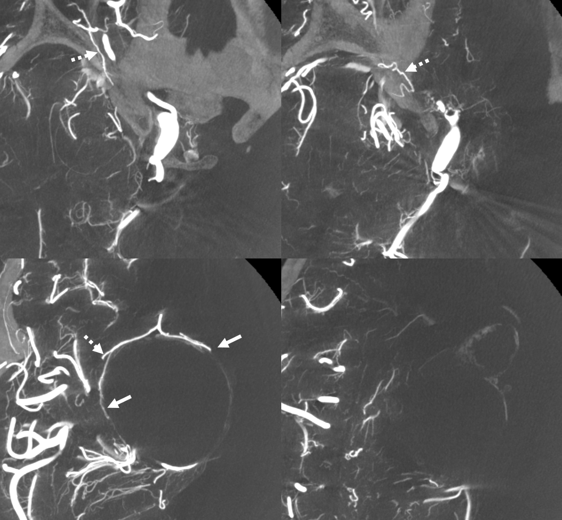
Sagittal MIPS show hazy eggshell wall enhancement
Video — Siemens Q, FOV 22 micro, 10 sec DCT, 3 cc/sec for 36 ccs 2 sec delay. Thin axial MIP
A better image is obtained a bit later on Siemens Icono 7 Sec dual volume DSA DCT unbinned micro FOV = 22 cm, injection 3 cc/sec for 30 ccs 3 sec delay 100% contrast
Video — same acquisition as above
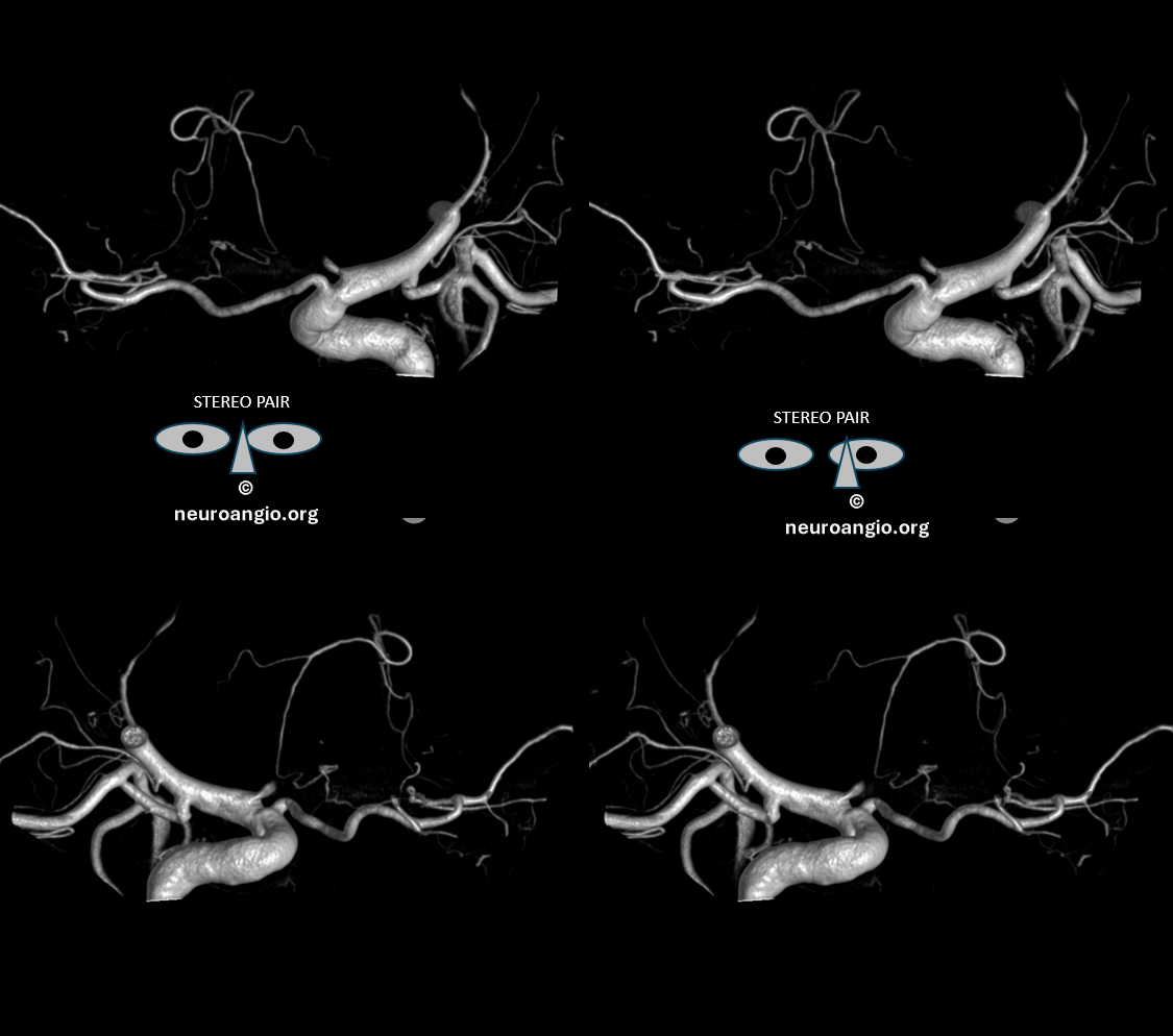
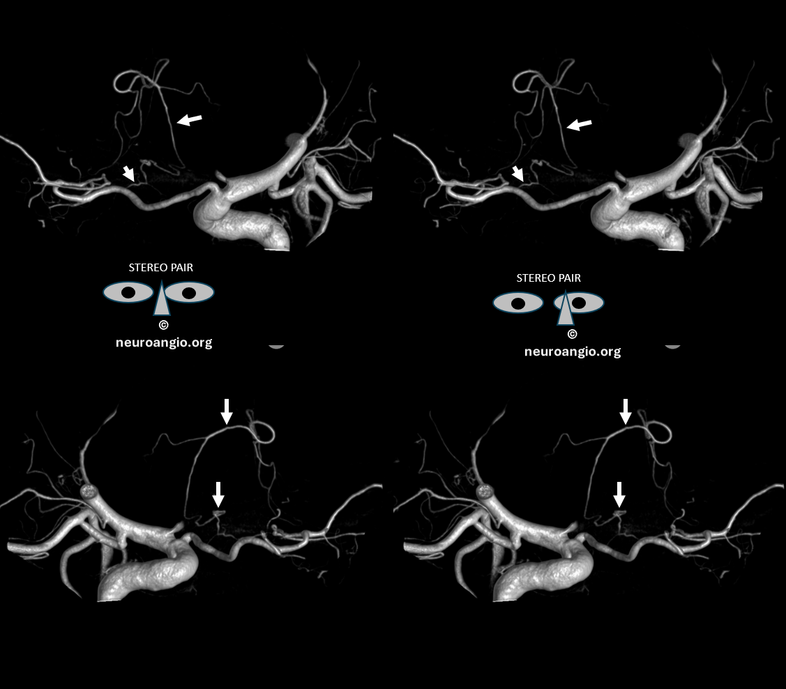
Stereo Pair volume rendered images
with arrows
Video
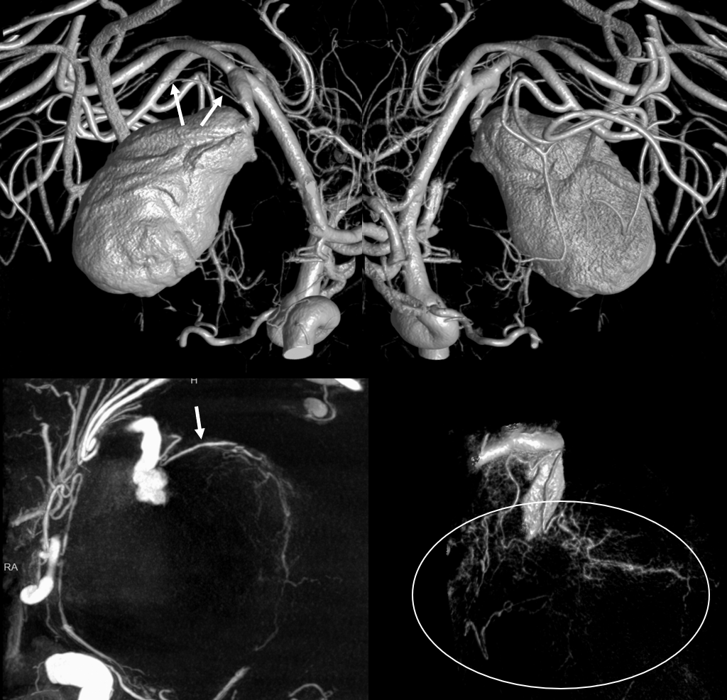
Case 2 — Giant MCA Aneurysm Link to Full Case

Vasa Vasorum (arrow and oval) origin from the MCA just proximal to neck
Video
Video vasa vasorum only
See full case link for treatment details — below is end result — coils occlude aneurysm and extend to parent vessel to close vasa vasorum also
Case 3 – Vasa Vasorum in ICAD — thickened atheromatous cholesteromatous (we made that word up just now — thank you — we are not Shakespeare — but play one here and there) wall can be full of vasa vasorum. Which can be distinguished from calcifications by obtaining a “mask” image. Full case is here