
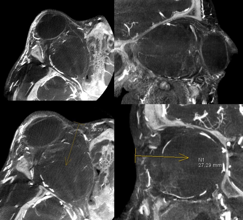
Rare case. Large retro-orbital malformation. Presented with gradual proptosis. Post canthotomy and lateral wall reconstruction

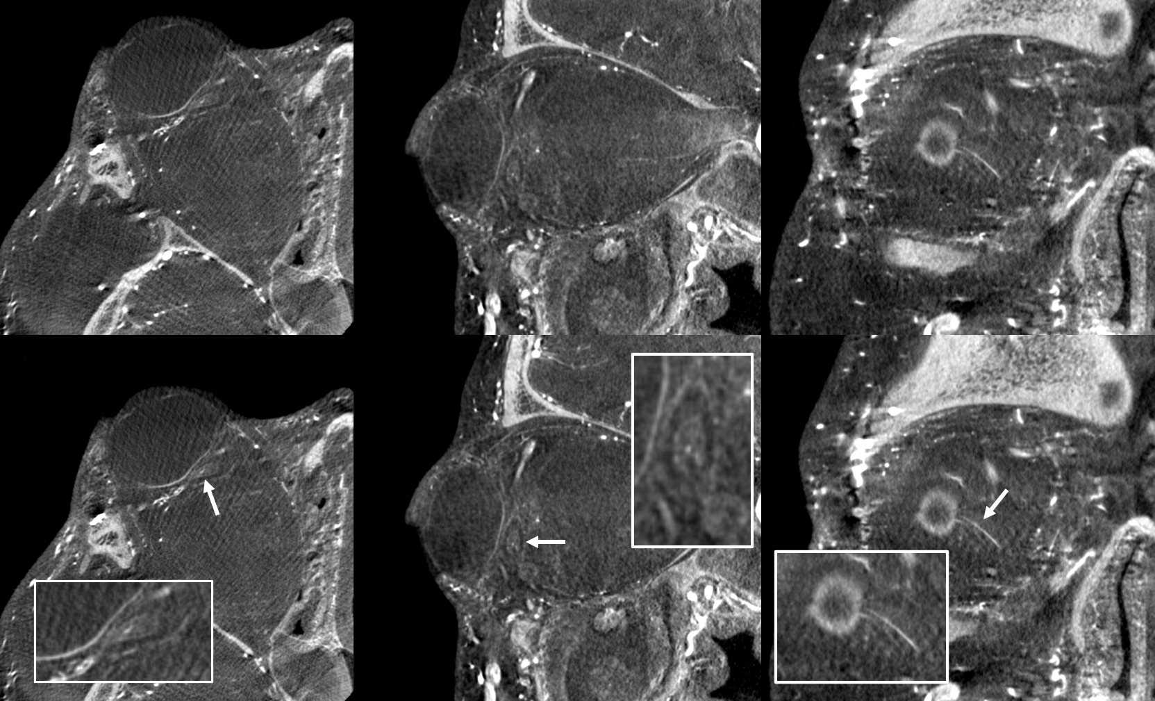
Usually this is done with ultrasound. But there are advantages of HR-CBCT (Dyna). Not much to see here of course
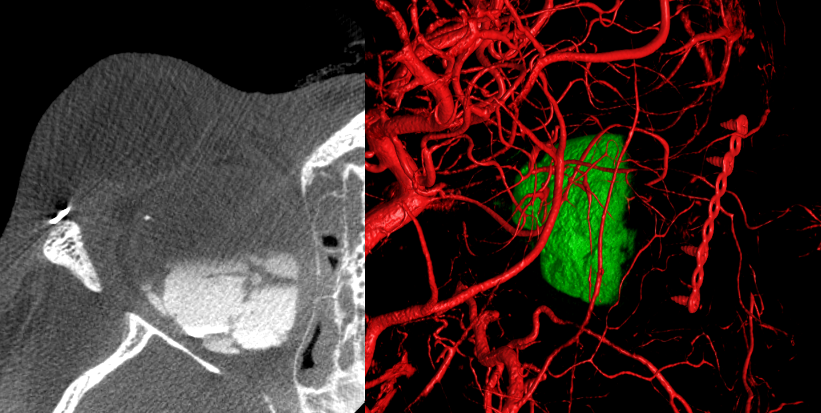
DYNA is another story. For example, it turned out to be the best way to see the optic nerve — the portion where central retinal artery is. MRI was OK, but this is better. Displaced medially and inferiorly
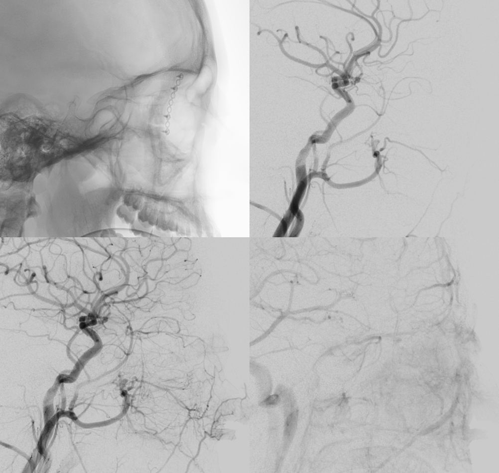
Needle guide approach.

15 units of bleo given (15 U/2.5 cc saline + 0.5 cc contrast). 3.5 mls of yellowish fluid came out of the 25 gauge needle. First time we did it bloody fluid came out and we gave 3 unis bleo/foam/air, having aspirated 9 ccs. This time we gave 15 units. Layering nicely. Fusion of bleo/contrast post injection and pre-injection RT CCA DYNAs
We are not uploading photos of the eye because of privacy etc. if you do this kind of treatment, you can imagine what it looks like.