
The material/cases in this section were collected and organized by Dr. Guglielmo Pero. A neuroangio team effort!
Acom aneurysm in a young patient. 3D acquisition in 5 seconds, 22 cm FOV, no secondary reconstructions, The origins of subcallosal and Heubner arteries are evident. All the reconstructions of this case have been done with Horos from the Philips datasets.
Post Artisse
Vaso-CT acquired with the 22 cm field of view, in 20 seconds and 20 ml 50% contrast media (300 mg/ml) manually injected with 20 ml syringe. Reconstructed with 50% FOV and 5123 resolution, after Artisse implant. Thin sagittal MIPs (0.1 mm)
Thick MIP reconstruction. Subcallosal and Heubner arteries are still visible — as they should be!
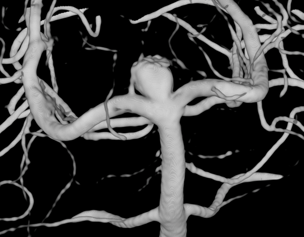
Basilar Bifurcation
Basilar tip aneurysm. A Percheron artery originating from right P1 is already evident.

3D (5 seconds) acquisition. In this case, all 3D and Vaso-CT acquisitions have been made with manual bilateral vertebral artery injection.
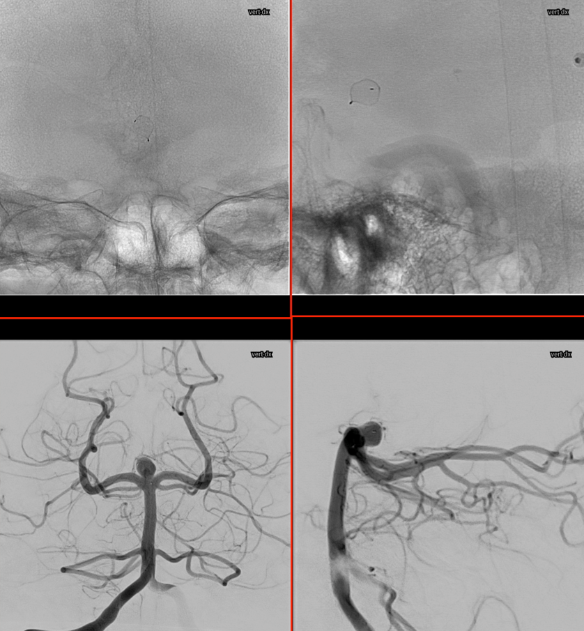
Below is Vaso-CT before Artisse. 22 cm FOV, 20 second acquisition. Bilateral pure contrast (300 mg/ml) manual injection with 20 ml syringes. Thin MIP reconstruction. No further reconstruction. The Percheron artery is well recognizable.
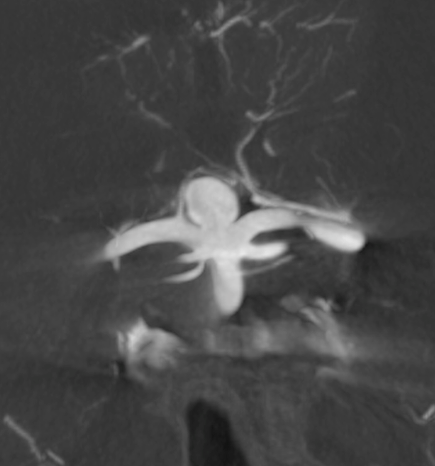
2 mm MIPs reconstructions
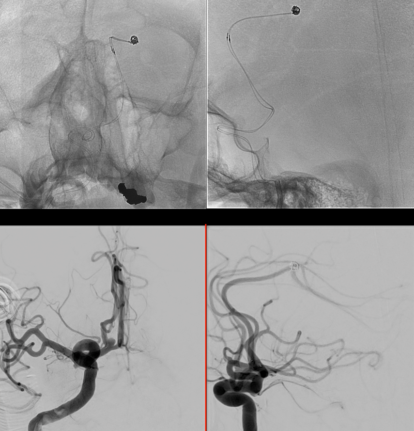
During Embo
Before detaching the Artisse. Same acquisition protocol as before Artisse positioning; bilateral 50% contrast (300 mg/ml) manual injection; followed by 50% FOV 5123 matrix. Note the artifacts caused by the radiopaque markers and pusher of the Artisse.
Post
ACOM and Pericallosal Aneurysms
3D acquisition 5 seconds, 22 cm FOV, 20 ml manual injection of pure contrast (300 mg/ml) with a 20 ml syringe. VR reconstruction made with Horos from the original dataset.

Two-microcatheter coiling
Post Artisse
Vaso-CT acquired with 22 cm FOV 20 seconds protocol, manually injected 50% contrast (300 mg/ml) with a 20 ml syringe, and thick MIP reconstruction after 50% FOV, 5123 matrix secondary reconstruction of the original dataset