The material / cases in this section were collected and organized by Dr. Guglielmo Pero. A neuroangio team effort!
A patient presenting with a giant serpentine MCA aneurysm. The plan was to do a high flow ECA-MCA bypass and then coil occlude the MCA

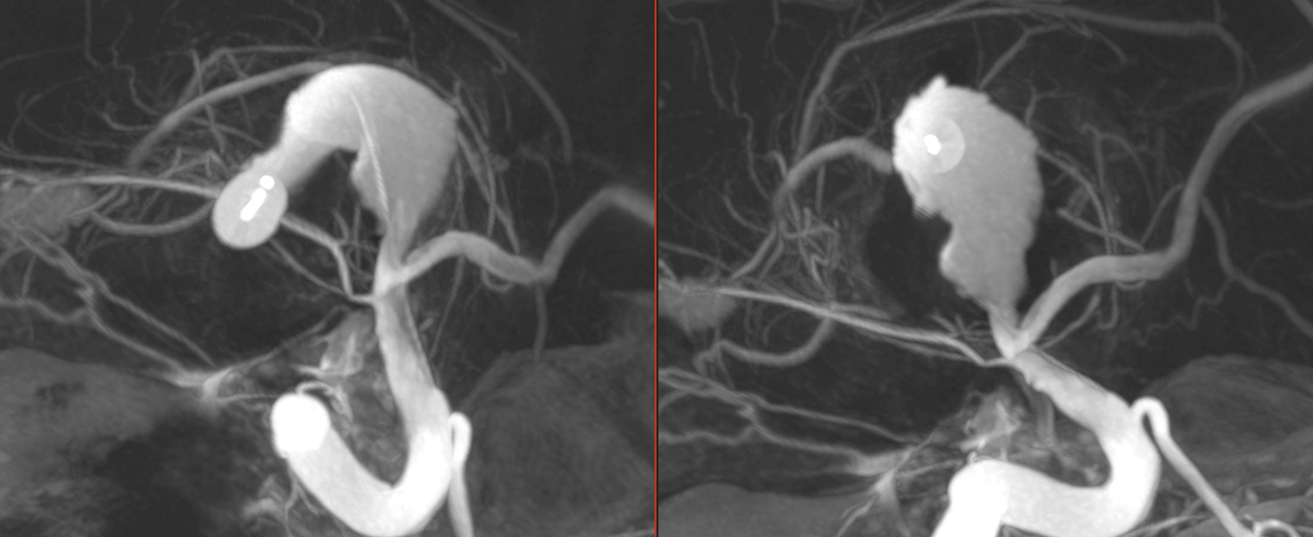
Immediately after bypass

The question is where are the lenticulostriate and insular perforators?
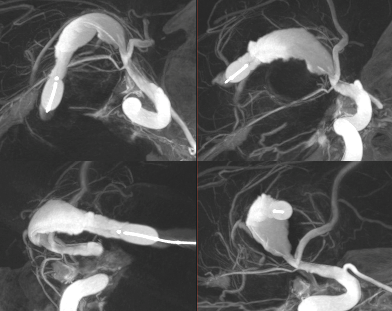
A dual lumen compliant balloon was inflated in the distal segment of the serpentine aneurysm and a Vaso-CT (22 cm FOV, 20 seconds acquisition) was acquired manually injecting pure contrast (300 mg/ml) in the ICA with a 20 ml syringe. It was done to be sure that no perforating arteries originate between the anterior choroidal artery and the bypass. Note the thin metallic filament of the non-radiopaque part of the microguidewire.
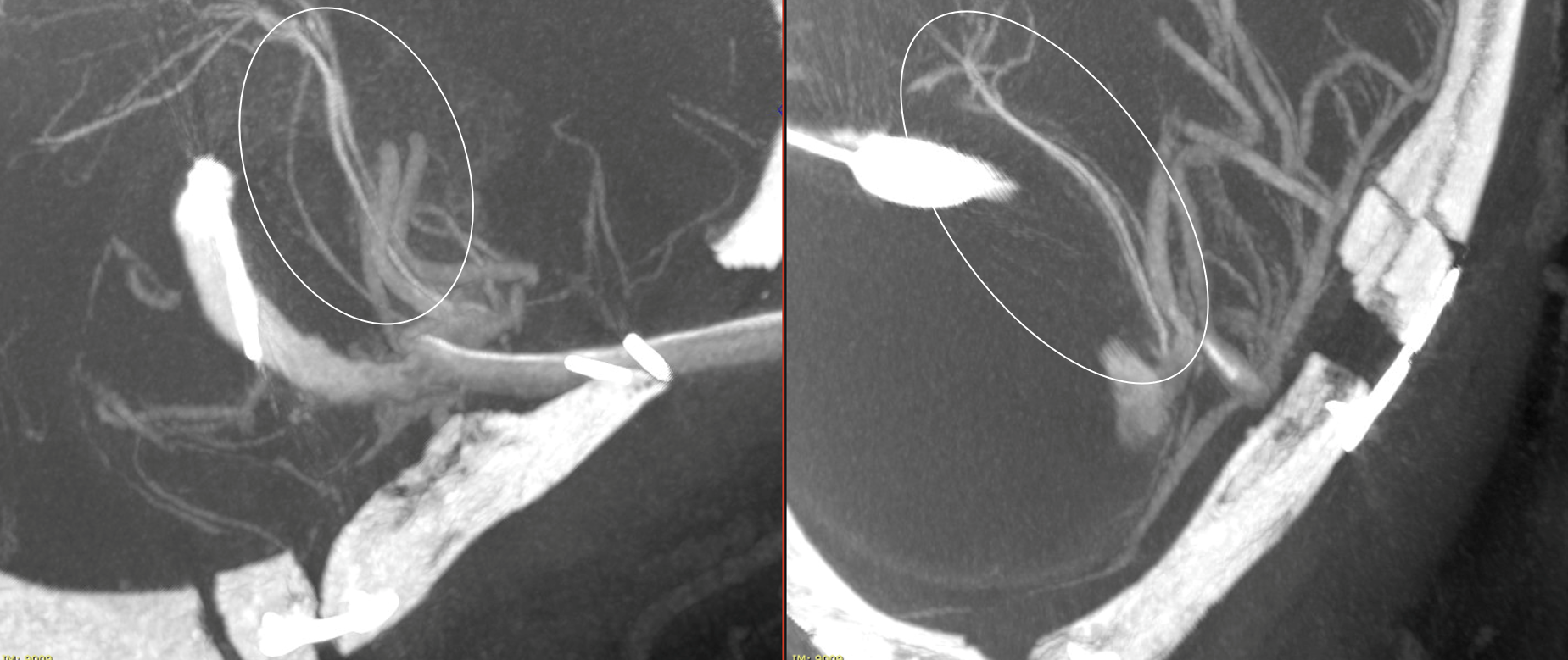
Another Vaso-CT was acquired manually injecting pure contrast (300 mg/ml) in the bypass (the injection was visually modulated to avoid reflux in the ICA), maintaining the balloon in place. It clearly shows the M2 perforators originating after the conjunction of the bypass.
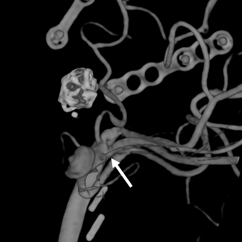
3 months follow-up control with 3D reconstruction of the bypass (3D 5 seconds protocol with 19 cm FOV, pure contrast (300 mg/ml) manual injection), confirming the origin of the M2 perforators (arrow) What else is new in the image since the bypass?