
Atheromatous lesions often increase wall thickness, with various tissues present. The biology is beyond complex and certainly beyond the scope of what we are about. However, the thicker the wall, the more likely one is to see vasa vasorum. Here is a really nice case of that
History is progressive weakness
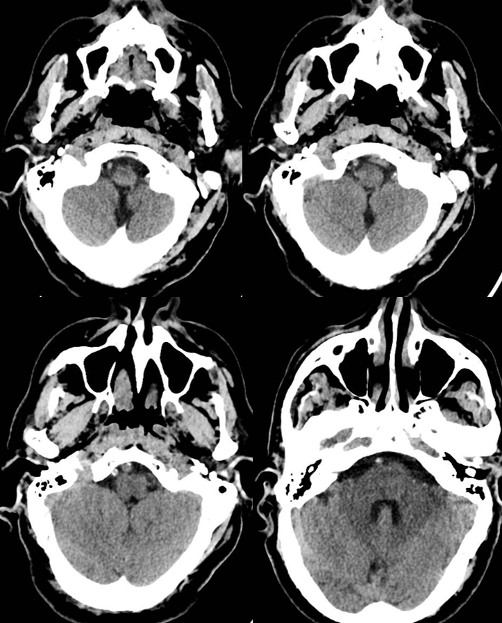
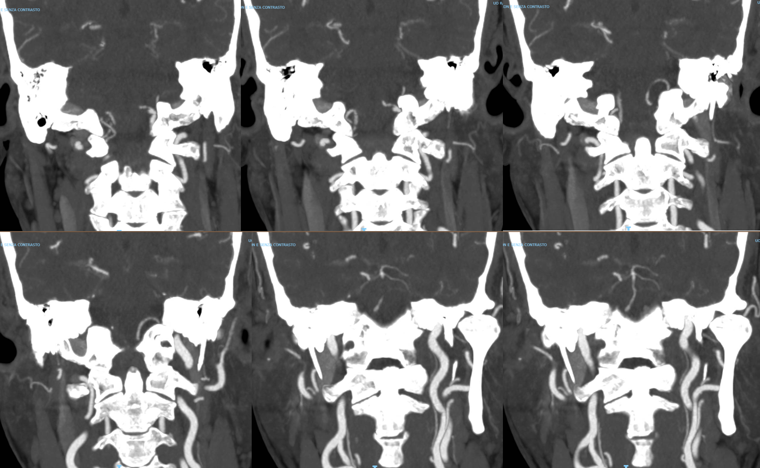
VB junction occlusion
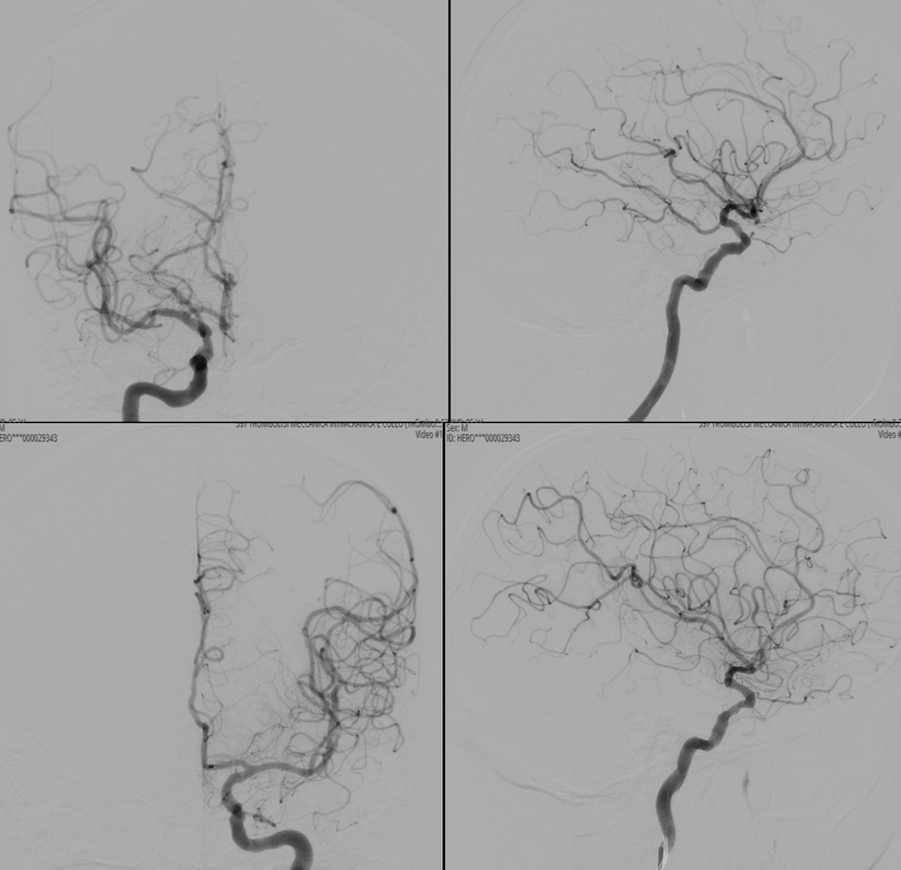
Leptomeningeal anastomoses are just as present and as variable as in the supratentorial compartment

This video below is the fusion of Vaso-CT (protocol 22 cm FOV, 20 seconds acquisition, 20 ml pure contrast (250 mg/ml) manual injection, reconstructed with 50% FOV and 5123 matrix) of the left (blue) and right (red) vert.
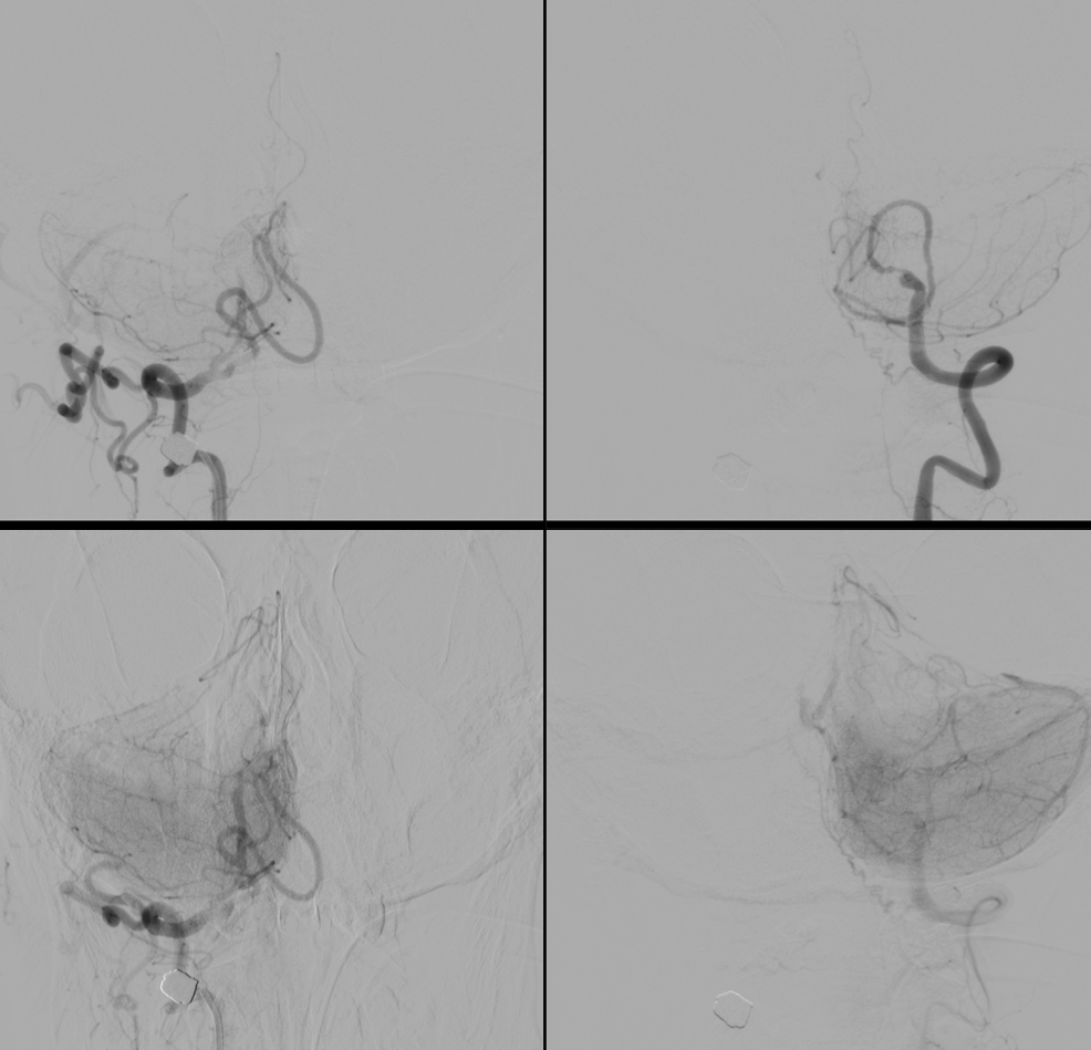
In faint blue we see the walls of the occluded part of the left vert (much larger than the right vert). Viewing the walls blue means that they are vascularized with the injection of the left vert (vasa vasorum). A mix of calcification and vasa vasorum is possible — see below. The basilar artery is almost white because it receives contrast with the injection of both vertebral arteries. Bone is also shades of grey.
Snapshots. The course of the closed vert shows that recanalization is better from the left.

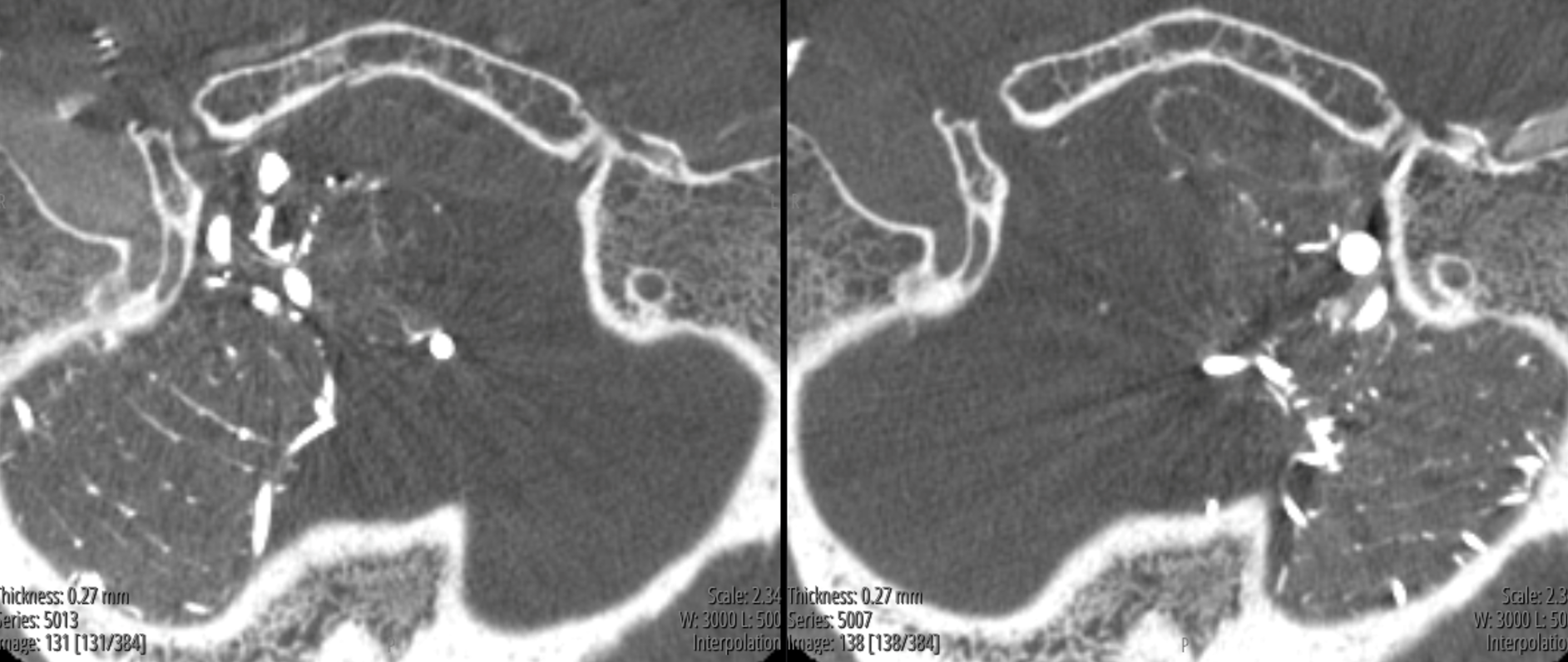
The simplest way to tell calcium from vasa vasorum is to get a noncontrast “mask” image. In this case, the below image shows right vert injection which serves as the mask for the left vert, on the left, and left vert injection image on the right. There is some calcification in the wall of the occluded distal left vert, but the wall is much more visible with the left vert injection, due to vasa vasorum.

Video
Post

See more Vasa Vasorum Cases here and a Giant Aneurysm Vasa Vasorum Case here