Another awesome vasa vasorum case by Guglielmo Pero MD
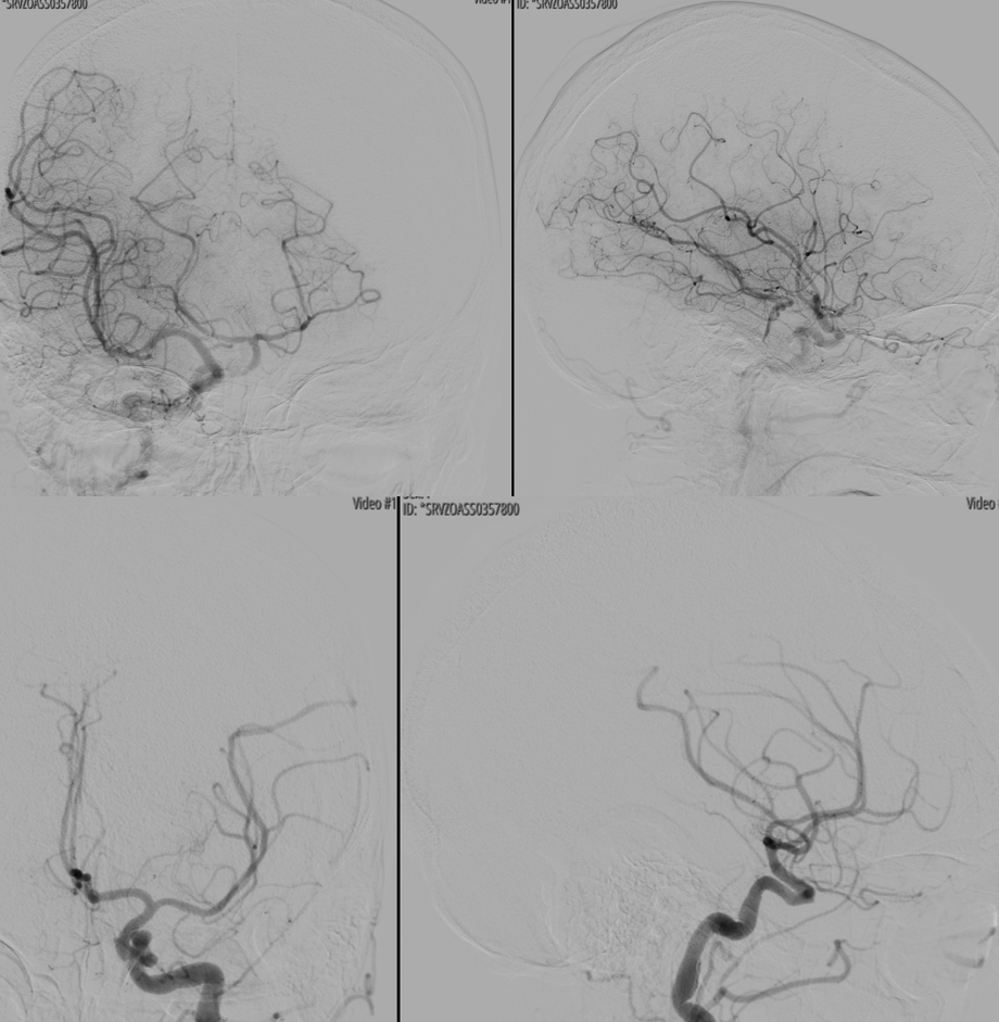
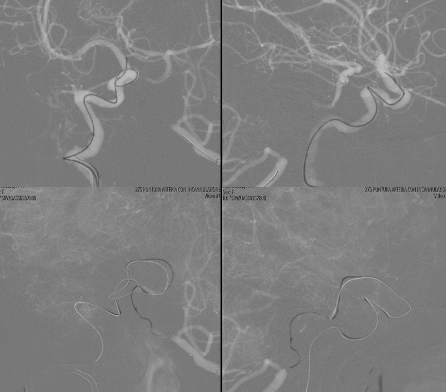
First angio
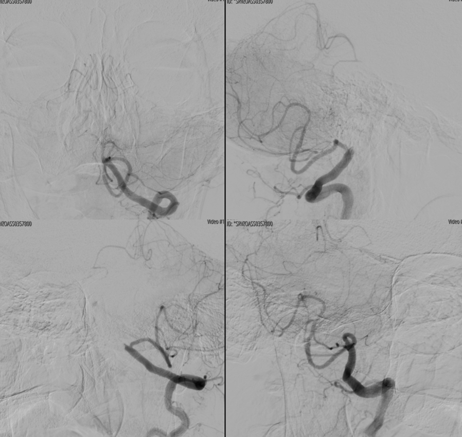
Small right PCOM
Unsuccessful Recan attempt

More
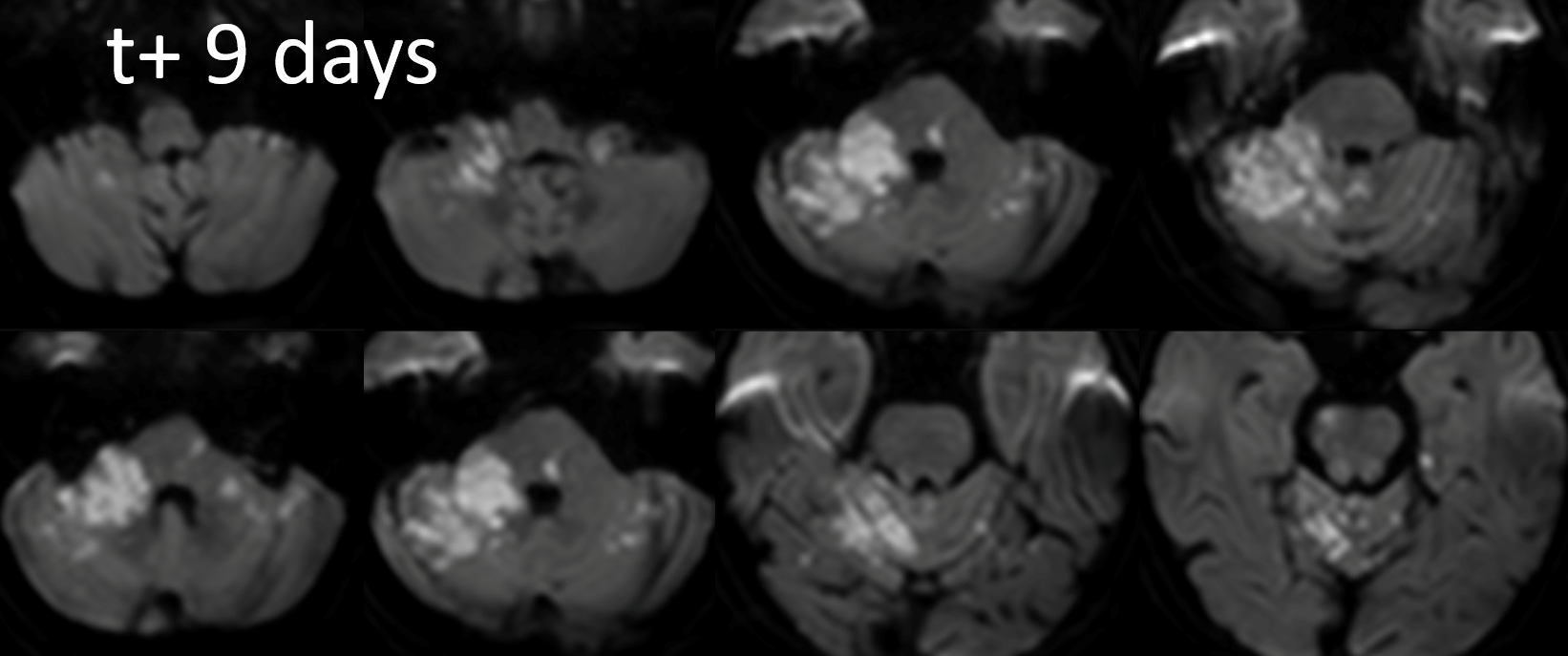
Back with a vengeance
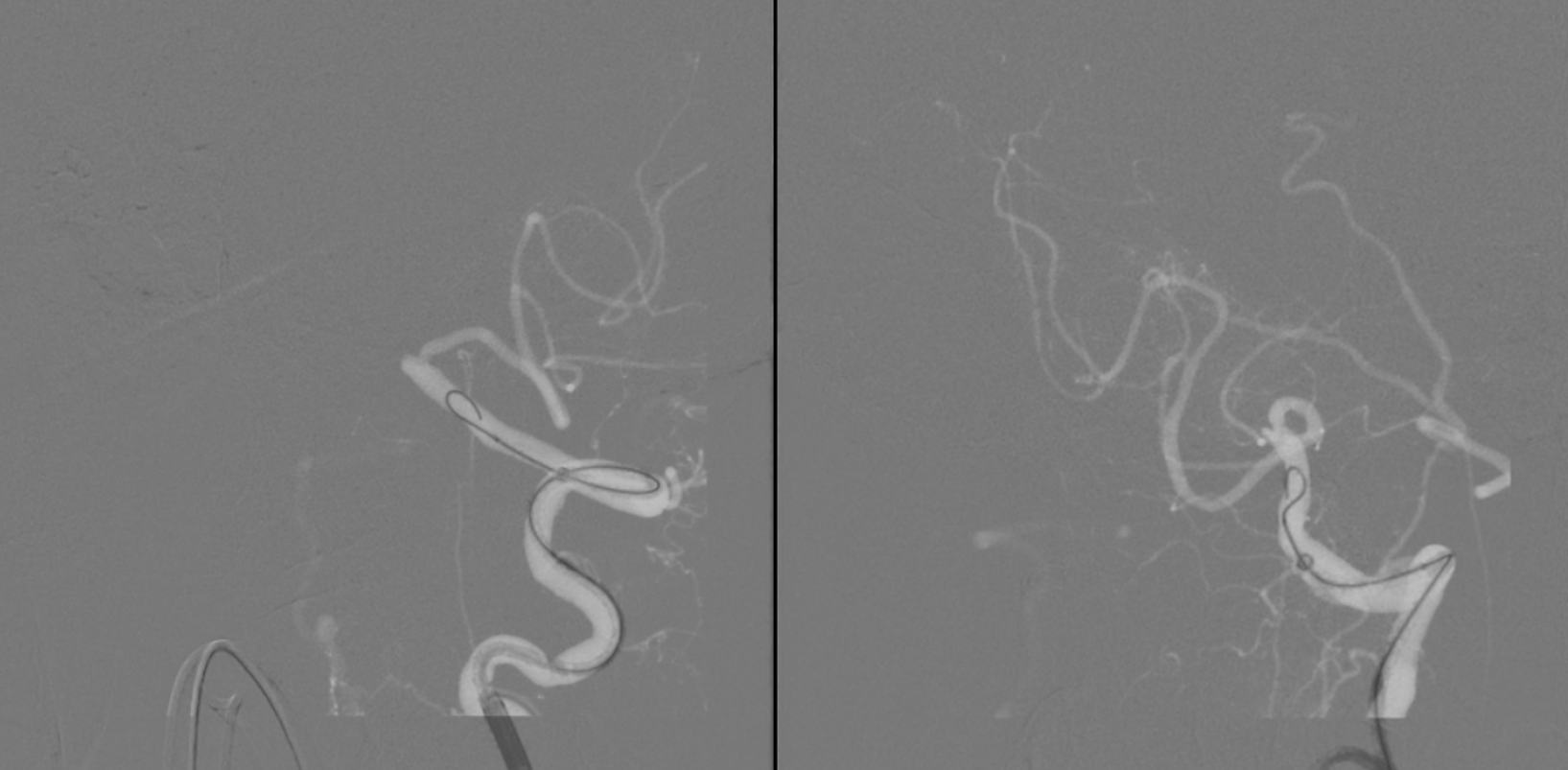
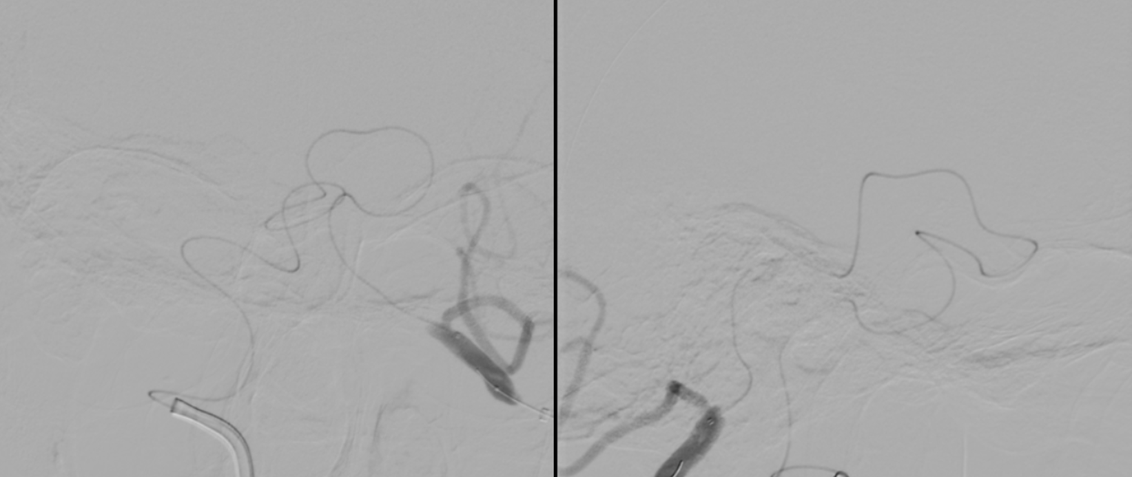
At first wire goes into subintimal channel — see how it stays on the side and does not advance in the movie below?
Now the good way
Once you are through, follow the wire with microcatheter, replace wire with snare, catch the wire coming up from below, and bring it up through the stenosis… sounds easy, huh…
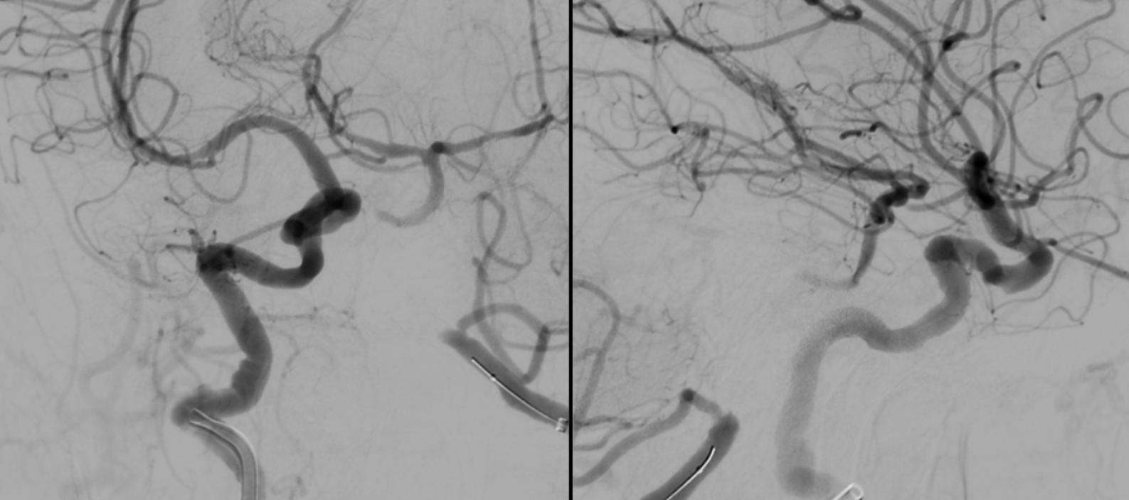
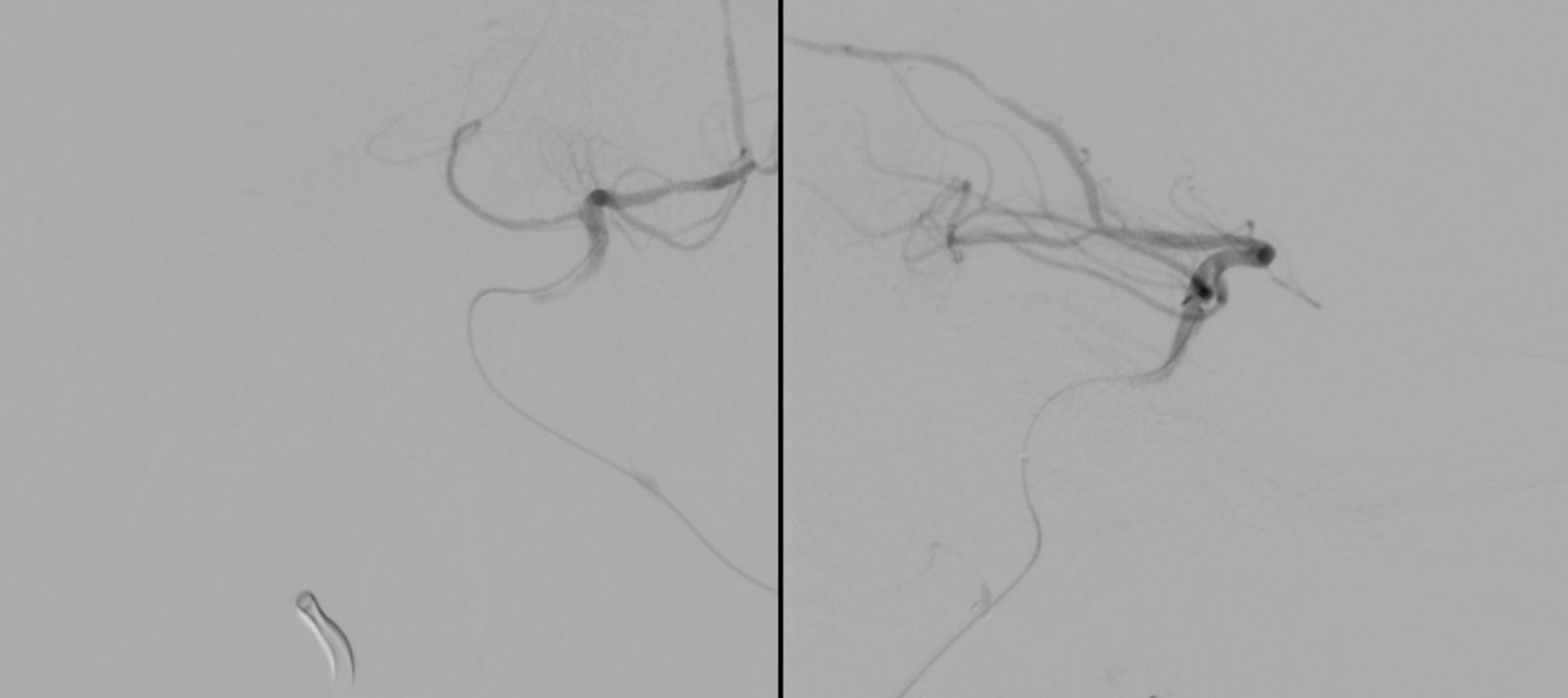
Now, image through a microcatheter from below
Stenting. Lots of calcs
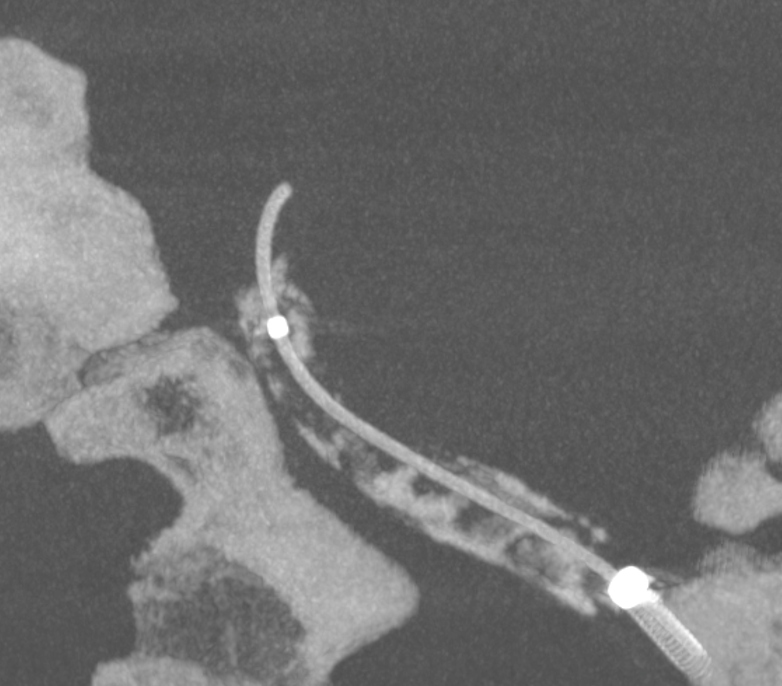
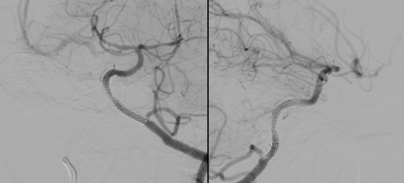
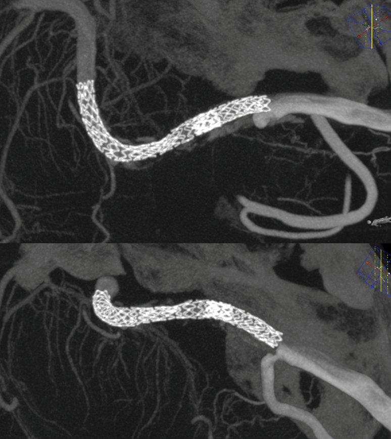
“Dry” noncontrast Vaso-CT (22 cm FOV protocol, 20 seconds acquisition, reconstructed with 50% FOV and 5123 matrix), showing the stents in the artery and the calcific plaques.
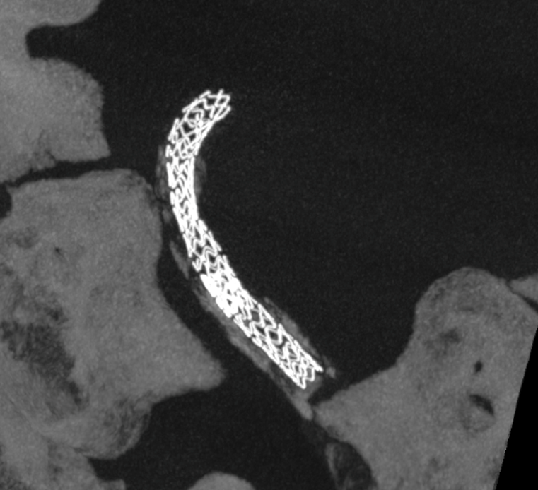
Laser-cut balloon-mounted stents make it easier to leave the PICA alone – critical given what you see below. The best choice in our opinion.
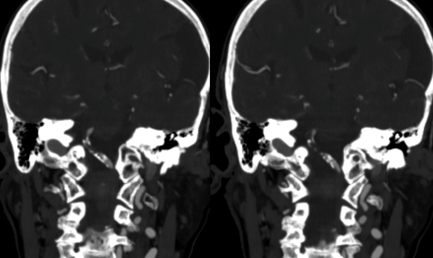
Vaso-CT (22 cm FOV protocol, 20 seconds acquisition, 50% contrast manual injection – 250 mg/ml, reconstructed with 50% FOV and 5123 matrix). We can see a stenosis at the origin of the PICA due to snowplough effect of the inflated balloon on the plaque — this is quite acceptable. With complete coverage of the PICA the result could be bad indeed. Precision matters a lot.