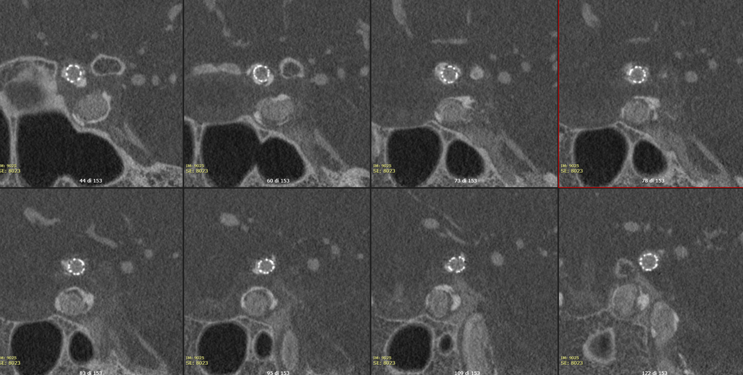
The Vaso-CT (22 cm FOV, 20 seconds acquisition, pure contrast – 300 mg/ml -manual injection 20 ml with a 20 ml syringe) shows the highly irregular atherosclerotic plaque with small residual lumen

If you want to use a balloon-mounted self-expanding stent here, your call. In our book, this is a balloon-mounted case any day. Yes, yes, the brain is not the heart. But for this problem, there is a reason balloon-mounted stents now rule, and they tried self-expanding ones at the beginning also…
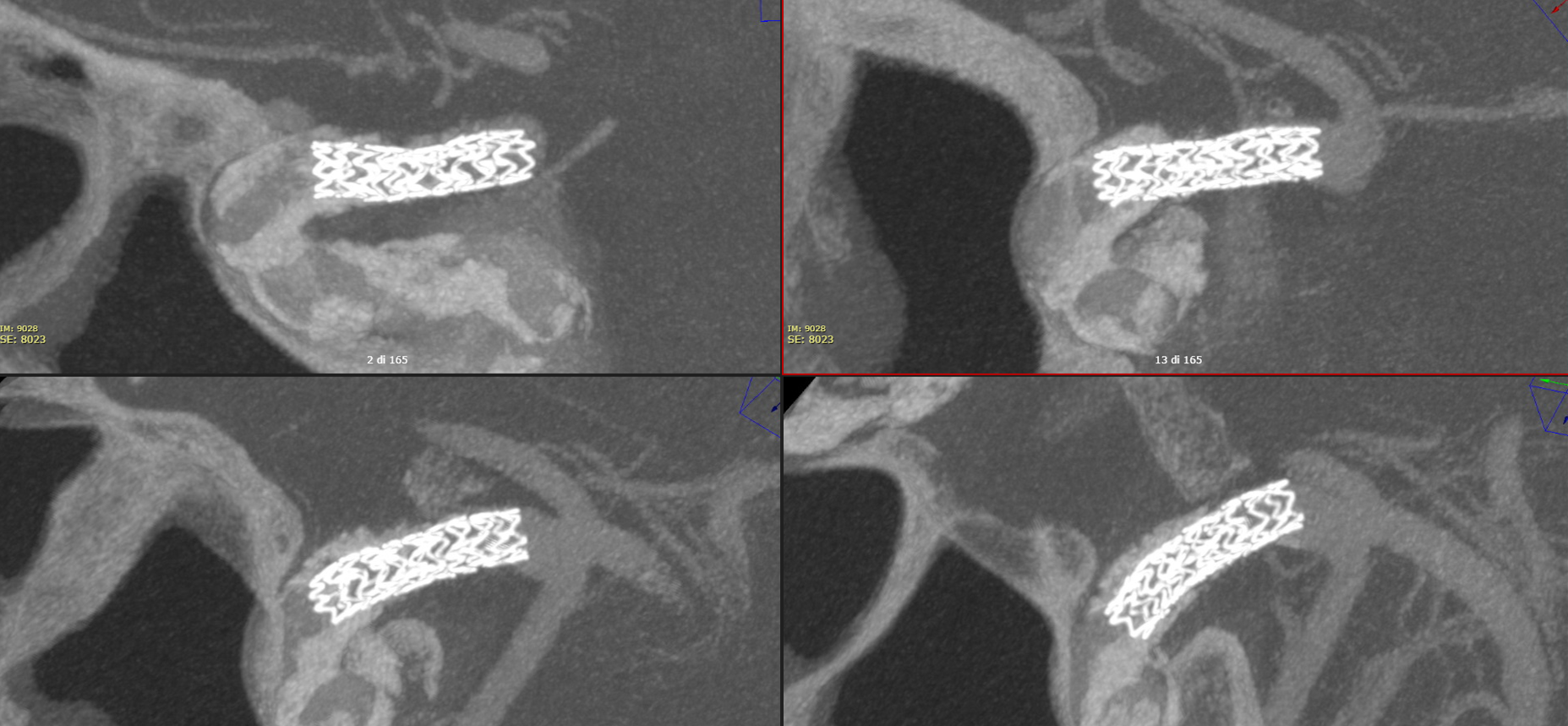
The Vaso-CT (22 cm FOV, 20 seconds acquisition, 20 ml of 50% contrast – 300 mg/ml – manual injection with a 20 ml syringe) clearly defines the lumen of the artery, the plaques and the stent. Note how the plaques were not visible in the pre-stent Vaso-CT (done with pure contrast injection) because covered by the high density of the pure contrast. Thin MIP reconstructions.
Thick MIP recons