![](https://www.neuroangio.org/wp-content/uploads/Archives/Spinal/Hemangioma-PSA-Identification/Hemangioma-PSA-Identification-09.png)
Preop embo of T7 hemangioma
![](https://www.neuroangio.org/wp-content/uploads/Archives/Spinal/Hemangioma-PSA-Identification/Hemangioma-PSA-Identification-01.png)
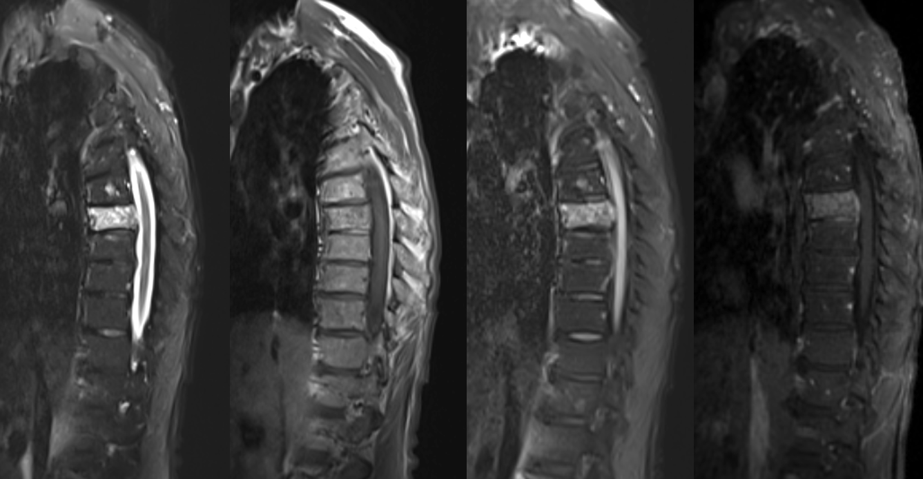
Importance of identifying spinal supply nearby
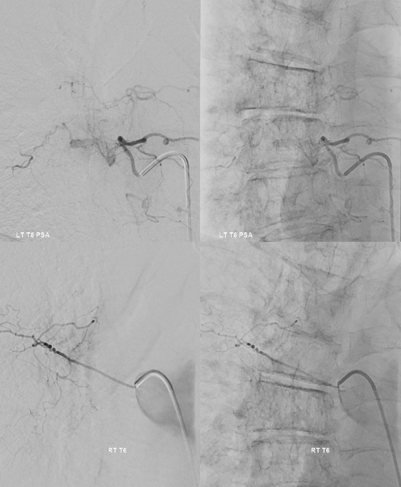
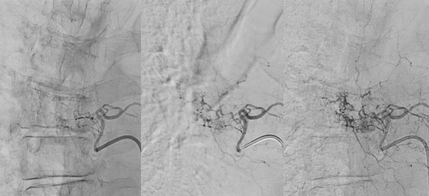
![](https://www.neuroangio.org/wp-content/uploads/Archives/Spinal/Hemangioma-PSA-Identification/Hemangioma-PSA-Identification-11.png)
Right T7 Below
![](https://www.neuroangio.org/wp-content/uploads/Archives/Spinal/Hemangioma-PSA-Identification/Hemangioma-PSA-Identification-03.png)
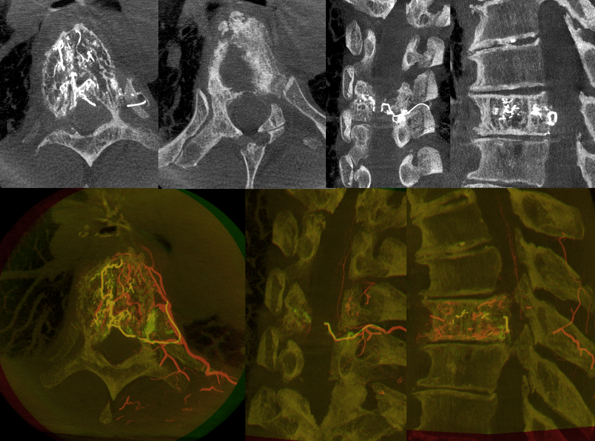
Right T7 DYNA CT — what is seen here that was not seen on 2D-DSA?
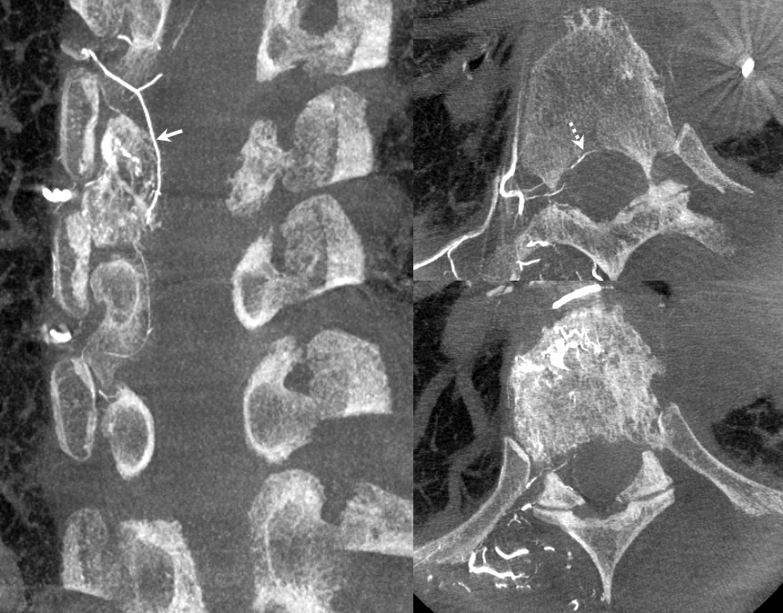
![](https://www.neuroangio.org/wp-content/uploads/Archives/Spinal/Hemangioma-PSA-Identification/Hemangioma-PSA-Identification-04.png)
Left T7
![](https://www.neuroangio.org/wp-content/uploads/Archives/Spinal/Hemangioma-PSA-Identification/Hemangioma-PSA-Identification-02.png)
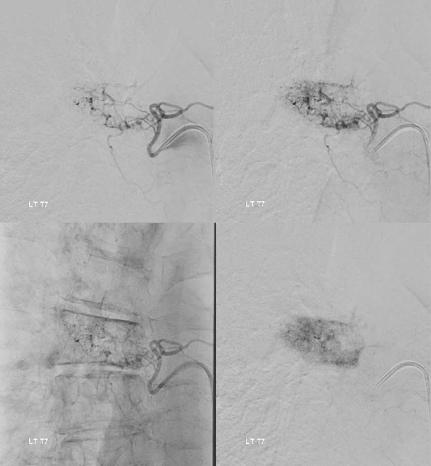
Left T7 DYNA — what is really seen here? Incredible…
![](https://www.neuroangio.org/wp-content/uploads/Archives/Spinal/Hemangioma-PSA-Identification/Hemangioma-PSA-Identification-06.png)
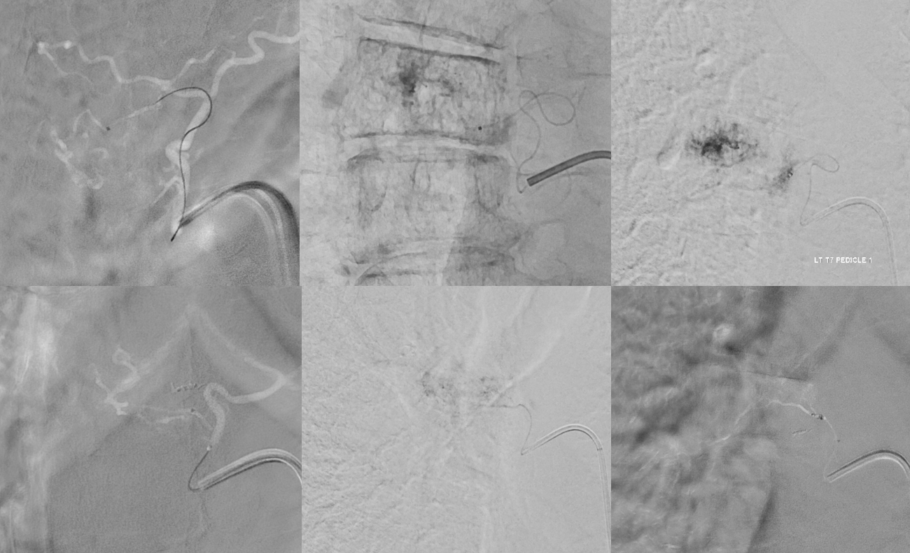
Can left T7 still be embolized? of course. Top row — posterior epidural / somatic branch subselective catheterization, followed by PVA embo — taking out the epidural component. Bottom row — anterior somatic branch — front of vertebral body subselective catheterization — more PVA embo from here
![](https://www.neuroangio.org/wp-content/uploads/Archives/Spinal/Hemangioma-PSA-Identification/Hemangioma-PSA-Identification-07.png)
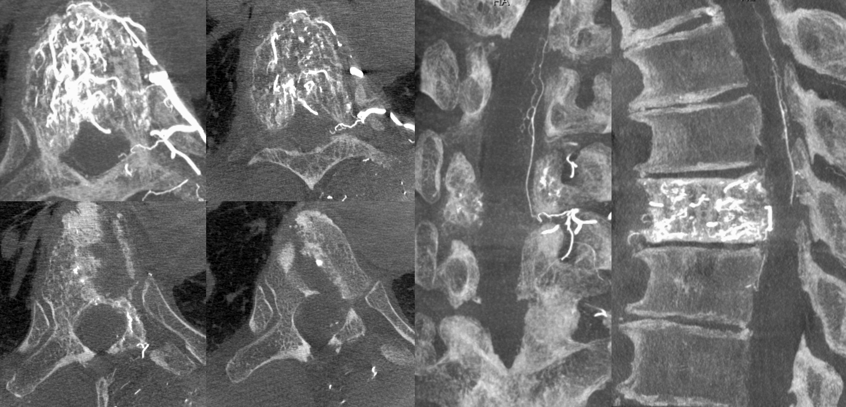
A micro DYNA was done from the posterior somatic / ventral epidural branch (top row above) — just to be sure. Top row below — MIPs. Bottom row below — “6D” MIPs showing co-registered images with the global left T7 DYNA
![](https://www.neuroangio.org/wp-content/uploads/Archives/Spinal/Hemangioma-PSA-Identification/Hemangioma-PSA-Identification-09.png)
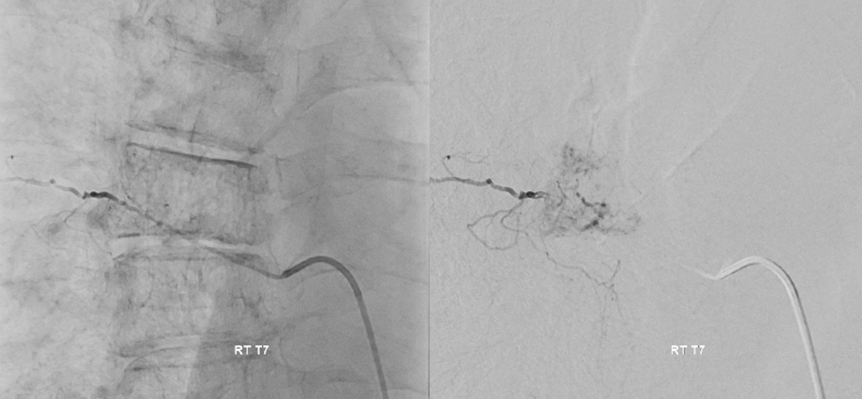
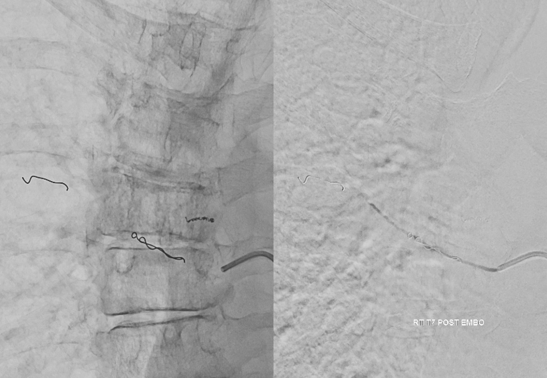
Post embo — partial embo but better. Now can you see the left PSA?
![](https://www.neuroangio.org/wp-content/uploads/Archives/Spinal/Hemangioma-PSA-Identification/Hemangioma-PSA-Identification-08.png)
Standard right side embo — pushable coil closure of the intercostal artery, followed by particle embo and closure of the segmental artery proximally with more pushables
![](https://www.neuroangio.org/wp-content/uploads/Archives/Spinal/Hemangioma-PSA-Identification/Hemangioma-PSA-Identification-10.png)
So, how about that for value of cone beam CT? See more cases in archives for pre-embo identification of cord supply…