Good example of something very vascular that picks up pial supply. Many times pial supply is not possible to embolize because there is too much brain supply also. Many times its possible but not done for any number of mostly bad reasons. Here is an example
Lots of pial supply — ACA here
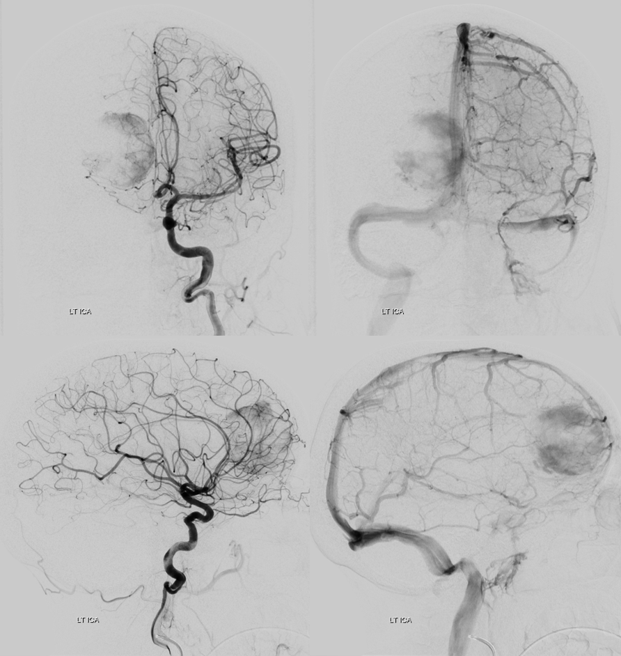
MCA pial supply also — plus the same ACA
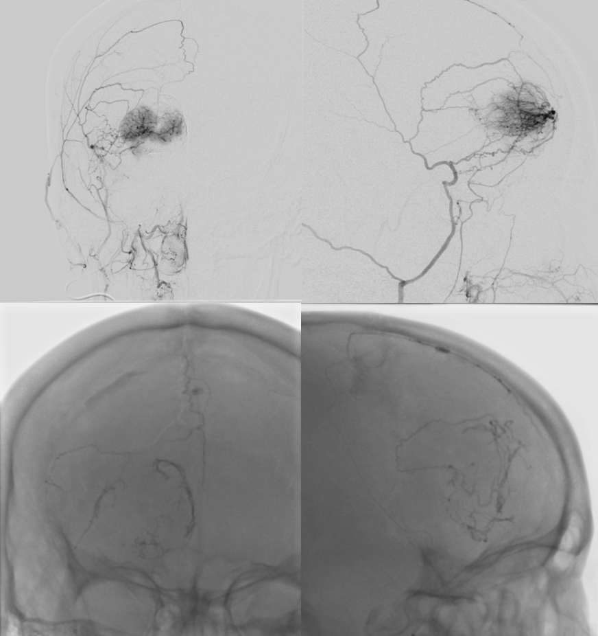
Headway 167 in A2 — all the way down. nBCA cast below 3:1 oil/glue
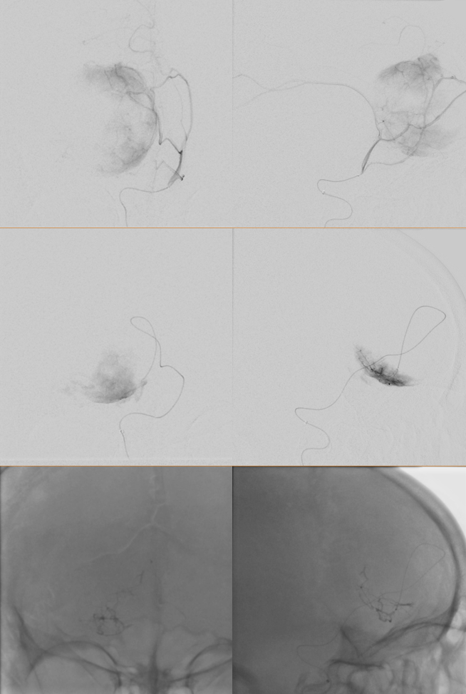
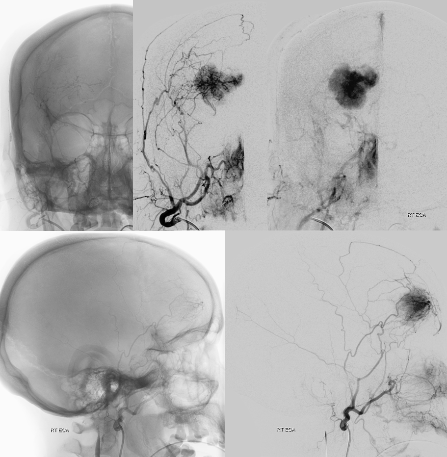
Another 167 duo — ACA — particle embo from here (45-150 / 150-250 um contours)
Magic 1.2FM in the distal superior division MCA branch — injection with contrast. Larger glue cast in the ACA
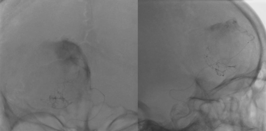
post pia supply embo. not too bad
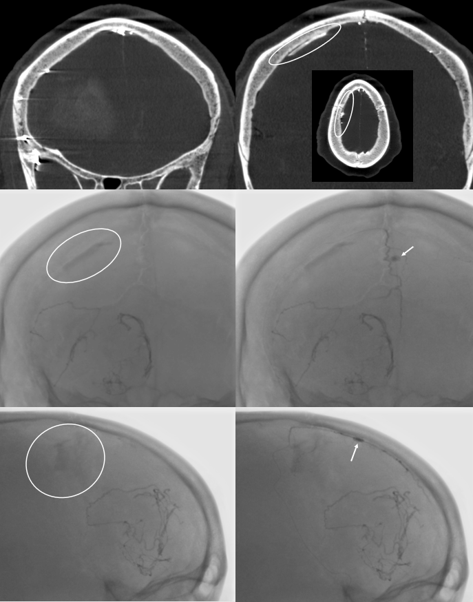
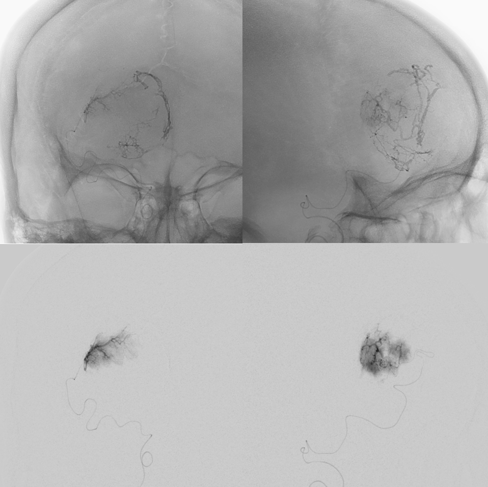
ECA — baseline after pial embo on top. Middle set — after embolization of some MMA supply with glue (arrows) and anterior meningeal SSS network (dashed arrows). There is some glue spillage in to SSS (arrowheads) and an oval over something not so good. What is this?
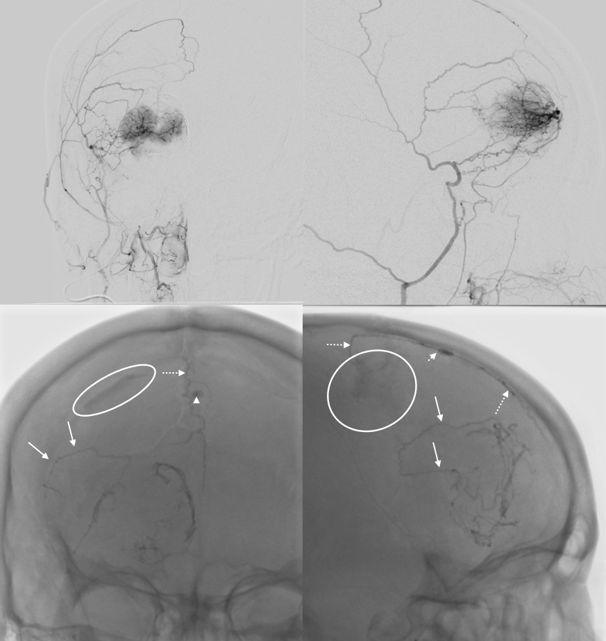
Without arrows
Coil off the upper MMA (arrows) , then particles from MMA position above the lowest branch. Why?
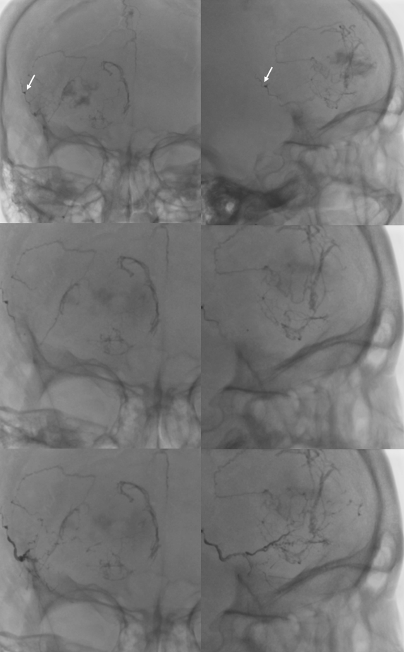
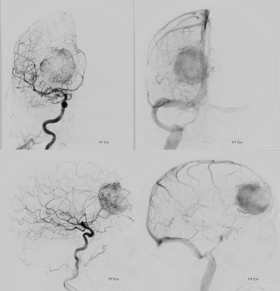
Home-made onyx 12 injection
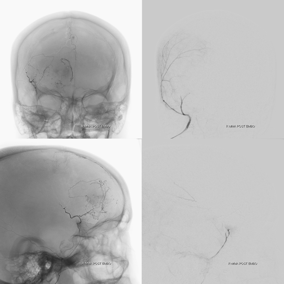
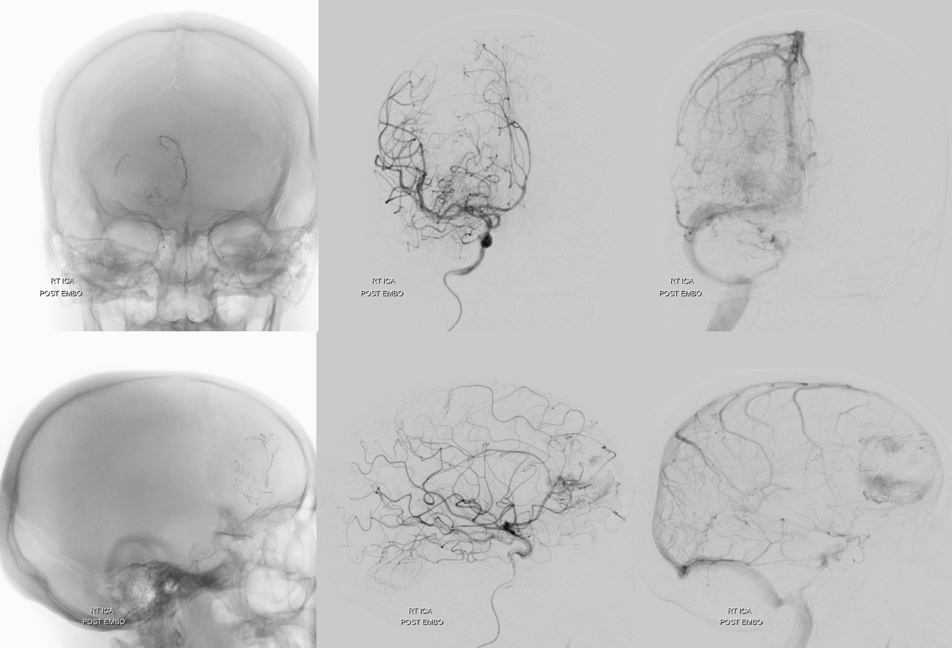
Post embo. Contrast staining in area of vasogenic edema posterior to tumor attests to its broken BBB
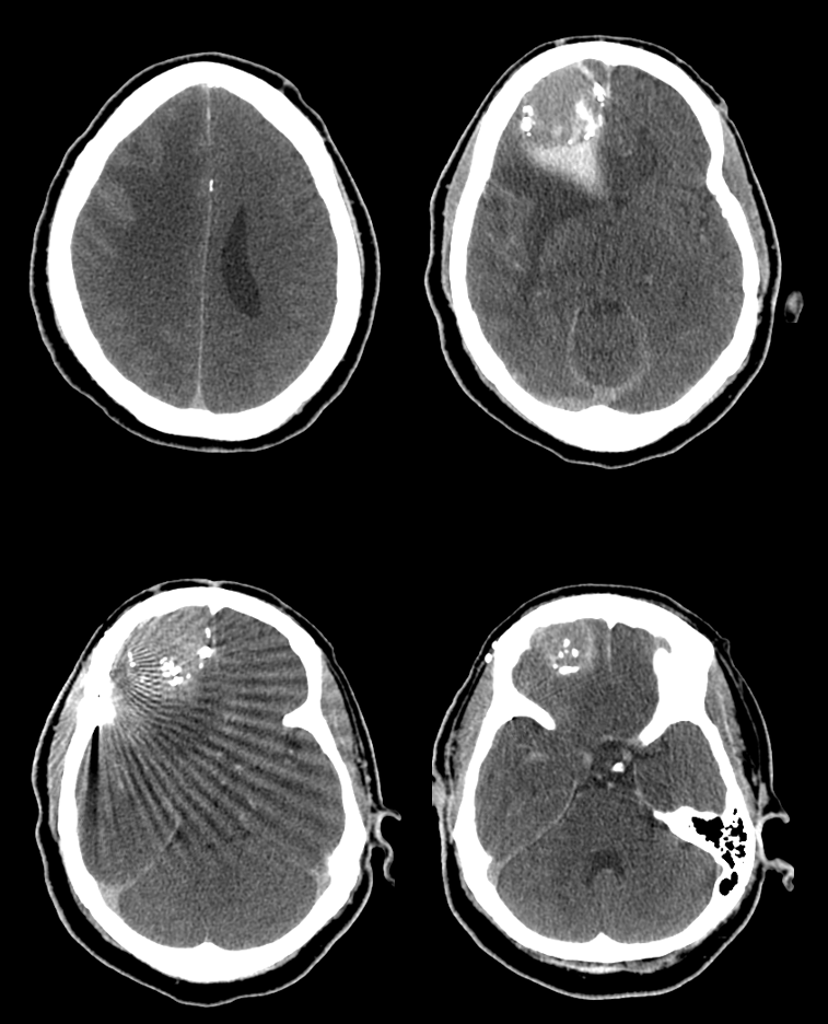
This is a DYNA CT of the same patient immediately after embo. What is inside the oval (see above?)