In keeping with the main theme of this work, the key task for the modern neuroangiographer who has come to rely on cross-sectional imaging for brain anatomy, is to know the location of the posterior fossa/ brainstem structures based on angiographic anatomy, particularly venous anatomy. There is so much variability in venous arrangements that, in my opinion, knowing where a given vein is with respect to the brain it drains and how it is related to the overall venous arrangement is more important than nomenclature. In this regard, the 4th ventricle is one of the key structures which can be located based on venous angiography.
One can use both morphometry (simple measurements), venous landmarks, or both. My general radiology mentor, Dr. Alec Megibow, holds that a radiologist with a ruler is a radiologist in trouble. Although he is certainly right, it’s nevertheless OK to admit trouble and use the ruler.
In a typical adult, the 4th ventricle is located more or less behind the pons; its craniocaudal extent corresponds roughly to the vertical length of the pons, which is going to be about 1 good old American inch (2.54 cm) long. Conveniently, the pons is also about 1 inch (2.54 cm) thick (or somewhat less) on a mid-sagittal MRI image; which also proves that the American system is the better one.
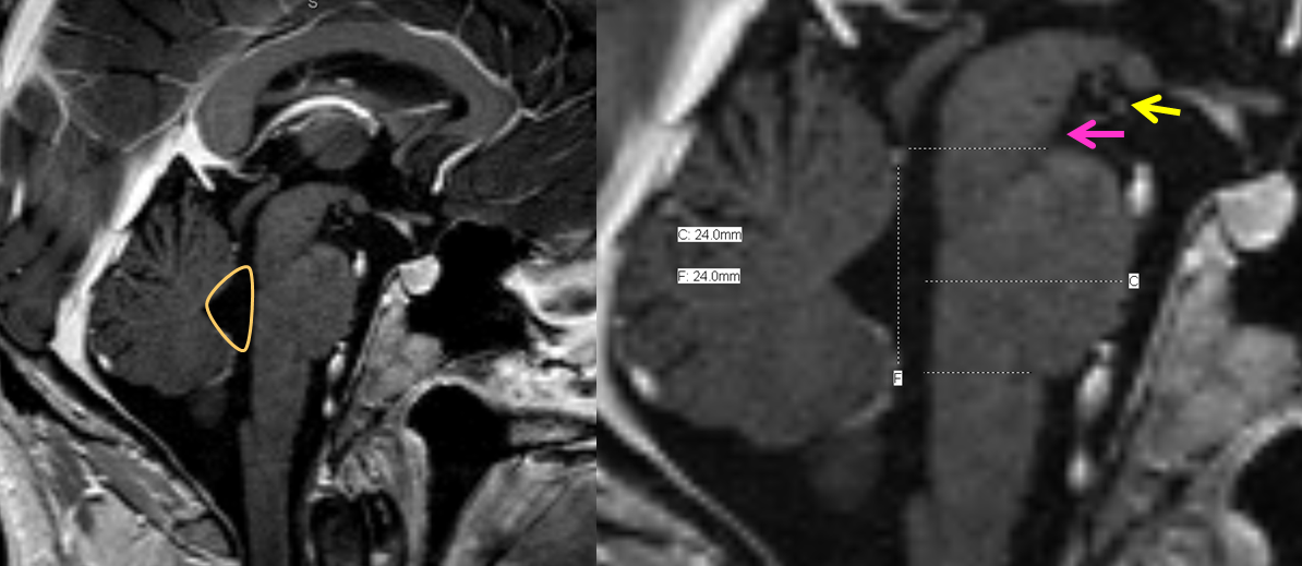
Now we apply this to the angiogram. The anterior (ventral) margin of the pons will be defined by the anterior pontomesencephalic/pontomedullary vein (blue arrow). The rostral (top) of the pons can be roughly marked by the interpeduncular vein (yellow), which will sweep posterior to the pontomesencephalic vein and will be located in the interpeduncular fossa (pink). The bottom of the pons is not consistently well seen on angiography because the transverse pontomedullary vein is rather inconstant, and there is not way to say angiographically whether it is in fact located in the pontomedullary sulcus or next to it. Therefore, one way to guesstimate the 4th is to draw two lines like so:
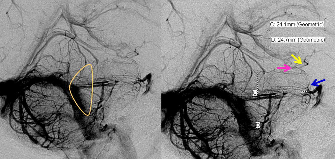
This is not perfect, for example the interpeduncular vein is somewhat below the inferior margin of the interpeduncular fossa, but that’s a good start. Notice that location of the IVth ventricle can be determined without the need to identify the vein of the Lateral Recess.
When the anterior draining group (anterior mesencephalic/ pontine/ medullary/ pontomesencephalic/ pontomedullary veins) are well-developed it is possible to see the inferior extent of the pons by the ventral course of the pontine segment of the vein as hugs the belly of the pons. The lesson here is that the pontomedullary sulcus and, thus, the inferior margin of the pons, sits rather low on the lateral view. In this case, the anterior group is beautifully displayed throughout its length, allowing for easy identification of the brainstem:
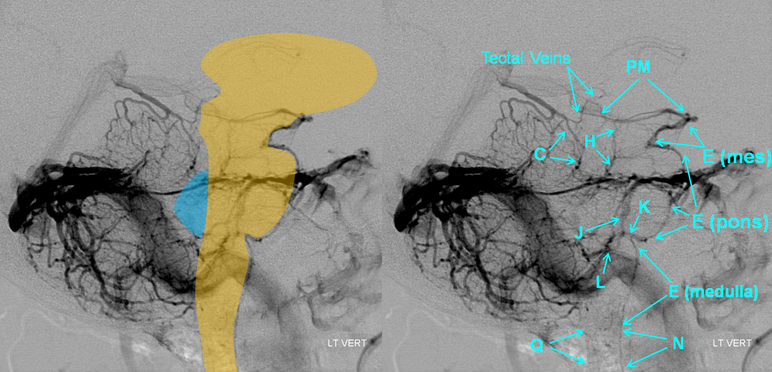
The reason for the prominence of the anterior group is the hypoplasia of the posterior portion of the Posterior Mesencephalic Vein (PM), so instead of draining into the Galen it empties towards the anterior pontomesencephalic (E) system. Also nicely seen is a longitudinal pontine vein (J) and the vein of the pontomedullary sulcus (K). The 4th ventricle will be located behind and slightly below the pons, underneath the Precentral Vein (C), with is caudal margin marked by the Vein of the Lateral Recess of the 4th Ventricle (L), a.k.a. the Vein of the Cerebellomedullary Recess of Rhoton. Also nicely seen are the tectal veins, marking the location of the tectal plate.
The same image without any labels, to best appreciate the natural beauty.
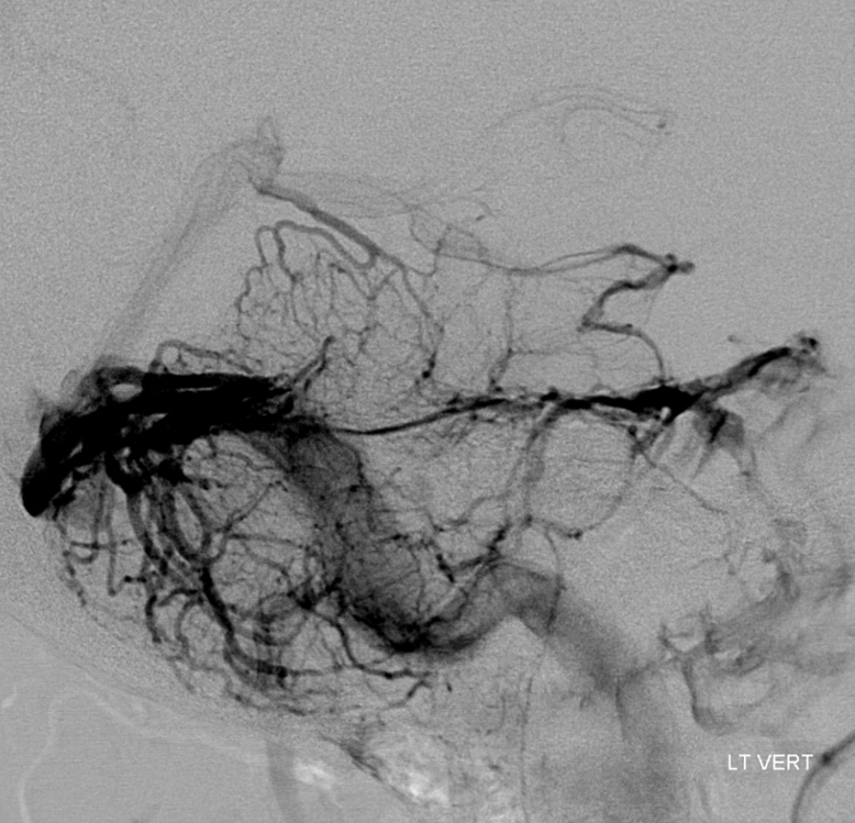
An image like this one makes things easy, but it is rarely this obvious.
Vein of lateral Recess of the 4th ventricle (L), a.k.a. the Cerebellomedullary Fissure
A relatively consistent landmark for the inferior aspect of the 4th ventricle is the Vein of the Lateral Recess of the 4th ventricle (L), as shown in figures from:
Yung Peng Huang and Bernard Wolf. Veins of the Posterior Fossa. Chapter 75, in Newton and Potts Radiology of the Skull and Brain. Volume 2, book 3. 1974
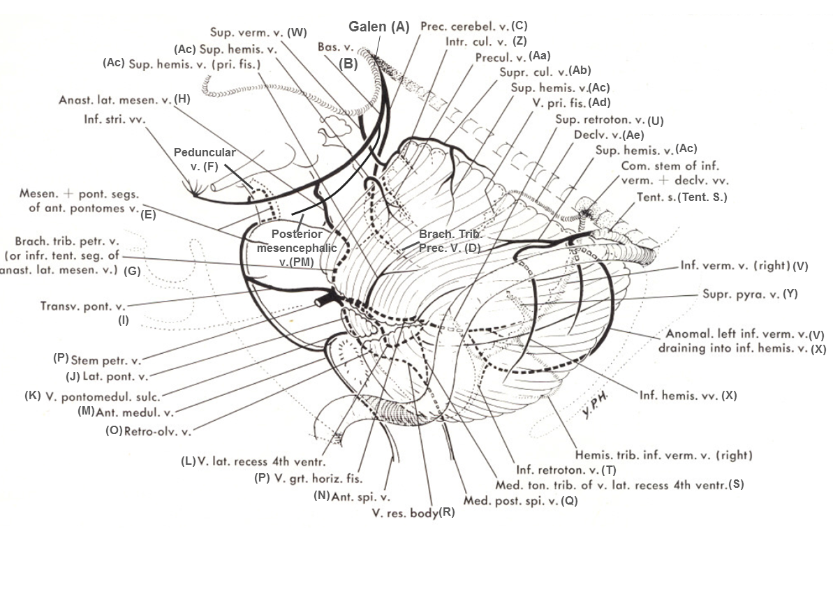
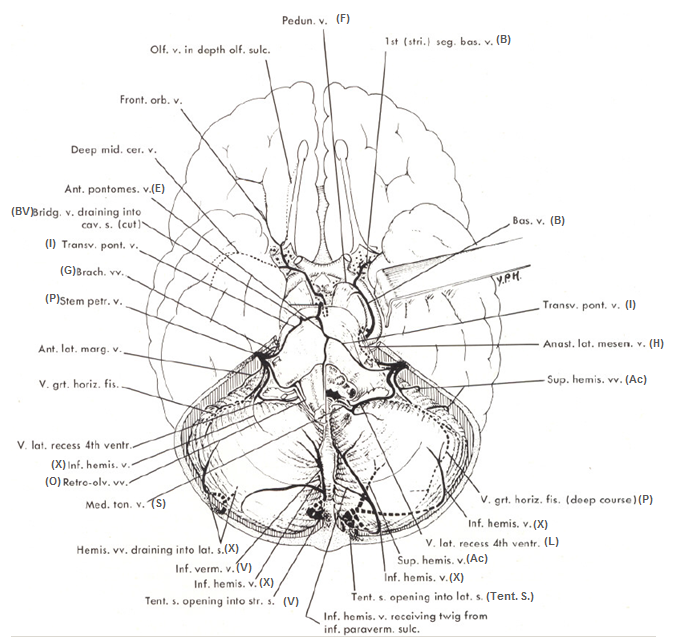
In the neurosurgical literature, as exemplified by Rhoton’s dissections, the same vein is called the Cerebellomedullary Fissure Vein. It serves as an angiographic landmark for the cerebellopontine angle.
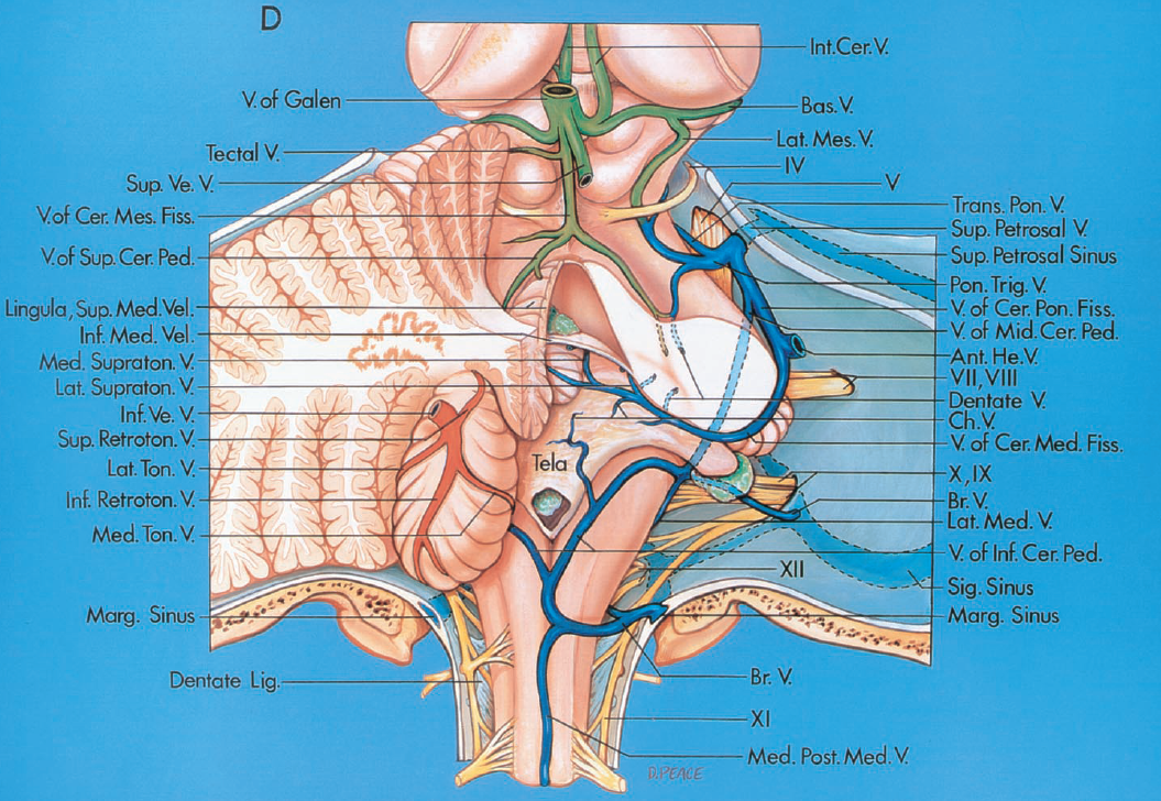
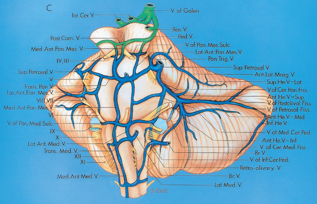
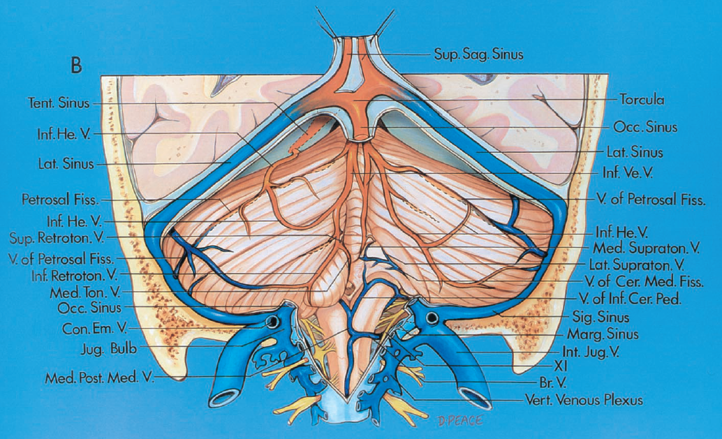
The following table serves as a correlation between the nomenclatures of Huang and Rhoton, to the best of my ability to read both of those great minds…
It is a large and relatively consistently seen vein, usually draining into the Petrosal Vein (P). It is usually prominent because it needs to drain the rather vascular 4th ventricle choroid plexus which can sometimes be seen as a blush on an angiogram.
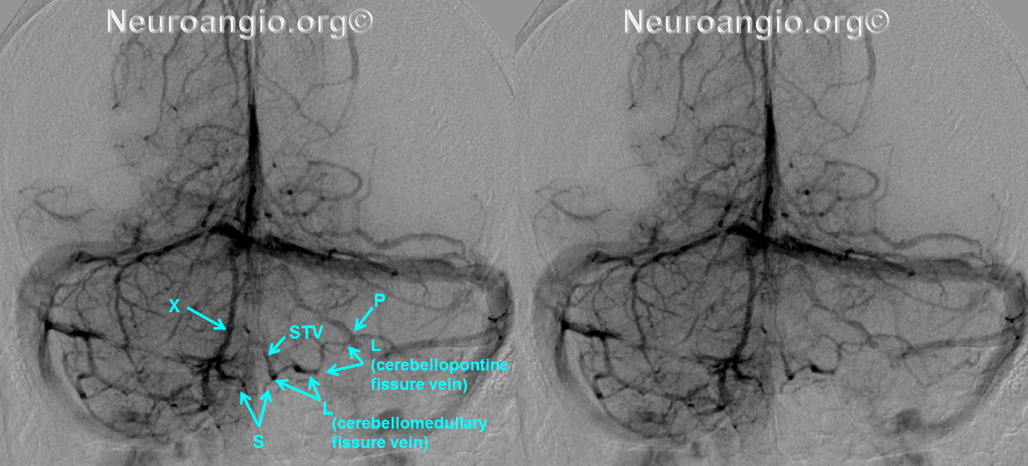
It is easy to identify this vein when one locates the Petrosal Vein. Any vein which courses inferior and then medial to the Petrosal vein must be either anterior or posterior to the medulla. Since such a vein is not likely to be prominent anteriorly, it is almost always going to be the vein of the lateral recess, as seen below:
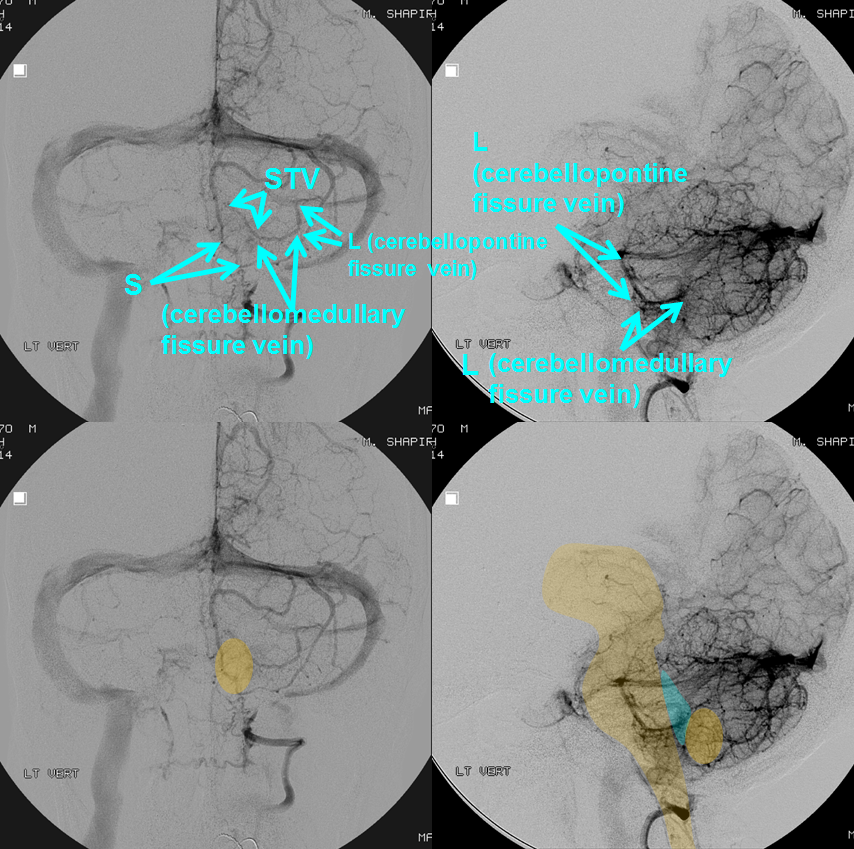
In the AP view, the Lateral Recess vein is the one which courses inferior and then medial with respect to the Petrosal vein (P). Keep looking for this vein in venous phases to learn to recognize “mechanically”.
Again, the vein has a characteristic hook-like shape on both AP and lateral views. The tip of the hook is in the ventricle and, as it comes through the Lushka it will form the bottom of the hook on both lateral and AP. Convenient. It will then ascend behind the flocculus, within the cerebellopontine fissure, where it is named “Cerebellopontine Fissure Vein” in Rhoton, to join the Petrosal vein (P). The flocculus separates this vein from the more anteriorly located Lateral Pontine Vein (J), also known as the Vein of the Middle Cerebellar Peduncle in Rhoton. There are of course variations which will be shown. So, the bottom of the hook marks the inferior extent of the 4th on a lateral view.
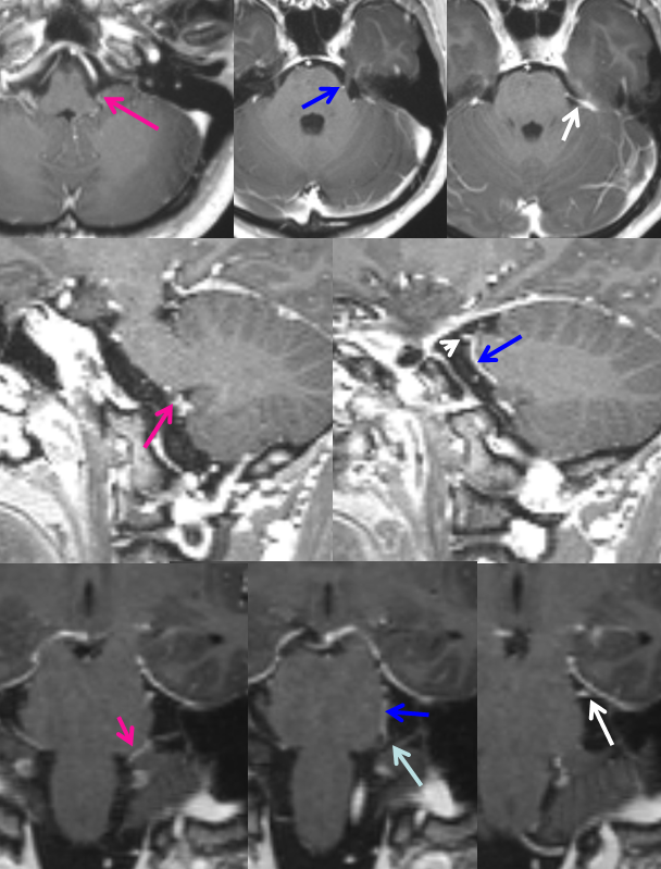
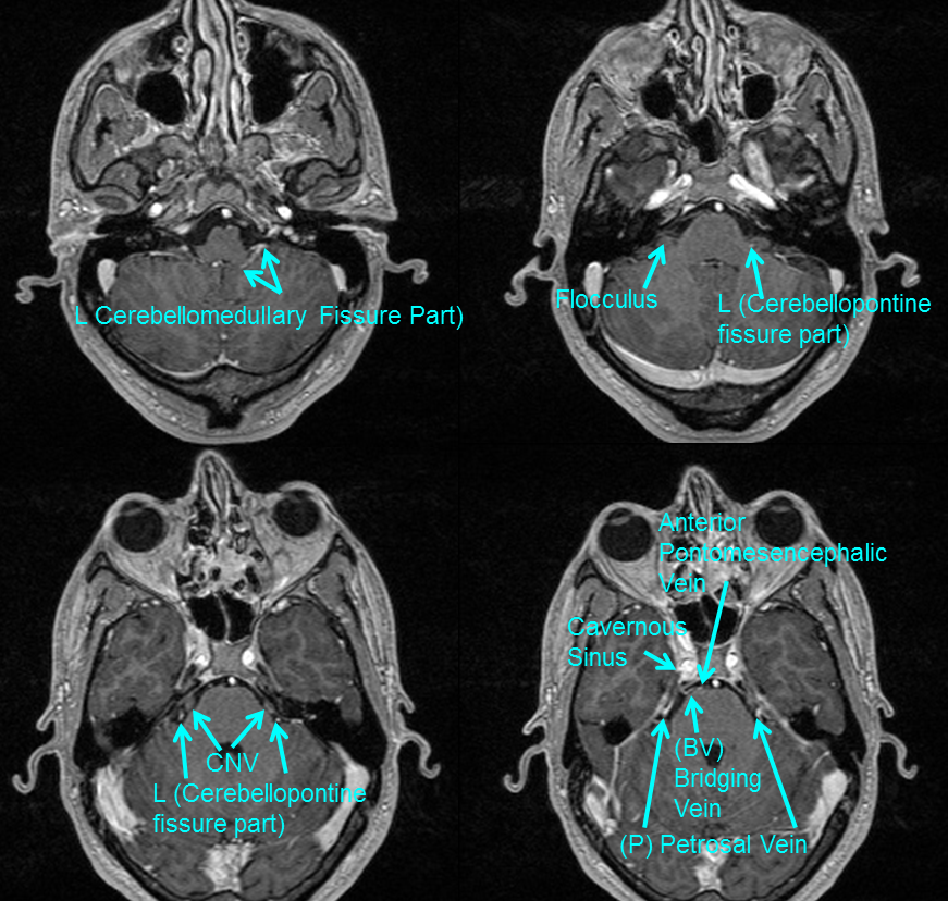
As with any posterior fossa vein, especially for those not yet comfortable with venous angiography, contrast MRI is an excellent way to see all kinds of veins. Any volumetric sequence (MP-RAGE, for example) will do a great job in a cooperative patient. Here, the vein of the lateral recess is marked with a pink arrow. It ascends on the lateral aspect of the pons, here being named teh Pontocerebellar Fissure Vein (Rhoton, blue arrow) to join the petrosal vein (white arrow, P). Notice how the vertical aspect of the lateral recess vein, a.k.a. the Pontocerebellar Fissure Vein is intimately related to the trigeminal nerve, usually running on its medial aspect, as in this case. Rhoton, in fact, in the Neurosurgery 2000 supplement manuscript on the retrosigmoid approach describes the phenomenon of venous compression upon the rootlets of the trigeminal nerve — the pontocerebellar fissure vein being one of the culprits.
Here is a CISS sequence example of the lateral recess vein, well-seen against the CSF. CISS and similar sequences are a great way to see veins in the various cisterns, provided one knows what to look for and does not confuse them with arteries or nerves. Notice how close the lateral recess vein (cerebellopontine fissure component according to Rhoton nomenclature) is to CN V
The choroid plexus of the 4th ventricle is, many times, found not only in the 4th ventricle but also outside of it, spilling out from the lateral recess / foramen of Luschka / lateral aperture.
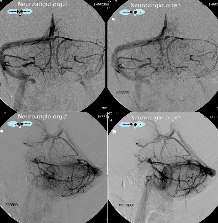
The lateral recess vein need not drain into the petrosal vein every time. In fact, there are rather commonly seen Bridging Veins (BV) in the area of the cerebellopontine fissure, connecting some anterior brainstem vein (such as lateral medullary/retro-olivary/lateral recess) to the inferior petrosal sinus. Rhoton showcases these veins more prominently than Huang, and I think that angiography as well as MRI show plenty of examples of such Bridging Veins (see dedicated Bridging Vein page), as in this example where the lateral recess drains via bridging vein on the left, and into the Petrosal Vein (P) on the right:
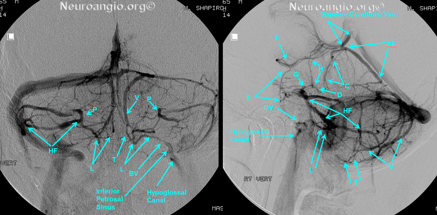
This is better appreciated in stereo:
Another MRI example of the lateral recess (L) vein, with its horizontal part (cerebellomedullary fissure vein of Rhoton) emerging from the lateral recess, then ascending on the lateral aspect of the pons as lateral recess (L) vein or, in neurosurgical literature, as vein of the cerebellopontine fissure, in close relationship to the flocculus and CN V, to converge onto the Petrosal vein (P). Notice homology of the Petrosal vein draining into the superior petrosal sinus with the Bridging Vein (BV) draining into the cavernous sinus.
Veins within the 4th ventricle
Rhoton’s diagrams depict and name several veins within the 4th ventricle and its lateral recess, such as the “Choroidal Vein” and “Dentate Vein” as seen below:

These veins are, as a rule, below angiogrpaphic resolution. Though they may be occasionally identified on high-field MRI, they remain of primary landmark interest to the rare neurosurgeon still willing to behold them in vivo.
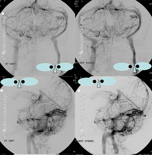
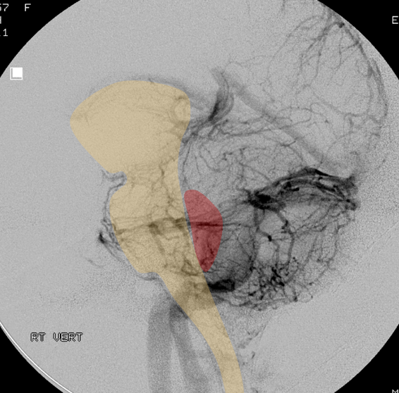
Here is a particularly well-developed lateral recess vein, including prominently the potion deep inside the 4th ventricle. The reason for this — the inferior vermian veins are not well-developed, such that its tonsillar territory is drained into the lateral recess vein partially (see below)
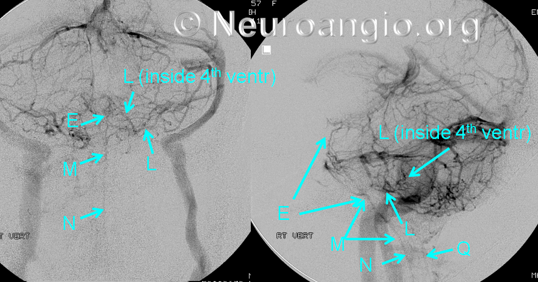
Here is a stereo of the same injection
An overlay of the brainstem and the 4th for clarity
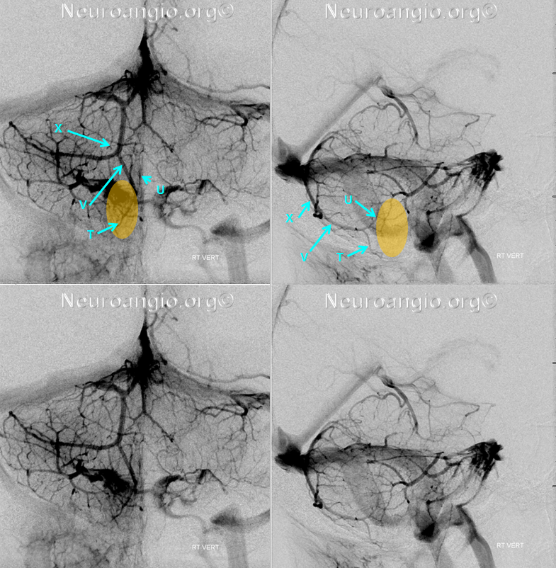
Tonsillar veins
The cerebellar tonsil is located lateral and posterior to the 4th ventricle. It is supplied mainly by branches of the inferior vermian vein (V) and the vein of the lateral recess (L) a.k.a. the cerebellomedullary fissure vein. The latter is usually responsible for the anterior portion of the tonsil, while (V) supplies the posterior part. Of course, variability is the rule, and when one is hypoplastic the other will take over the job.
The sum total of the story, in my opinion, is that tonsillar veins are variable and their nomenclature is confusing. Moreover, the veins are so variable that assigning a specific name to this or that tonsillar vein may not be helpful or accurate. The most important point, again, is to know where the vein is in relation to the brain, or the tonsil part of it in this case.
In this example, the lateral recess vein on the left receives the medial inferior tonsillar vein (S) and the anterior superior tonsillar vein (STV). The way to identify (S) is to look for a vein which courses inferiorly from the medial horizontal aspect of the lateral recess vein (the cerebellomedullary fissure vein). On the right, the inferior vermian vein is prominent, and receives (S) instead.

The cerebellar tonsil position can be usually determined based on location of the superior (U) and inferior (T) retrotonsillar veins, which together with the inferior vermian vein (V) look like a claw in the lateral view, as in the following example. I think that this “claw” is the most consistent venous part of this whole tonsillar business. The point, again, is to recognize from the venogram where the tonsil is.
Most of the time, the inferior vermian vein will be taking care of the bulk of the tonsil. Sometimes, the drainage is balanced, and the tonsil is neatly outlined by its veins:
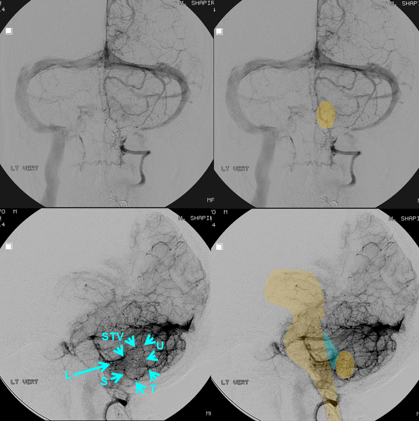
Now with appropriate labels:

Same case, in stereo:

In conclusion, remember that the choroid plexus is a relatively vascular structure, and thus the vein of the lateral recess (L), a.k.a. the cerebellomedullary fissure vein will be present in nearly every case. Look for it as a way to define the inferior border of the 4th ventricle in both AP and lateral views.
Here is another case of easily identifiable paired inferior vermian veins, supplying the tonsils:

This is better appreciated in stereo:

Unpaired vermian vein
Although there is one vermis and one superior vermian vein, the inferior vermian veins are typically paired. However neither the falx cerebelli need be there nor should one always have two vermian veins. In this case, there is clearly only one (V), with well-seen retrotonsillar superior (U) and inferior (T) vermian tributaries. The latter is connected to the posterior spinal vein (Q)
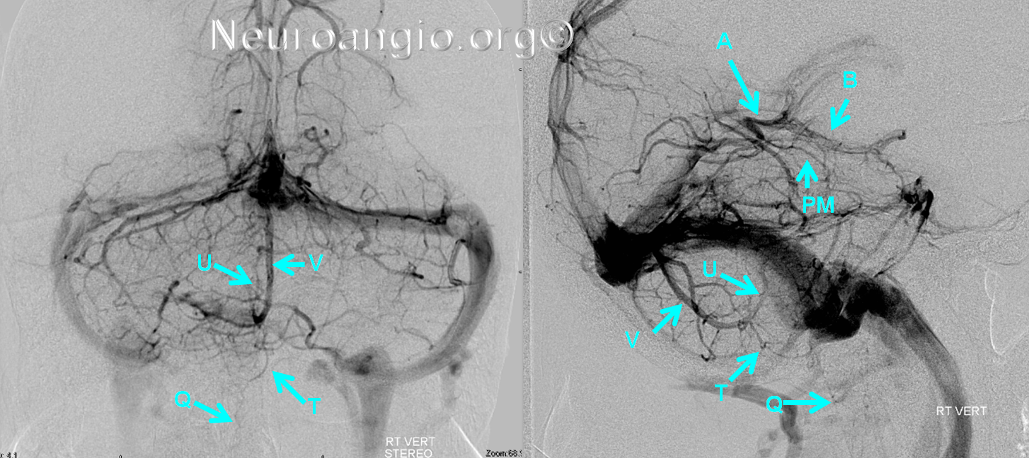
Again, best seen in stereo:
Caveat — appropriate injection
It is important to remember the basic rule that, in angiography, veins are only visible when the arterial territory which they drain is injected. In the posterior fossa, this is most important for the PICA territory, which is not always sufficiently seen from the contralateral vertebral injection. Even when enough contrast can be retrogradely forced into the contralateral PICA to sufficiently evaluate the arterial anatomy, the same contrast volume may not be adequate to see the veins. Remember that 75% of total blood volume is in veins at any given time. Since the PICA usually supplies the inferior cerebellum, the venous complex corresponding to the PICA is the inferior vermian veins, tonsillar veins, anterior cerebellar veins, and some medullary veins.
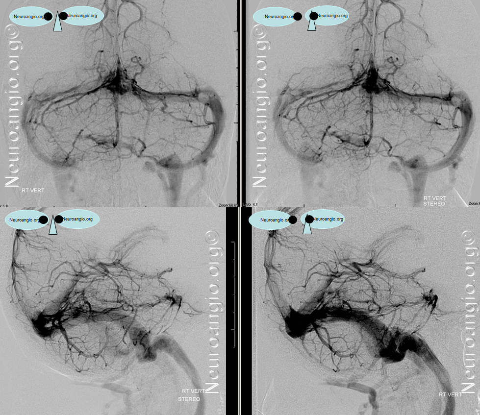
Here is an illustration of this situation, where right vertebral injection transiently fills the left PICA, yet insufficiently to see the left vermian vein (top row). Rather than concluding that the vermian vein is unpaired, one should either inject the left vert or allow for sufficient reflux into it from the VB junction to adequately fill the PICA territory. In the follow-up angiogram (bottom row) the left vertebral was injected with sufficient reflux to see both well-developed inferior vermian veins:
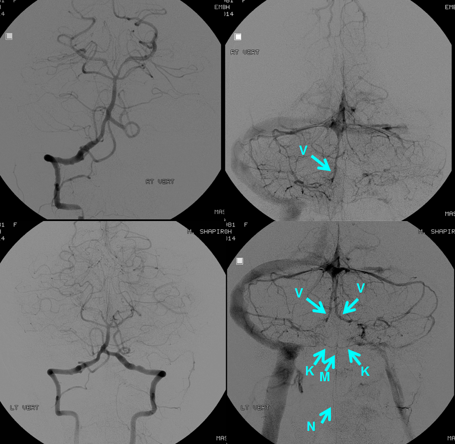
Remember rule #1 — if you don’t see a vein, it doesn’t mean that its not there, if the same vein could be draining the territory of an artery which was not injected to produce the picture you are looking at. Inferior vermian veins are a good example of this: only the left one may be seen from left vert injection, and the right one might be there, if you had injected the right vert. All sorts of fakeouts are possible with in-vivo angiography, depending on the arterial arrangement.
References:
Yung Peng Huang and Bernard Wolf. Veins of the Posterior Fossa. Chapter 75, in Newton and Potts Radiology of the Skull and Brain. Volume 2, book 3. 1974
Yung Peng Huang et. al. The Veins of The Posterior Fossa — Anterior or Petrosal Draining Group. September 1968. American Journal of Radiology. For a full text PDF click here.
Yung Peng Huang et. al. The Veins of The Posterior Fossa — Anterior or Petrosal Draining Group. September 1968. American Journal of Radiology. For a full text PDF click here.
Albert Rhoton, The Posterior Fossa Veins, Neurosurgery. 2000 Sep;47(3 Suppl):S69-92.
Albert Rhoton, The Cerebellopontine Angle and Posterior Fossa Cranial Nerves by the Retrosigmoid Approach. Neurosurgery. 2000 Sep;47(3 Suppl):S93-129.
Albert Rhoton, The Posterior Fossa Veins. Neurosurgery 2000 Sept 47(3), S69-92.
