
Case Courtesy Drs. Eytan Raz and Howard Riina
We have multiple examples of missed dural fistulas (spinal dural fistula parent page here). Usually, the solution is to repeat the angiogram. However, advanced MRI can be quite helpful these days. See below
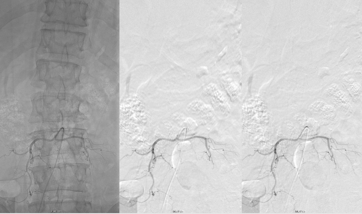
Initial angiogram is “negative”
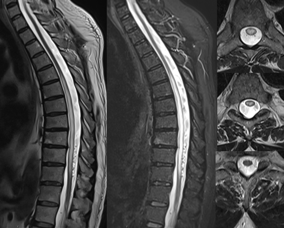
A follow up high res MRI is done

CISS coronal imaging is key — the pathologic vein is obvious. Left L3 location is likely

Sequential images. Radicular vein marked by arrows. L3 nerve by dashed arrows. Notice that the vein is quite away from — not on surface of — the nerve

L3 (conjoined) angio. Vein marked by arrow

Prior angio is below. what is the problem? Imaging was not done long enough to see the vein — note lack of vertebral body blush.
Back to current angio — coil off the muscular branch and place microcatheter in right position
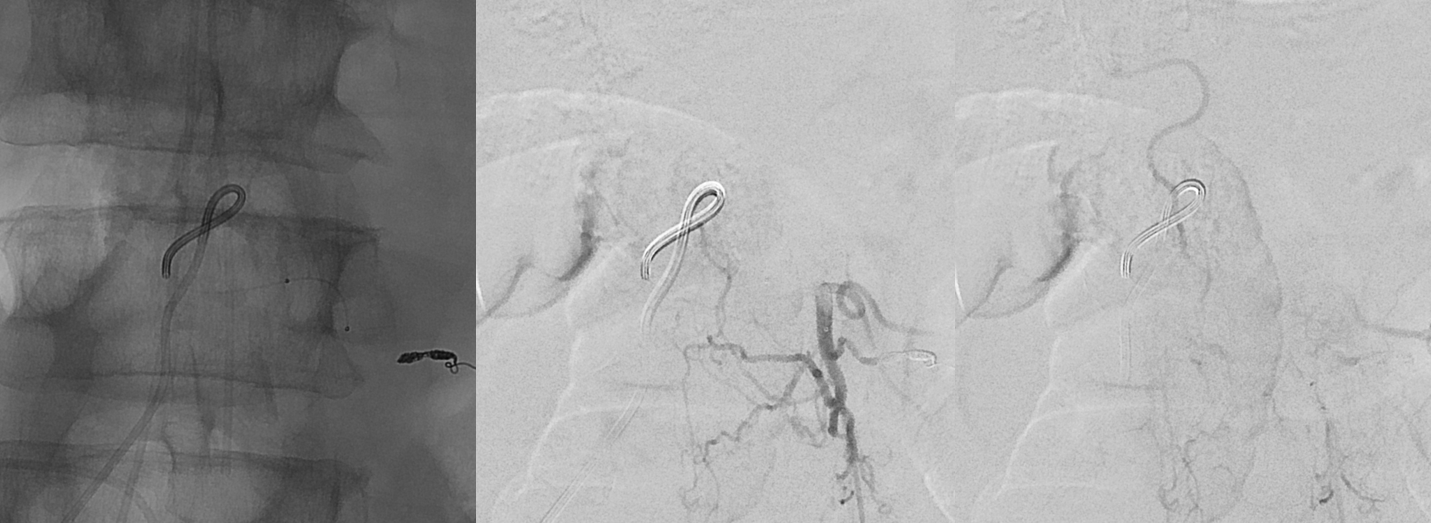
Glue cast on the rightmost image

Delayed post MRI
