
Case with Dr. Osamah J. Choudhry
Nothing Taboo about it. Been done and written up, here and here for example. But you have to have experience with embolization in other territories, with meticulous technique, before doing something like this. Its not for places where embos are not done for whatever reason, except when they are really necessary — except then there is no experience doing them.
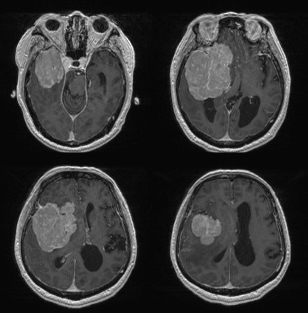
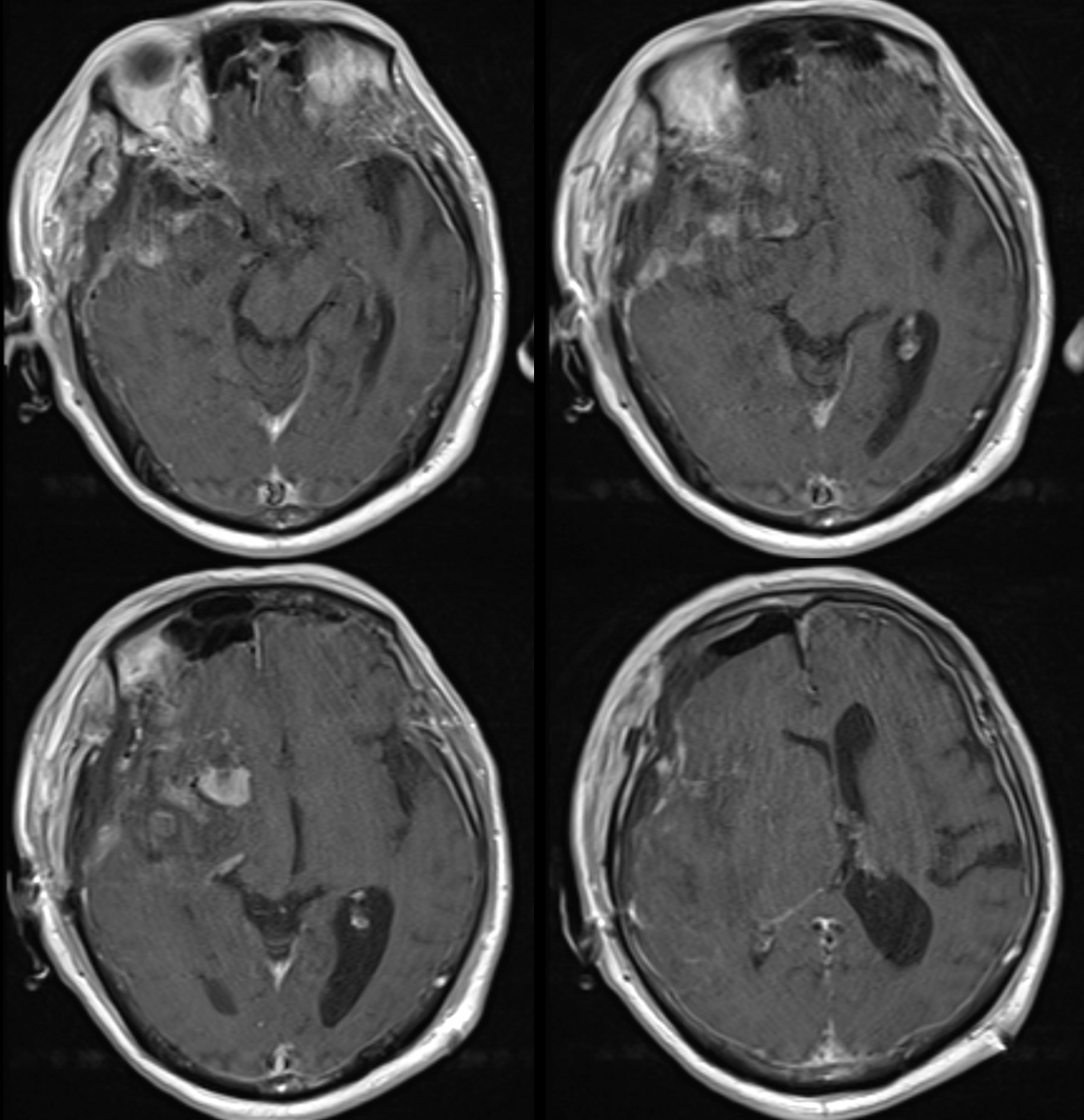
Giant sphenoid wing mening
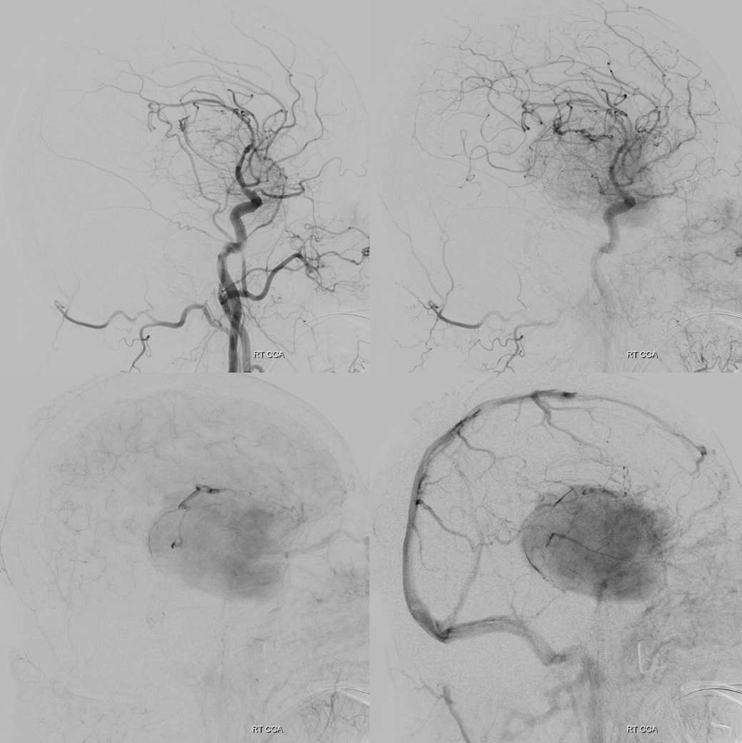
DYNA
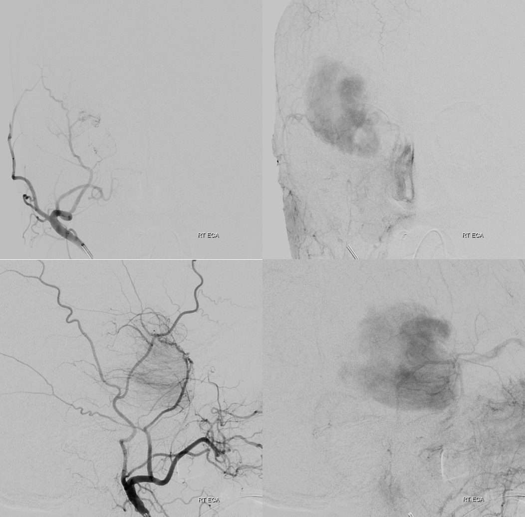
ECA Dyna
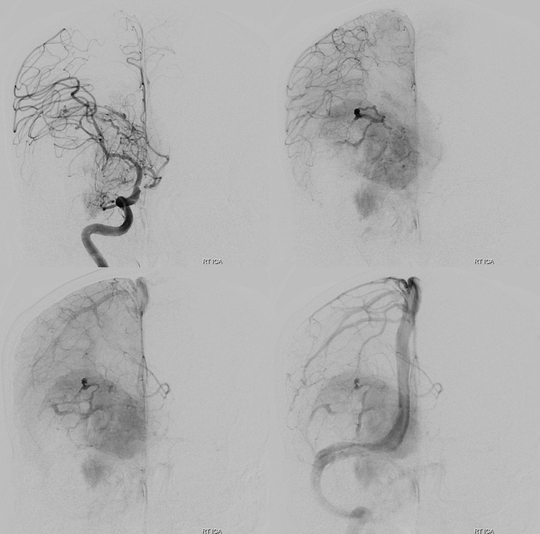
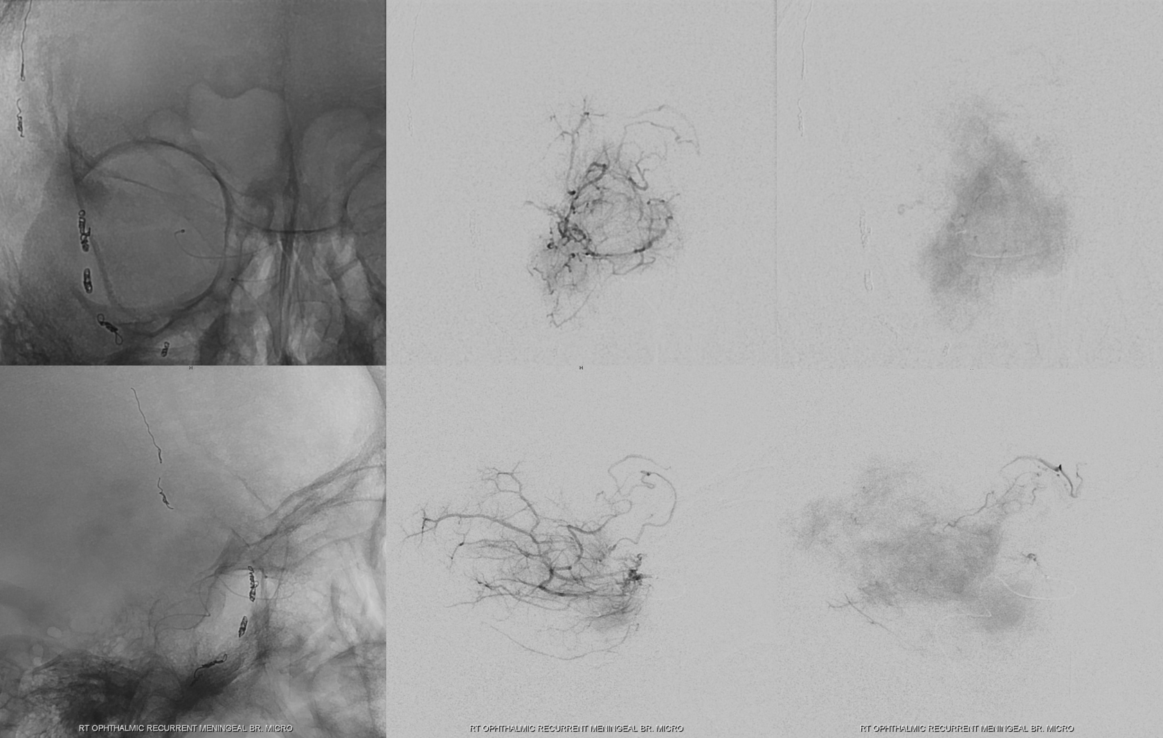
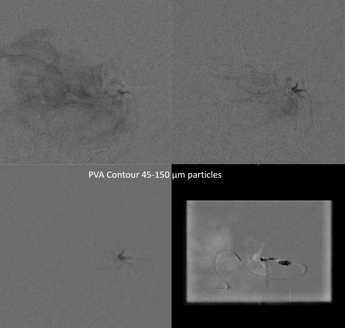
Video of particle embo of the MMA, followed by ophthalmic catheterization. I did cut corner here — the support is not optimal. Its a 5F vert quite low. Optimally need more support closer to the ophthalmic .
Single Tip Headway Duo and Synchro 2 soft. Headway single tip is the go-to for these kinds of jobs — it follows 014 catheters beautifully and takes up to 250 micron particles. Because we believe in small particle embo as the main method of tumor devascularization, we use 45-150 contours.
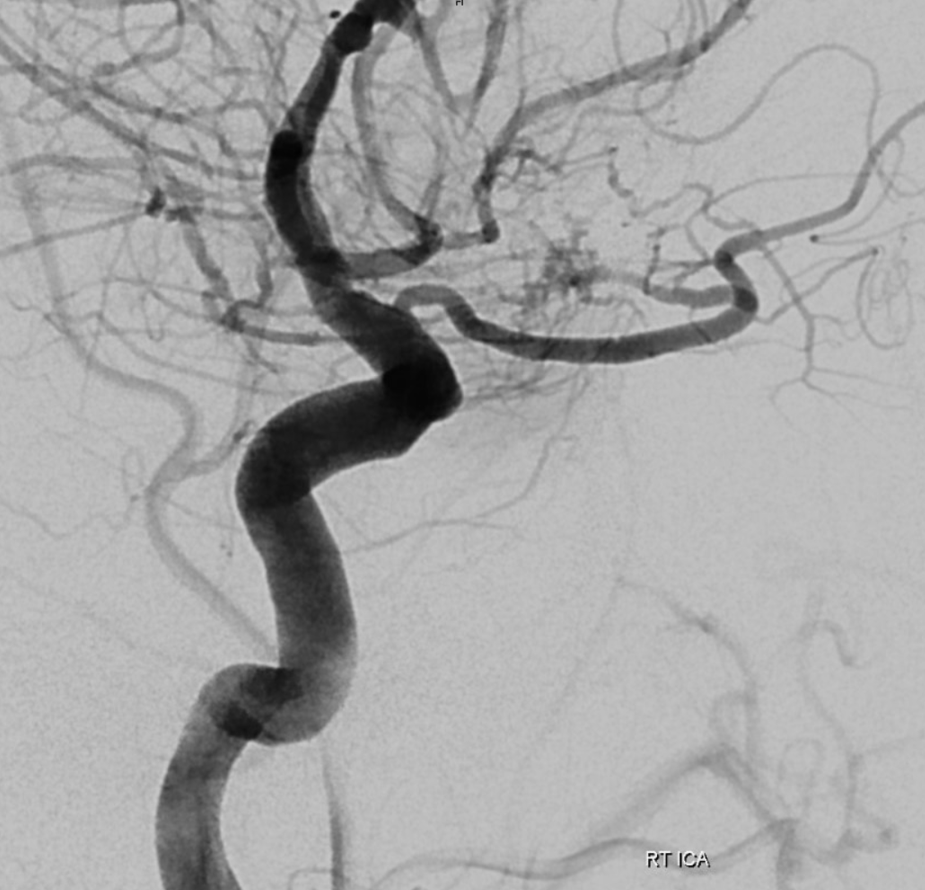
The movie below is MMA embo, followed by ophthalmic catheterization
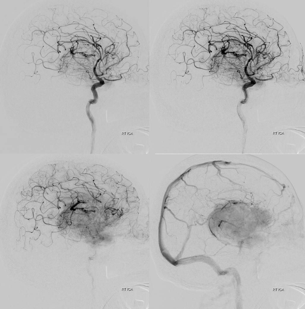
DYNA in embo position — the concern is actually not the central retinal artery — its proximal to the recurrent meningeal in this ophthalmic artery configuration — see ophthalmic artery page for details of over / under course and implications for central retinal artery origin. The concern is what supplies the optic nerve. Its practically impossible to see these branches, and if any supplies comes from the recurrent meningeal, well… Fortunately, that is not the embryologic way, as we understand it. So, with the DYNA not lighting up the optic nerve, i think its good to go.
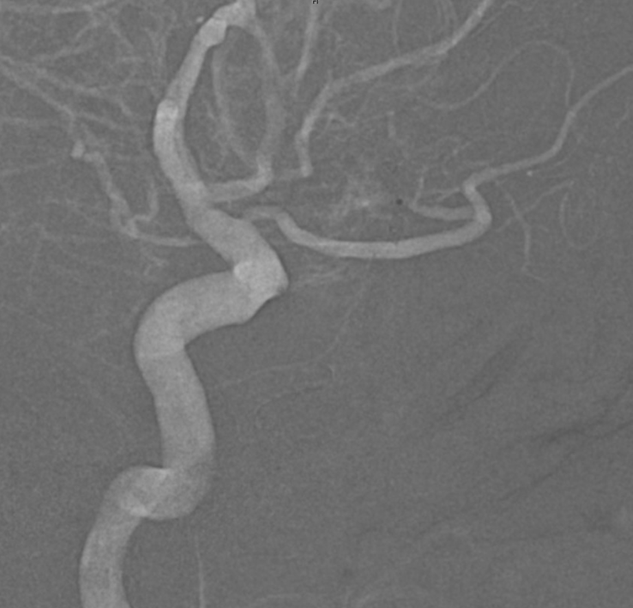
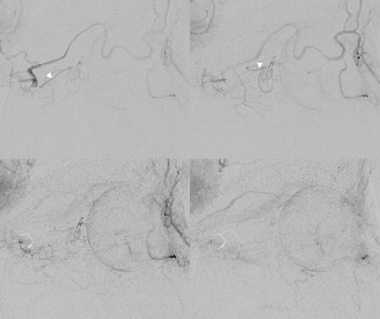
Pre-embo micro
Particles are safest, in our opinion. Pretty much the only concern is reflux. There is no “pull” at the end, no chance of dragging glue or onyx on the tip, no need to build plugs or seek wedge positions. Particles and a coil or two at the end. If you are worried about bubbles that always come out with coils, don’t do them.
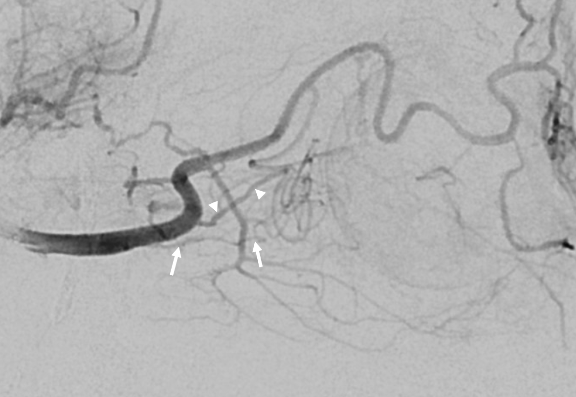
Amazing post embo DYNA distal to origin of the central retinal artery — see selective contribution of the ciliary arteries to the choroid — and no CRA in the middle of the nerve
DSA from the same position — a perfect demonstration that one can have a very nice choroid blush with no central retinal artery. Arrowheads on ciliary arteries.
More proximal post-embo DYNA shows the CRA also — see its proximal origin
CRA = solid arrows. Central Retinal Vein = dashed ones
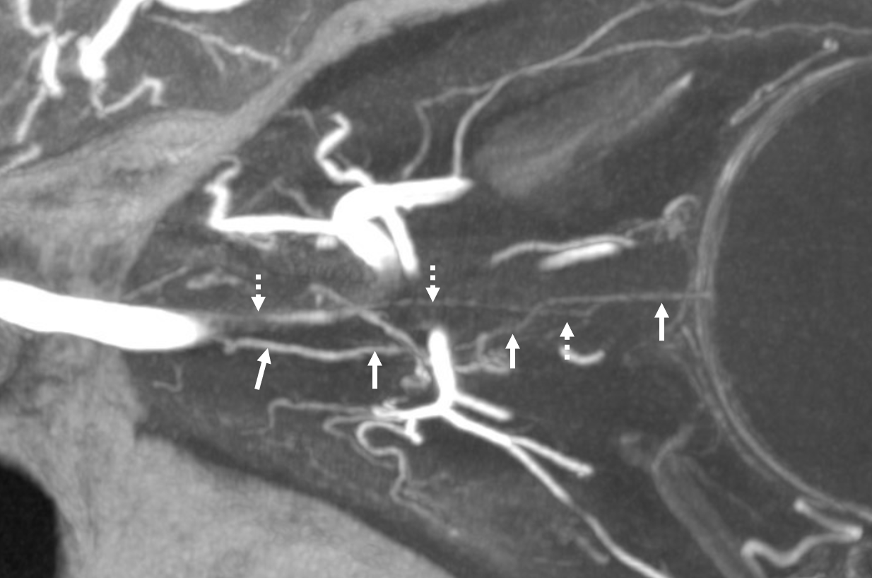
CRA = arrows; ciliary artery = arrowheads.
Just a cool picture
Post resection — the piece over there left on purpose where it was stuck to the M1
See Archives for more Tumor Embo cases