
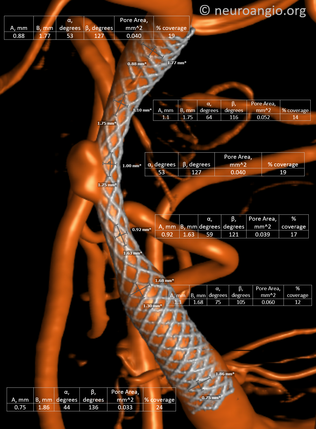
Something that can be easily done with modern cone beam imaging and a bit of junior high trig… If you dig it, DIY!
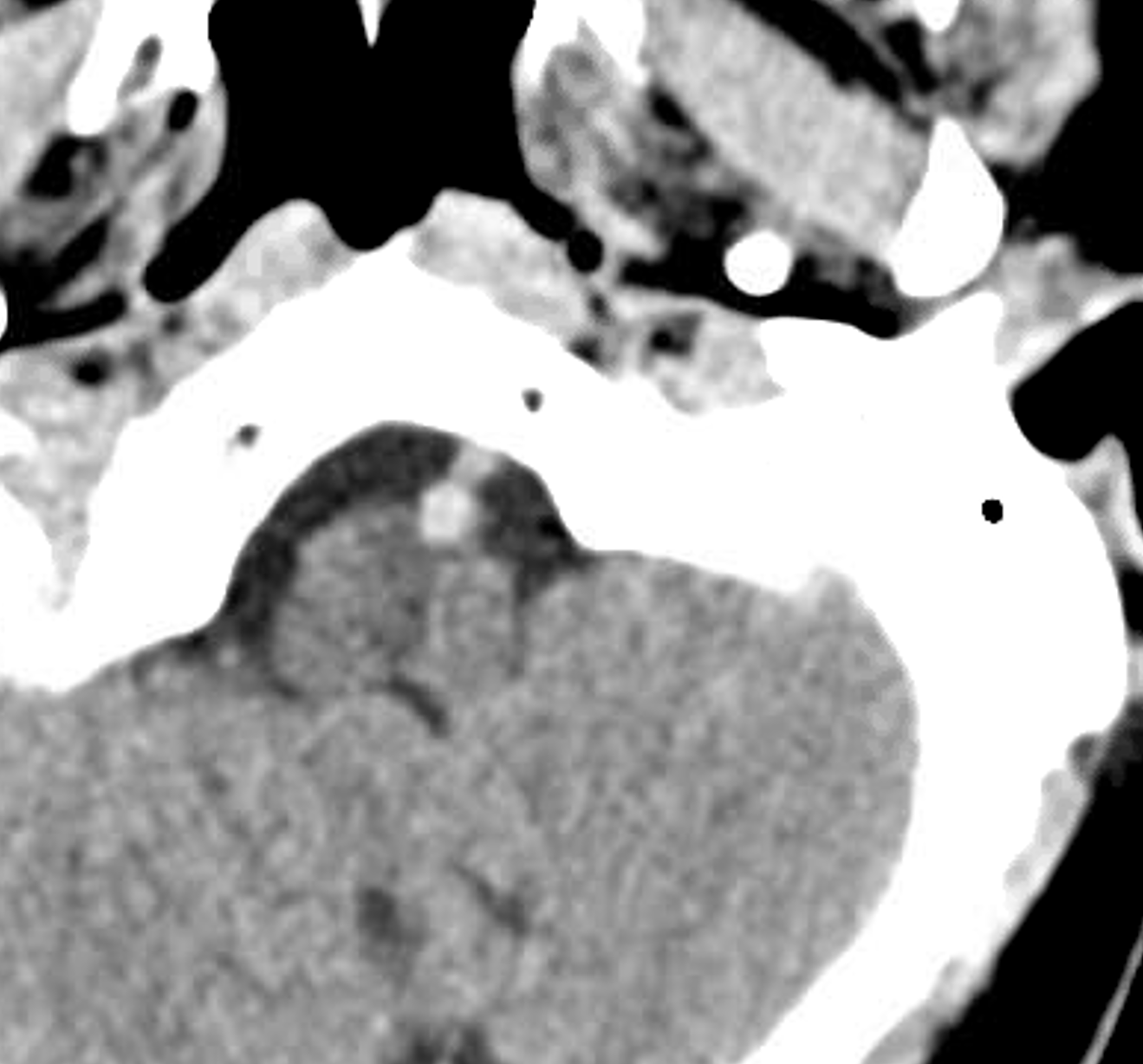
Presentation is sudden headache
Noncon head CT
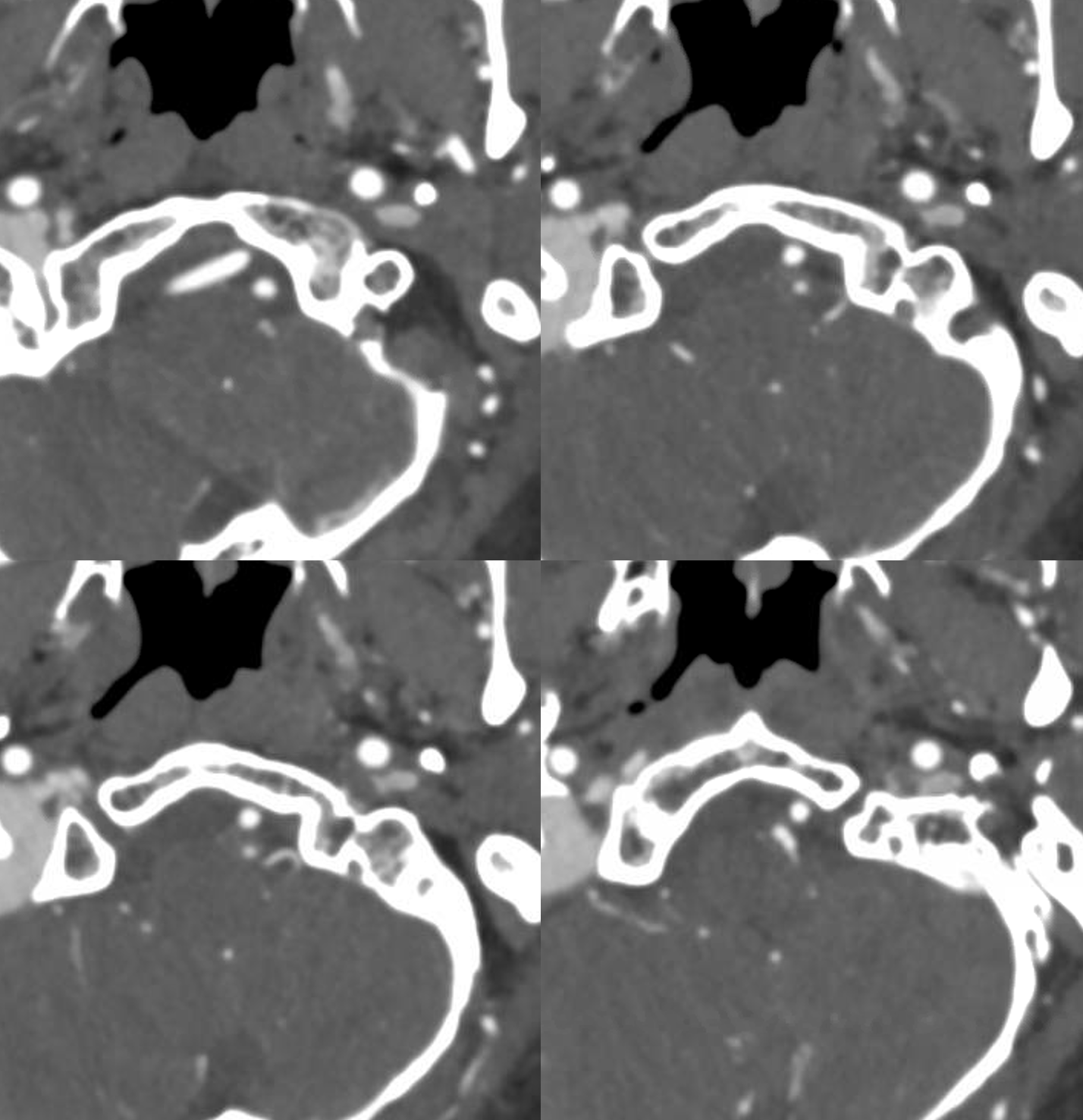
CTA source
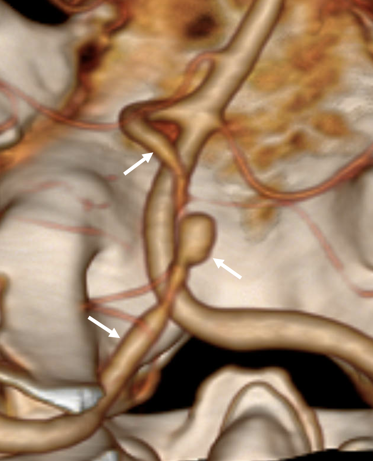
CTA Volume Rendered
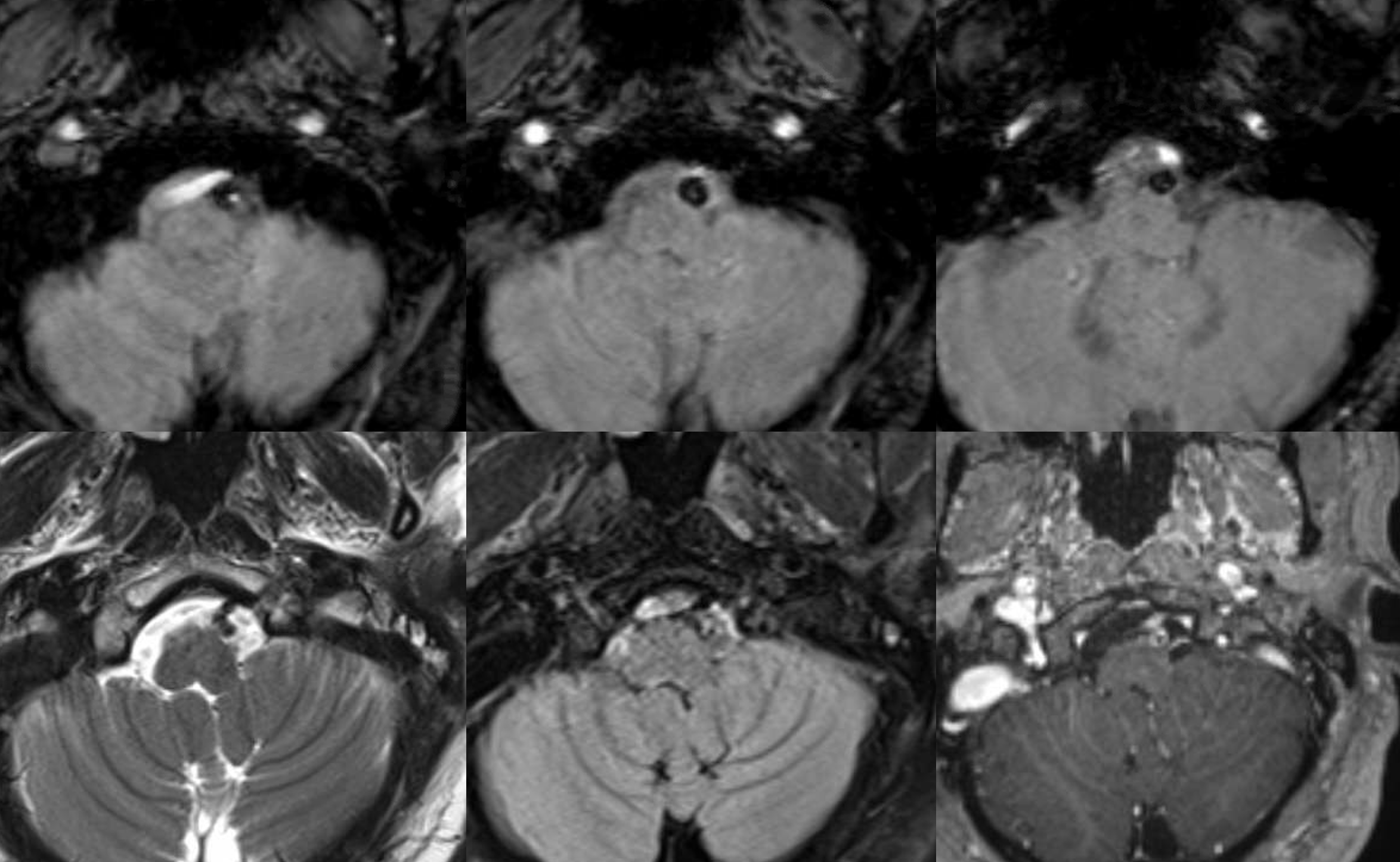
MRI. Best seen on gradient echos — dissection with blood products in the wall
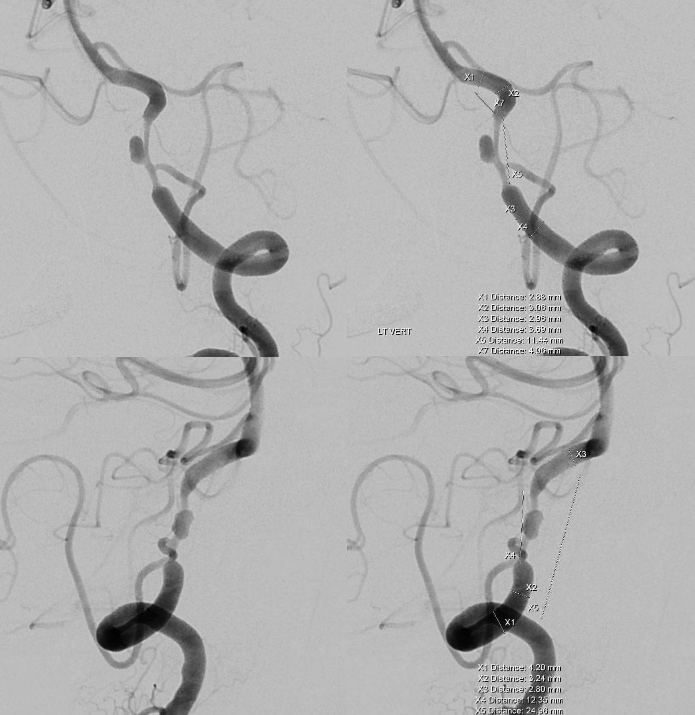
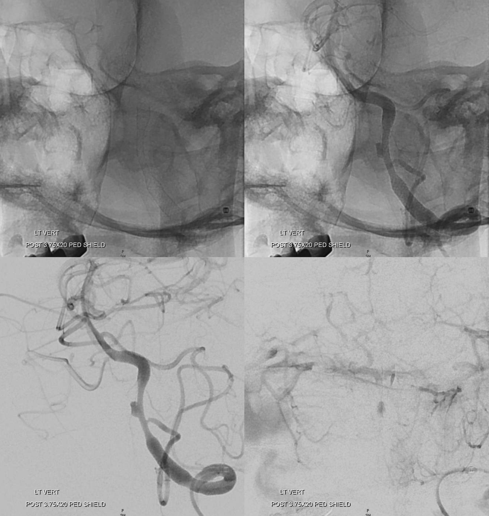
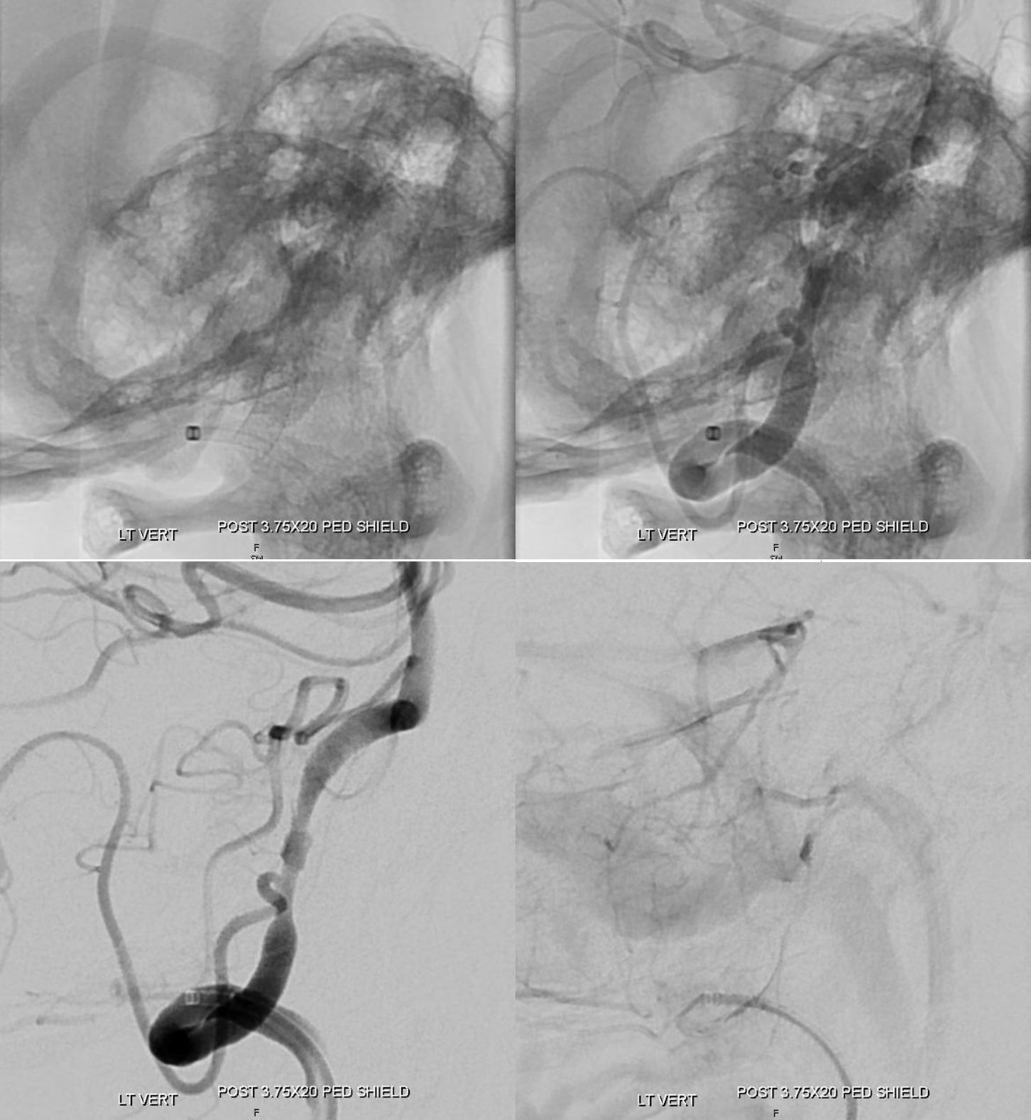
DSA
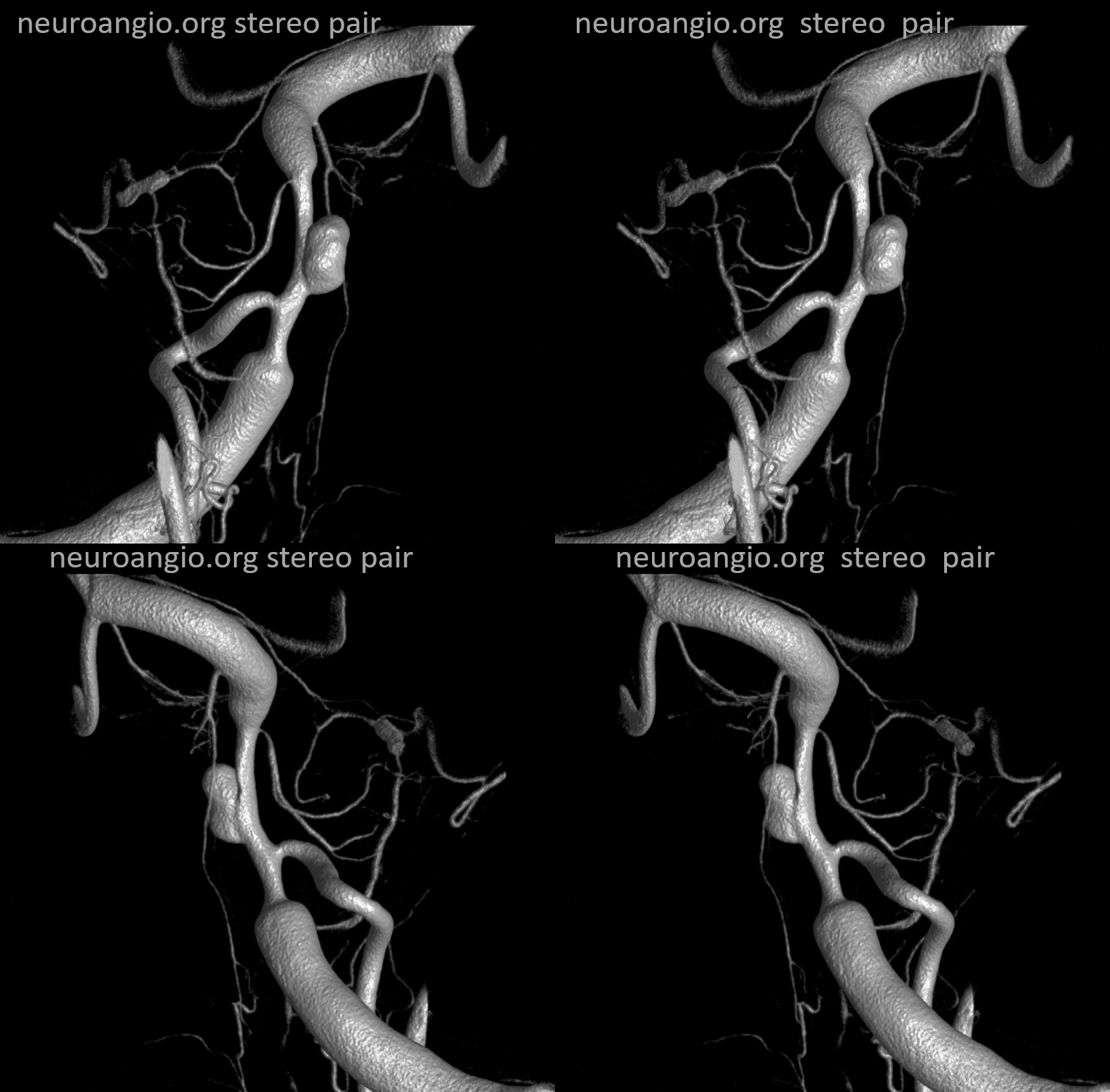
DYNA Volume Rendered stereos — prominent lateral medullary perforator in addion to the PICA
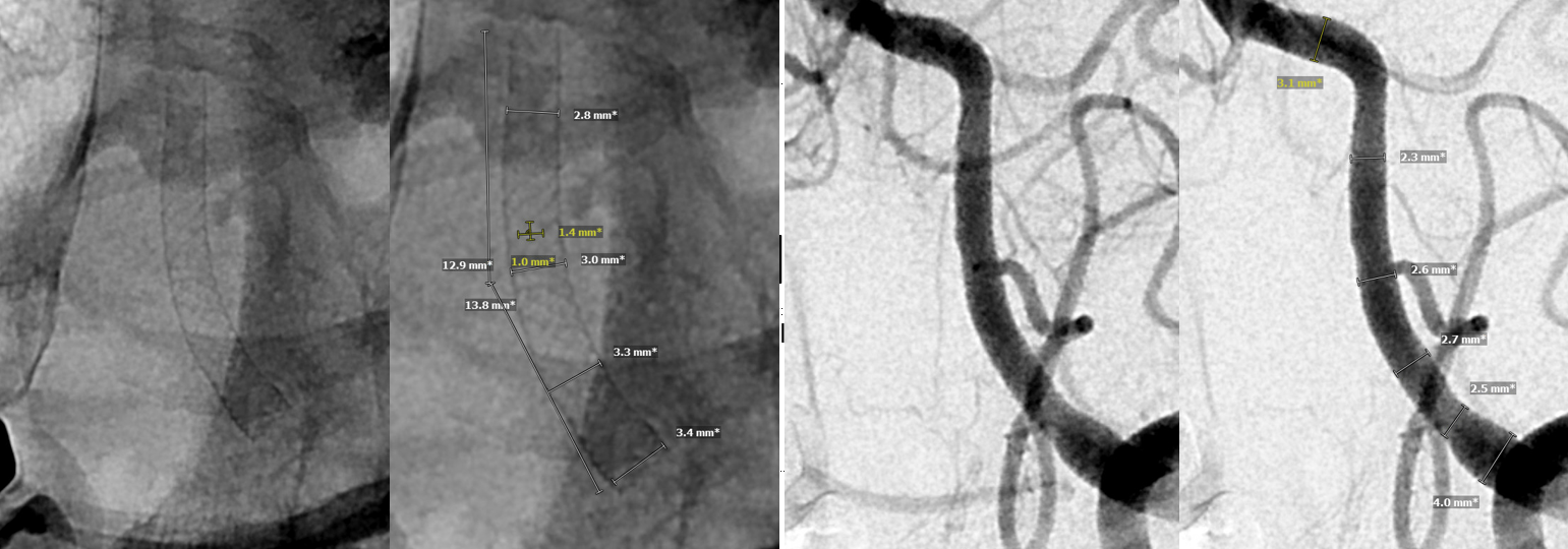
Post Pipeline Shield — single device
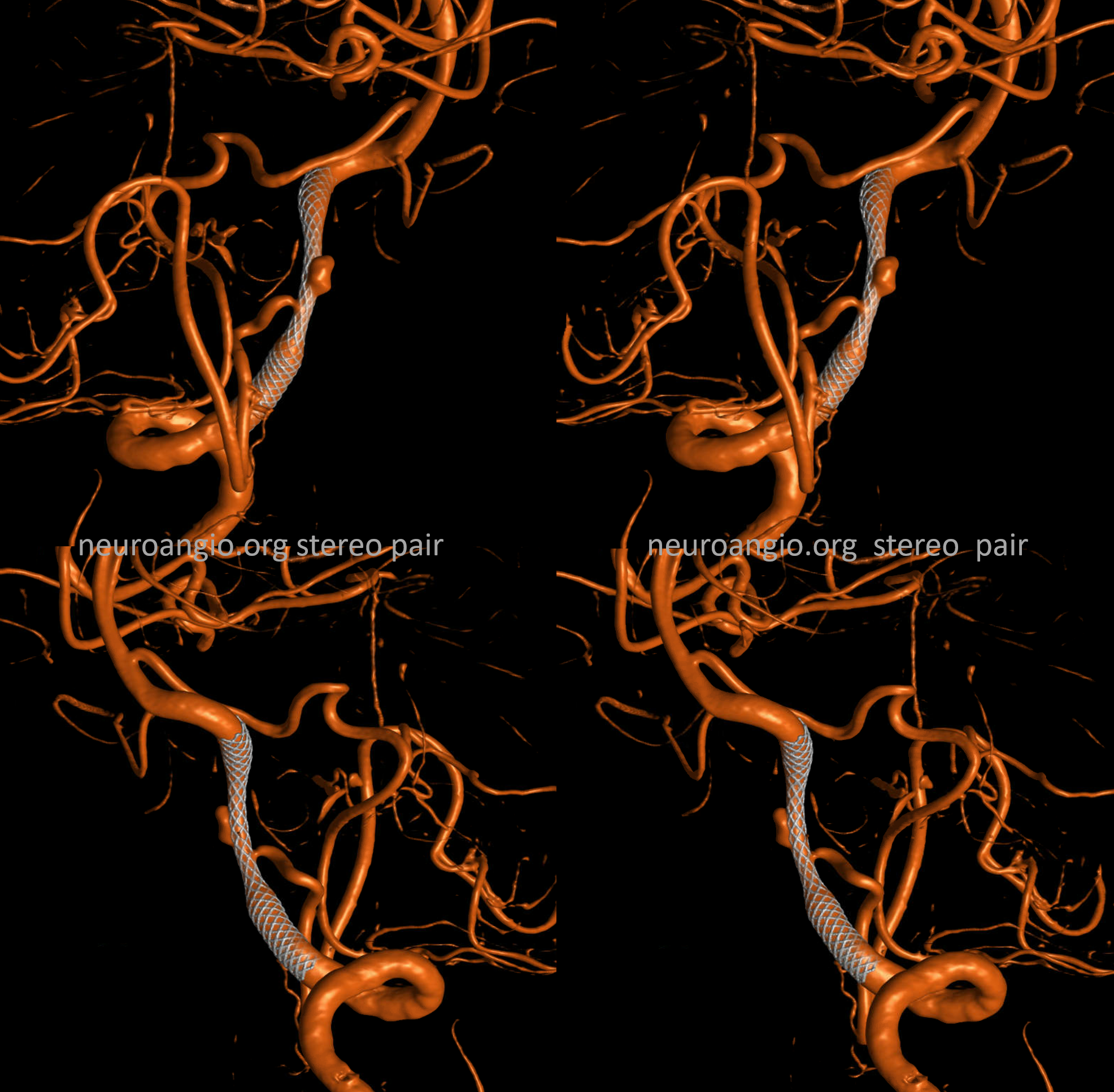
See the Cr-Mo wires?
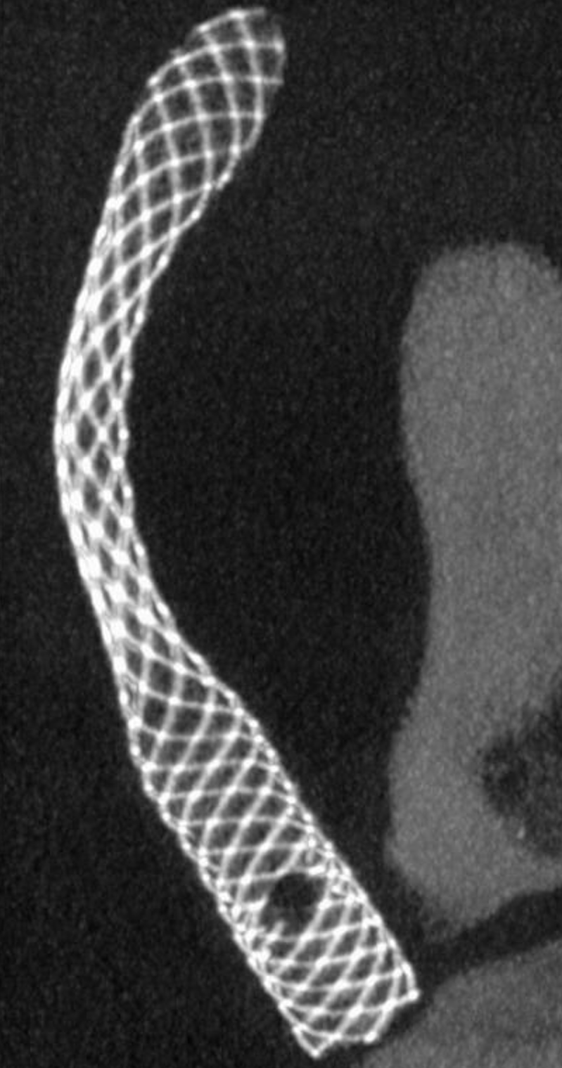
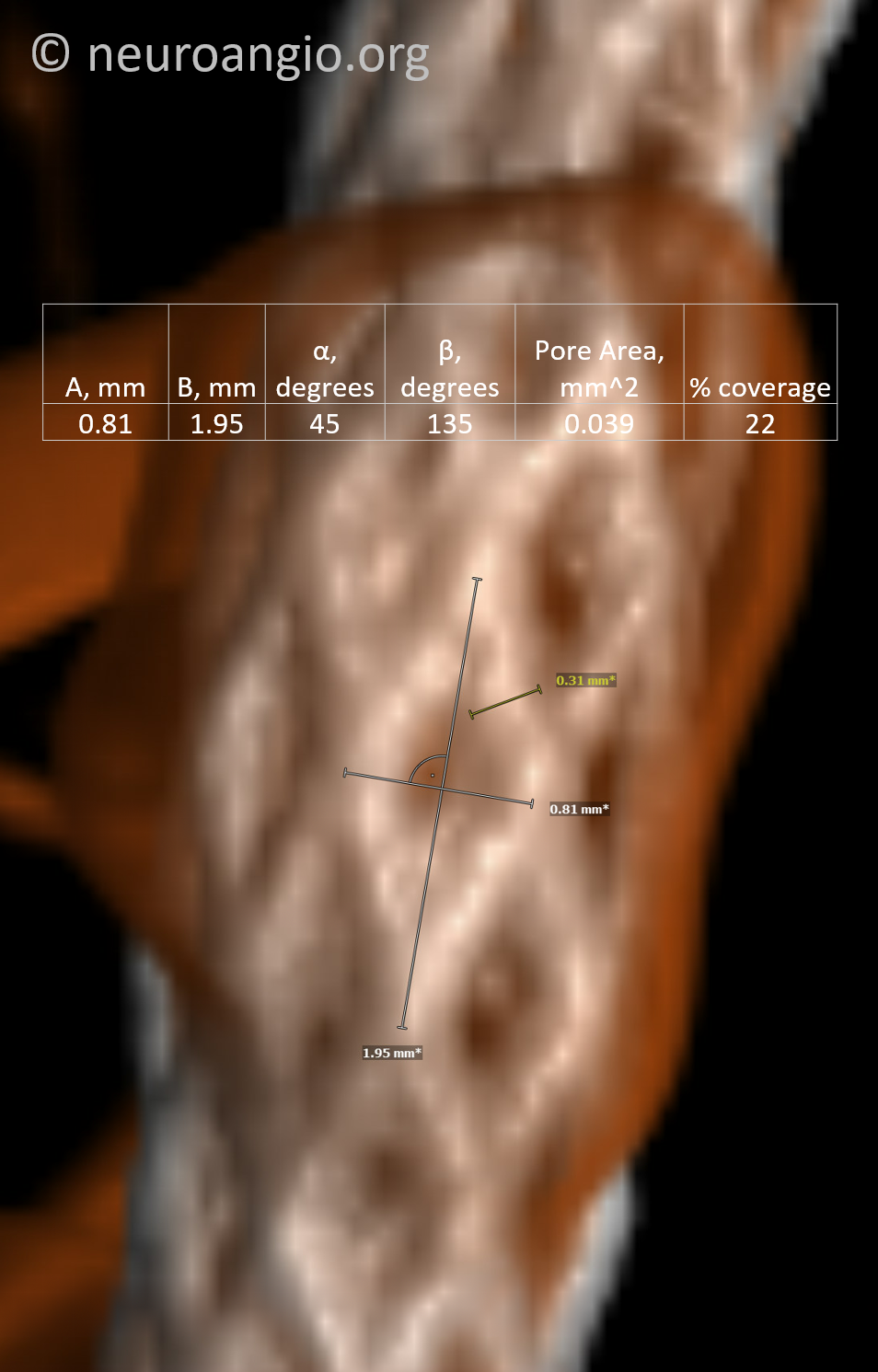
Once more… A and B are the width and height of the rhombus. That all ya’ll need
At the aneurysm. Note how much the thickness of Pt wires is exaggerated on imaging. The real thickness is 30 microns — 1/10th of the measurement. Which is why A and B (above) are measured the way they are
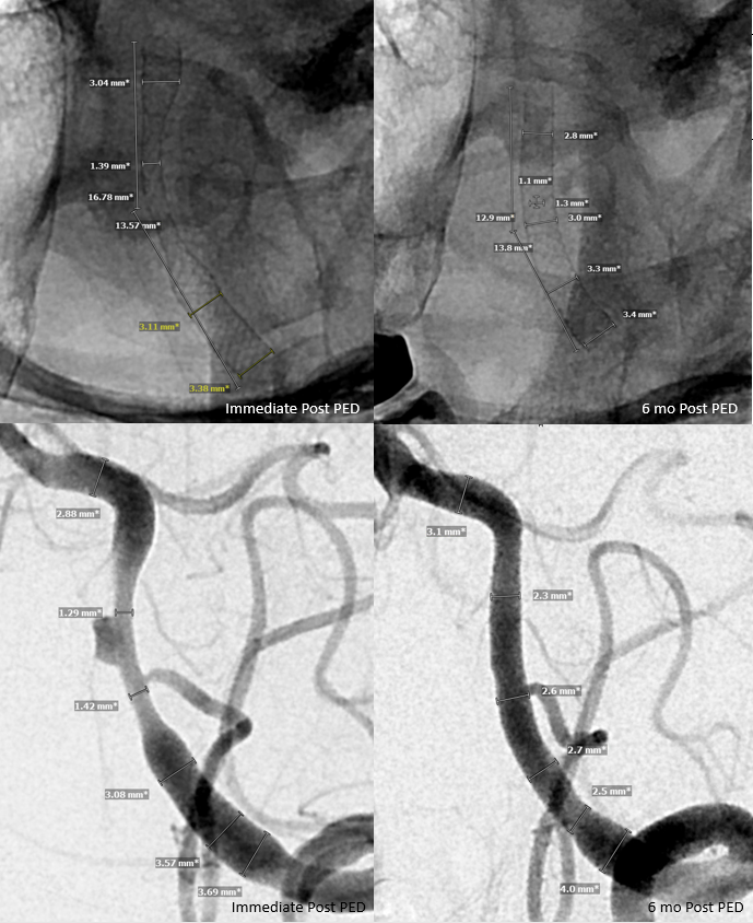
FOLLOW UP
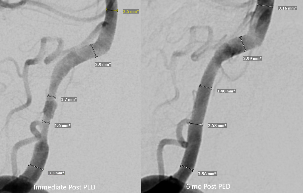
Of course, nothing like follow up to show what works. 6 months post — see remarkable arterial remodeling and cure
Comparision — device foreshortening is associated with expansion at the previously dissected, now healed segment
Dig it? DIY then! And see Pipeline Device Properties page for more info