12a — “Collateral Branches” Supplying the Intraorbital / Intracanalicular Portions of the Optic Nerve
14a — Superior Hypophyseal Branches supplying the optic chiasm, intracranial / intracanalicular portions of the nerve
As in anything anatomical, basic knowledge and diagrams detached from reality are not helpful. For the optic nerve and the eye, simple thinking reduces everything to the “central retinal artery”. As if nothing else exists. BUT… It only supplies a portion of the optic nerve. It is NOT the only supply to either globe or retina. It’s relationships with other globe players, such as ciliary arteries, is complex and individualized — with important clinical implications.
So, we will here show what we know about optic nerve supply OTHER than the central retinal. Now, if you are interested in optic nerve head and this area, study Sonah Hayreh. References here. Also study our seminal and so far only publication on cone beam CT visualization of the central retinal artery, published in Radiology
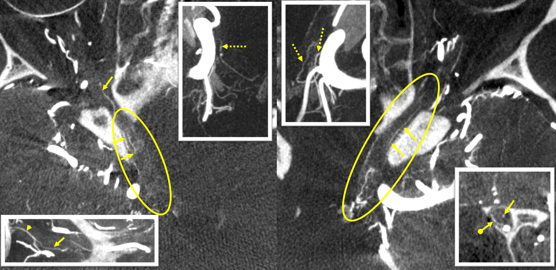
Superb Nerve Visualization Post Tuberculum Sella Meningioma Resection
Another example where anatomy is accentuated by pathology. Visual deficits post resection.

Bilateral CBCTs post op. There are no proximal / cave superior hypophyseals — presumably missing after surgery. There is collateral support of these from PCOM on the right and a more distal superior hypophyseal on the left (dashed arrows). There is unusually good visualization of the intracranial optic nerves (ovals), probably because their surface vessels are dilated to minimize vascular resistance in setting of collateral support. Support also comes from the intraorbital ophthalmic artery, in form of a recurrent branches (arrows) of the central retinal artery (arrowhead). Note also that a branch is present inside the optic nerve (arrow, inset, left), which is highly unusual.
Without labels

Fusion of bilateral cone beam CTs — there is incredible optic nerve and chiasm visualization (ovals), as well as supply to the pituitary stalk (arrows)

Pre and post verapamil infusion — green is post

Pre and post verapamil infusion images:

Canalicular Segment
Segment inside the optic canal. Usually has at least one recognizable on high resolution CBCT branch, which projects posteriorly. It is frequently involved in supply of meningiomas, and as such is at least partially dural and thus supplies the optic nerve sheath. It is probably part of the “collateral network” described by Hayreh, also providing supply to the optic nerve. Shown by arrows in these CBCTs of 2 normal optic apparatus patients presenting with typical aneurysms.

The same branch (arrow), enlarged markedly, to supply a meningioma (ball arrow). Curiously, the very long central retinal artery (dashed arrows) also originates from the same branch — which is both a variation and a cautionary tale for those who think that central retinal origin can be predicted based on general anatomic information. It is a dangerous assumption, and best decisions are always made on case-by-case basis.

Movie — follow the optic nerve head to trace CRA to the canalicular branch.
Same case — left ICA — more distal origin, no longer canalicular, but proximal intraorbital branch (arrows) supplies the meningioma, in addition to the expected anterior (ball arrow) and posterior (dashed arrow) ethmoidal arteries

Another case
Intracanalicular branch (arrow) in setting of meningioma, predominantly supplied by the deep recurrent meningeal artery (dashed arrow)

Extreme Visualization in Cavernous Dural Fistulas
The most exuberant enlargement of canal branches occurs in dural fistulas, usually cavernous. This suggests that the branches involved have some dural preference. There is likely a mix of predominantly dural branches and some piodural ophthalmic equivalents. All of these regress when fistulas are successfully treated. Below is an interesting example of a left cavernous dural fistula with prominent transosseous supply. This is sometimes called “osteodural” by some, although the name and entity are controversial, with disagreements among the various osteodural proponents. Our position is that the only thing that matters is where the shunt point(s) is/are. If they are in the dura, its dural. If in bone, its ostial. We believe it is rare to be unable to distinguish the two, though of course there are plenty of exceptions.
Fistula, before and after treatment

A lot of supply came from the right side. DYNA of RIGHT CCA. Proximal common branch of central retinal and ciliary arteries (dashed arrow) also sends posteriorly a branch (arrow) that would be called recurrent by Hayreh. Also present is another recurrent branch (ball arrows) from the region of the short ciliary arteries

Without arrows

Left CCA injection shows multiple vessels in the optic canal and orbital apex, a mixture of arteries and veins.

CBCT Video. Right CCA. Siemens Icono. 7 Second DSA/DCT 22 cm unbinned, 4 cc/sec for 40 ccs with 3 sec delay, 100% contrast concentration. 512×512 secondary reconstruction.
Left side
Fusion 6D
An example of exuberant transosseous supply, mainly by the superior pharyngeal division. Also noted are several branches in the superior orbital fissure (arrow). As you can see, it is silly to imagine that each of these branches has a name, distinct course,etc. These, in my view, represent pre-existing vessels which enlarged to supply the fistula, rather than “new” arteries. The discussion of hypertrophy vs hyperplasia is as important as it is bereft of high-quality biomolecular data in this disease.

Fusion MIPs. Note bilateral pituitary stalk supply (this is normal)

At the end of treatment, a faint separate shunt can be seen in the right cavernous sinus. Naturally, it was left alone. There is a good chance it will close by itself. Won’t it???

About 3 months later, the patient reports redness and proptosis of the right eye. Oops. Images below show interval development of a definitively right-sided fistula — the miniscule version of which was seen at the conclusion of the first embo. You decide for yourself what should or should not have been done differently. Bottom row shows post-treatment images (closed more selectively by embolization of the intercavernous sinus and the posterior right sinus)

Now we know that all the branches in the LEFT optic canal are arteries. Note that the optic nerve is relatively bereft of vessels — which means that the enlarged ones are mostly dural.

Without ovals

Right CCA CBCT —
Left CCA