Classically, a pair of vessels extending longitudinally on the posterolateral surface of cord. Practically, these are a highly variable network — from nearly absent to as larger or occasionally larger than normal anterior spinal artery. Although usually not capable of cord resupply if anterior spinal is occluded, it is a mistake to think them unimportant. Occlusions, especially iatrogenic — by liquid embolic material penetration — can lead to profound and extremely disturbing / painful deficits (cord pain, sensory issues).
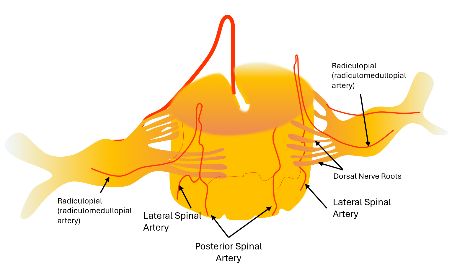
Below is one of our commissioned art pieces — key is to look at cord from posterior — to highlight PSAs. Arteries have different names in various sources and you need to know that. Ignore the dural fistula here 😉 – we get to that on the “spinal venous system” and “spinal dural fistula” pages.
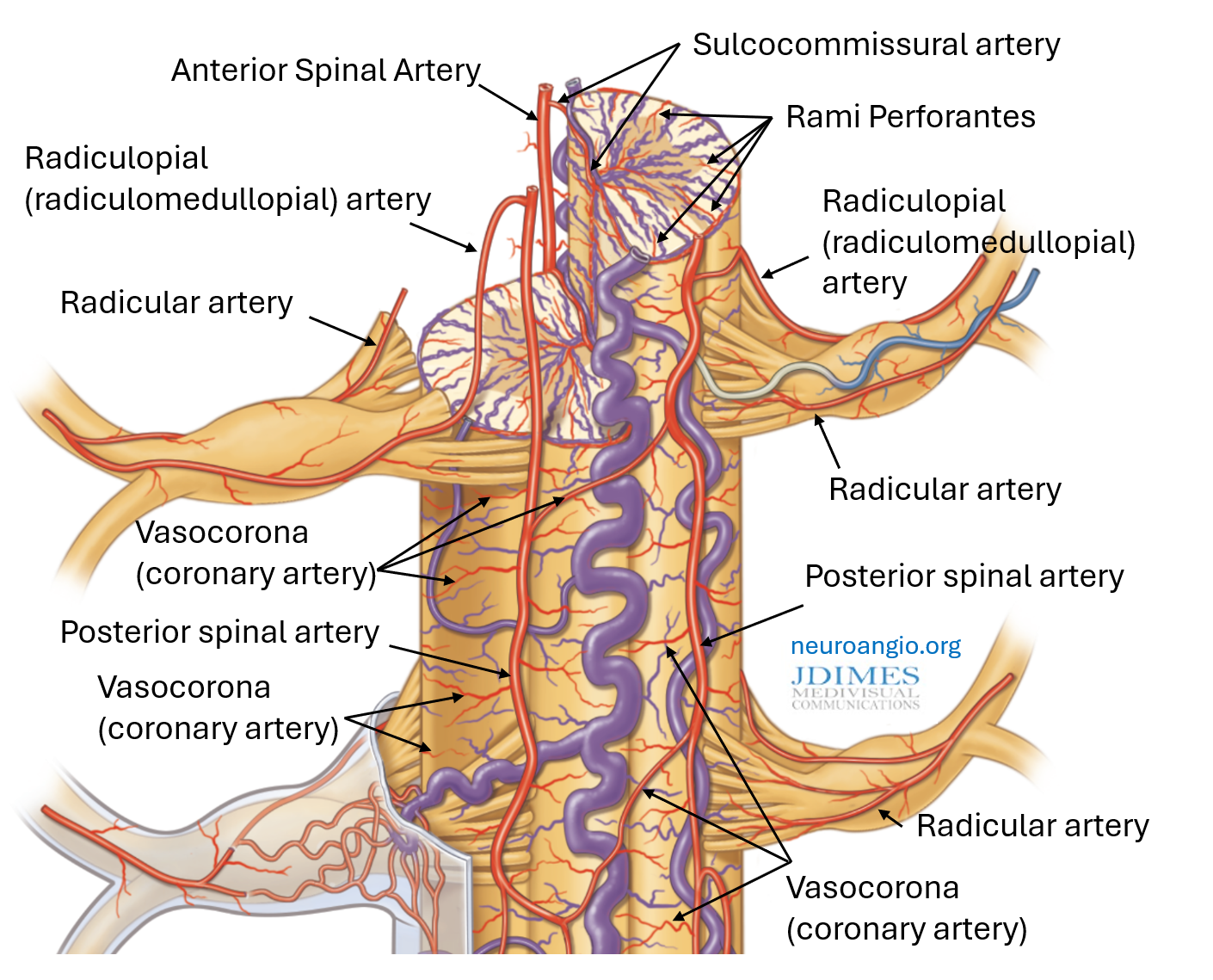
Basic building blocks of cord supply are limited. Nerve roots are supplied by “radicular” arteries, with dorsal and ventral branches associated with respective roots. When dorsal radicular branch supplies posterior spinal system it is called “radiculopial” or “radiculomedullopial”. Or other names… The idea is the same. Surface cord connections of any type — including between PSAs are “vasocorona” or “coronary” to add to confusion. Penetrating vessels into cord substance are “rami perforantes”. The big ones entering cord from the ASA via the ventral fissure are “sulco-commissural” — supplying either half of the central cord, especially gray matter. This is about it…
Here is what its really like

Radiculopial arteries
Invisible on modern non-invasive imaging. On 2D-DSA have a typical off-midline hairpin turn. Below is an old, good example. The ASA is dead center (white arrows), while the PSA is off center (red arrows). Technique is key — make sure the image is centered — the spinous processes are in the middle. The “hairpin” turn of radiculopial to posterior spinal transition is off-center and very tight — classic.
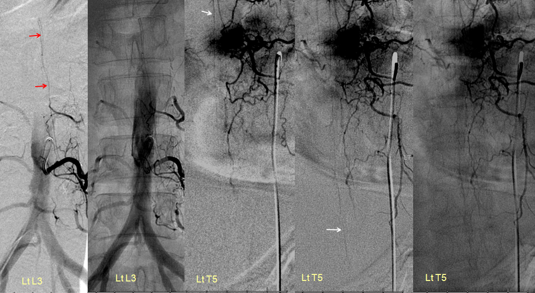
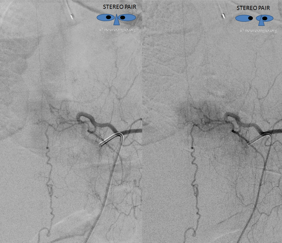
Another example — stereo pair
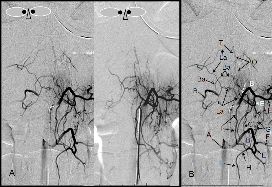
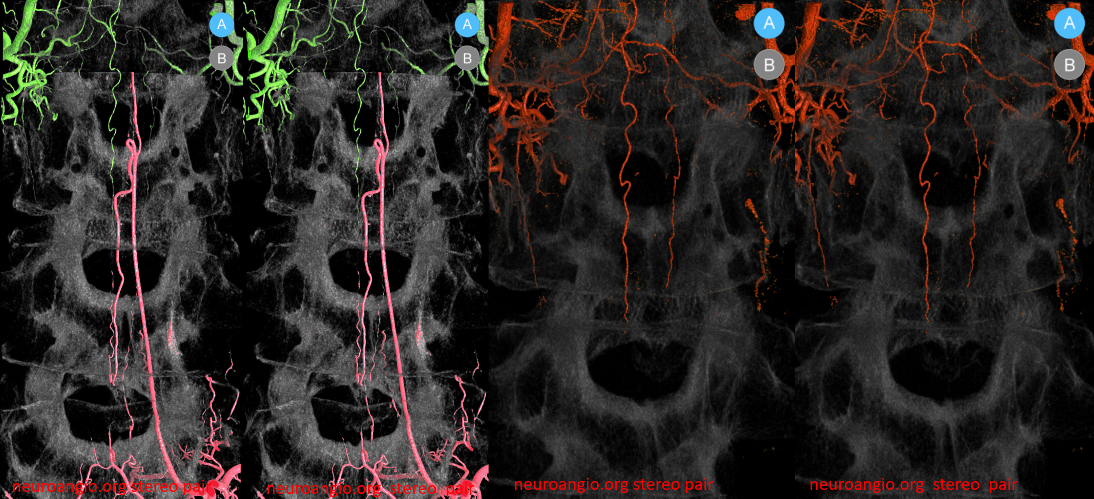
A — Stereo pair, and B – legend, demonstrating multiple transverse and longitudinal anastomotic connections. The pre-vertebral anastomosis, hallmarked by its proximal location relative to segmental artery ostium, is labeled “C”. Also present are pre-transverse (G) and post-transverse (I) anastomoses. A not so commonly encountered anastomosis between two proximal segmental arteries (Ba) is present, along the anterior circumference of the vertebral body. A prominent posterior spinal artery (T) and its radiculopial feeder (O) can be seen, in an off-midline position.
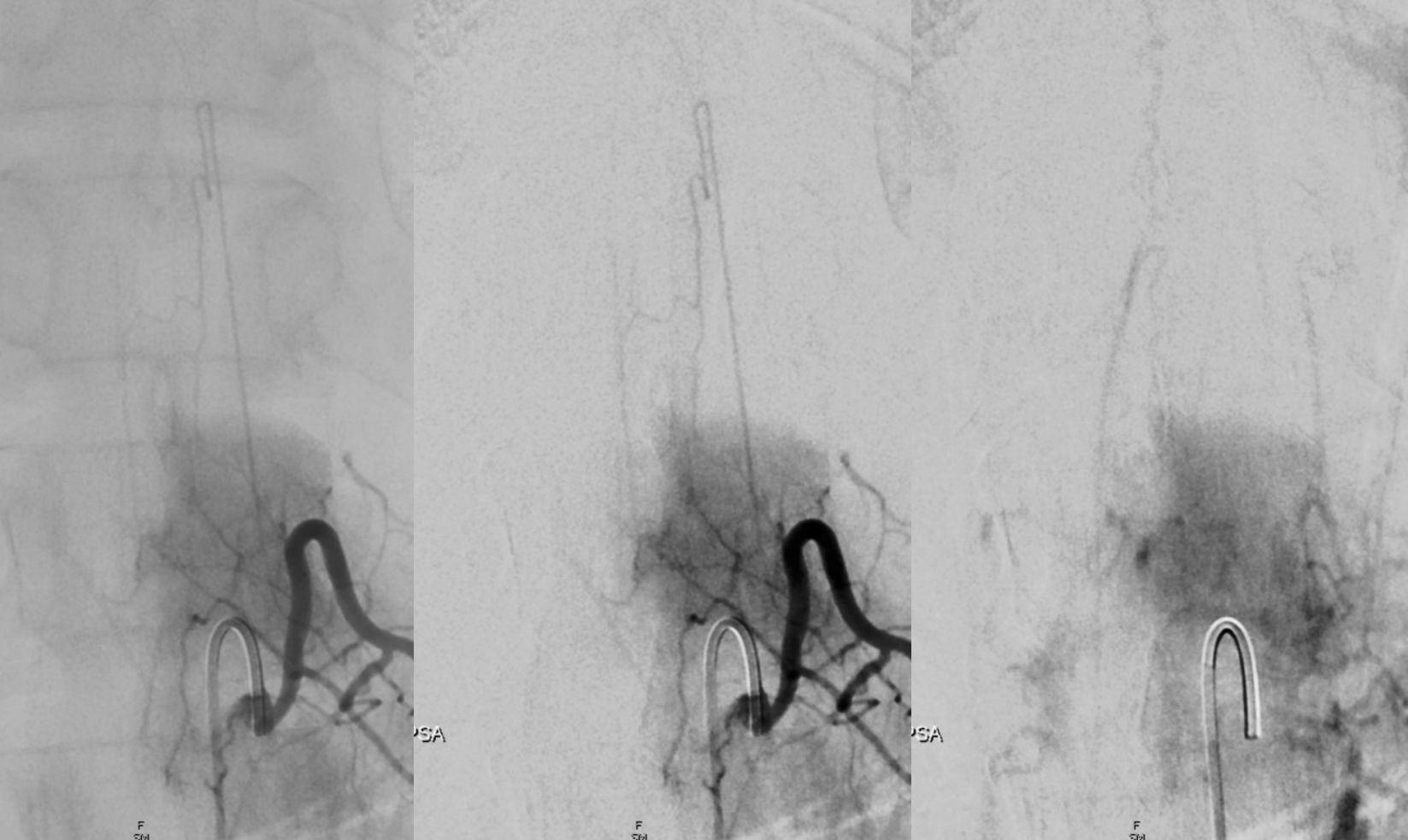
Another one. When injecting large posterior spinals, normal cord veins can be seen (rightmost image). This is very important. Seeing venous phase, usually after injecting dominant ASA (Adamkiewicz) is indirect proof of normal cord hemodynamics (lack of venous congestion). Cord venous phase is usually absent in spinal dural and spinal epidural fistulas.
Much better definition is usually achieved with high resolution Cone Beam CT — the same injection as above in GIF
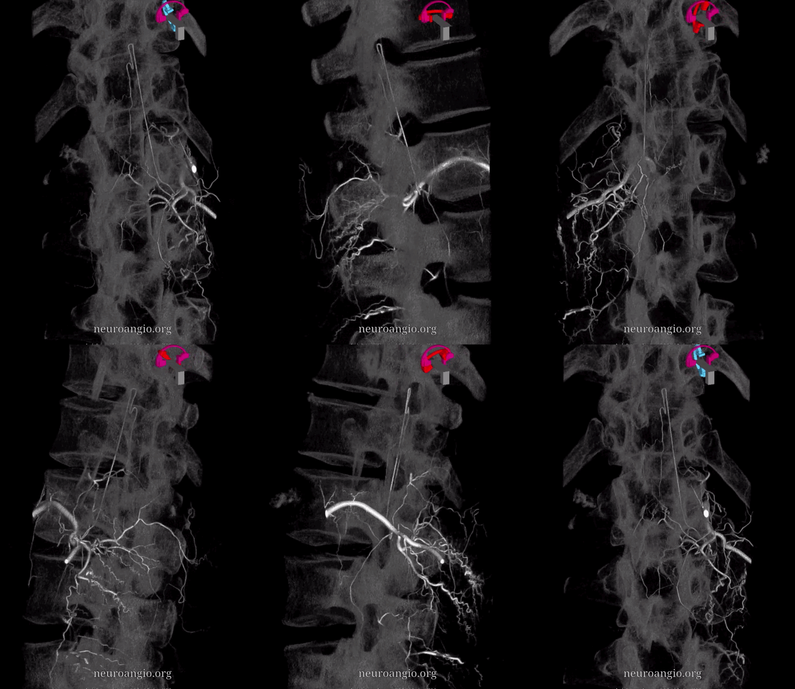
MIP Video
Sequential sagittal MIPs. In all of these, multiple vasocorona connections exist to the contralateral PSA
Cross-eye stereo pair
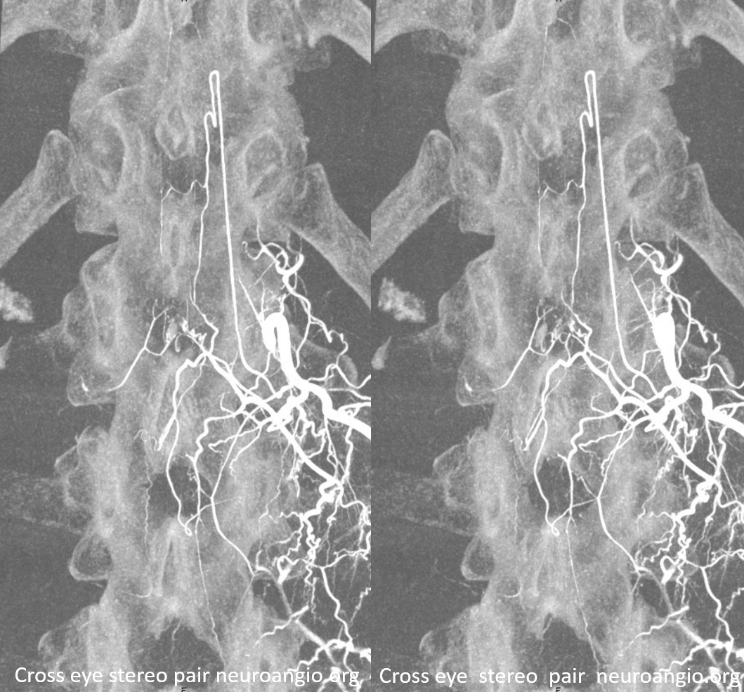
Same in Volume Rendered
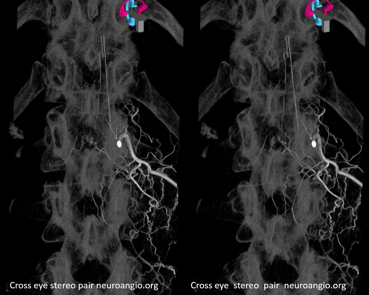
Finally, anaglyph stereo pair
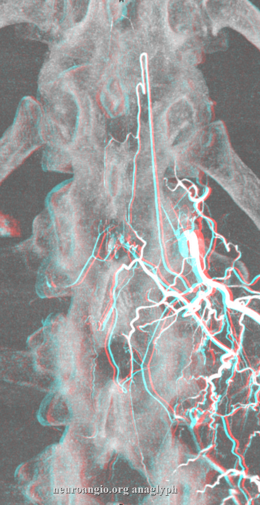
It is silly to try to estimate the number of radiculopial arteries. The more we look, the better our equipment, the more we see. Some are larger, some smaller. Below is an image of some small ones — at the limit of good technique resolution. Many are obscured by sloppy spinal angiography, and many more exist beyond what we can see.
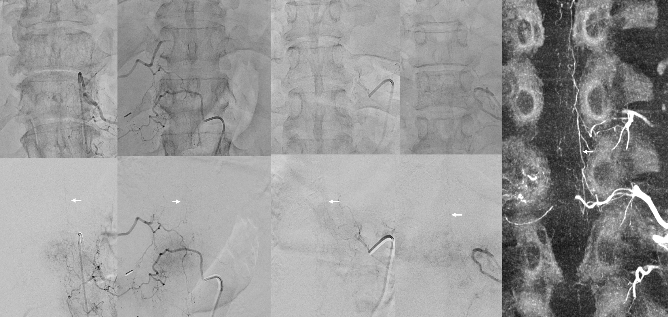
Below are some examples of smaller ones — 4 different levels, unsubtracted above, DSA below – -arrows – radiculopial. CBCT is the same case as rightmost pair obviously shows a very large PSA — much more than 2D would suggest
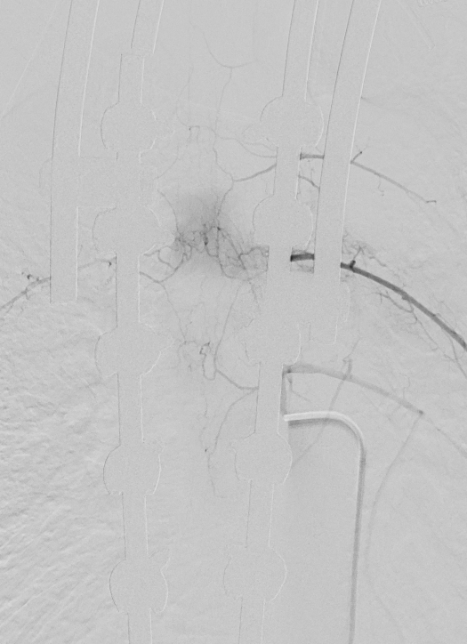
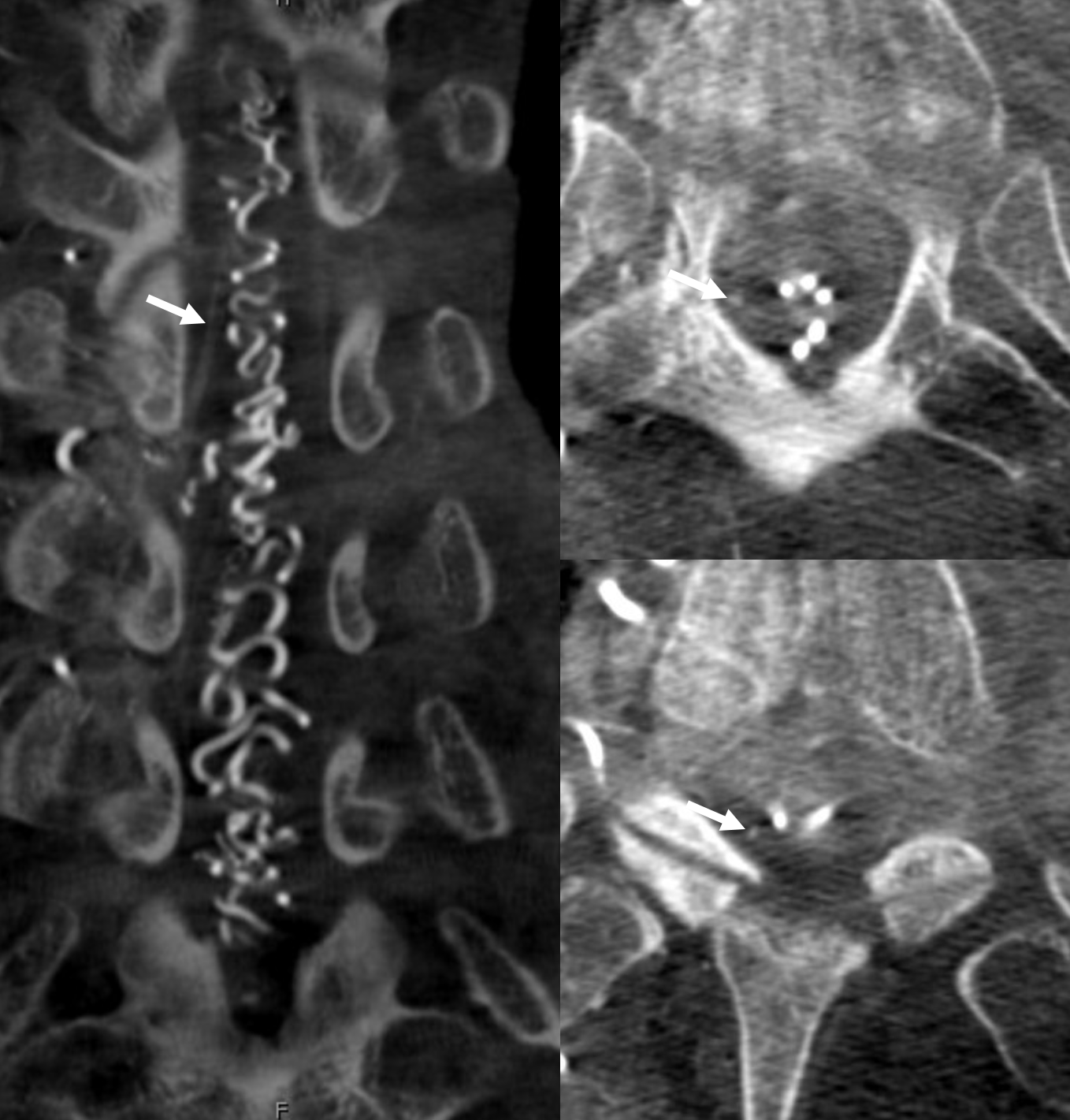
With good technique, its possible to get good CBCT images with spinal fusion hardware in place. Pretty neat. Bilateral PSAs for planned revision fusion
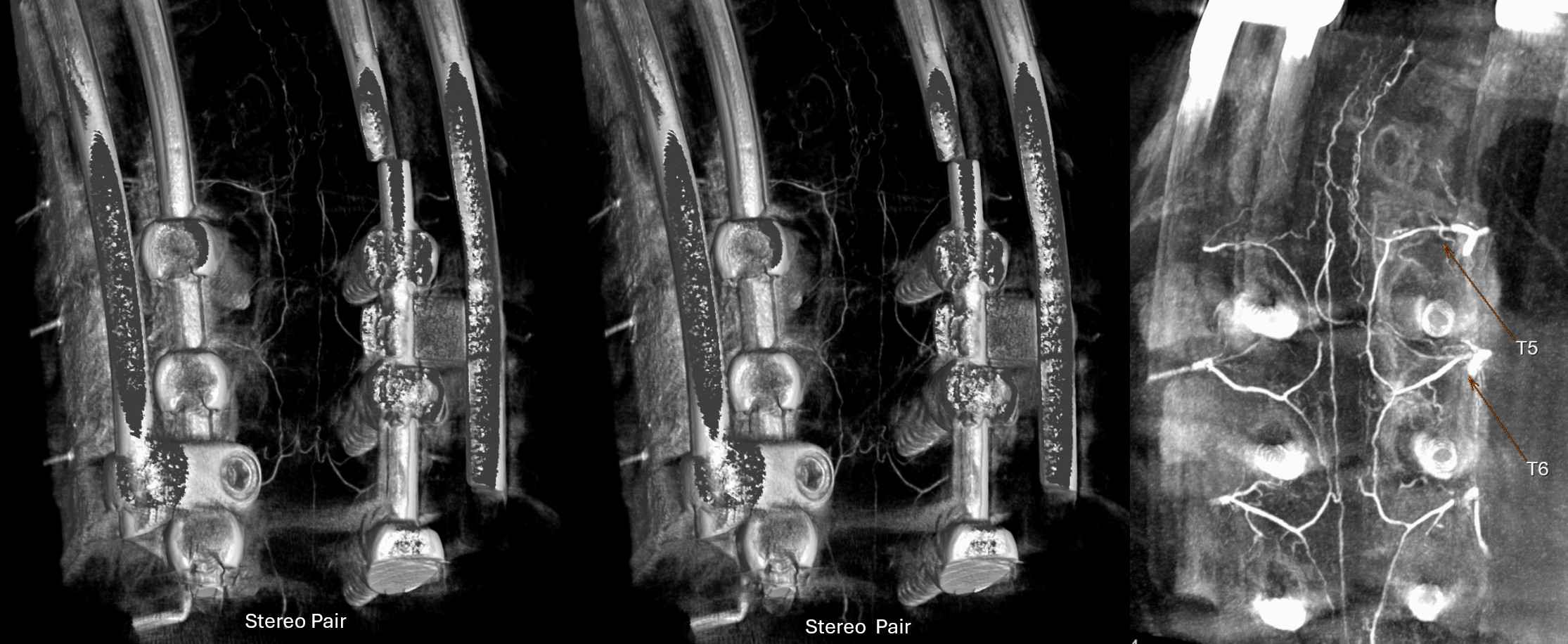
Same case — the retrocorporeal network is well seen as multiple segment arteries have not survived fusion. Can you see the PSA there? That’s homework.
“Basket”, Conus Medullaris Area, and connections to the Anterior Spinal System
It’s not so complicated — when cord is larger, it needs more blood. Which means that at both cervical and lumbar enlargements the posterior spinals (it is the same network as lateral spinals — see dedicated page) are larger. At conus medullaris / lumbar enlargement, macroscopic connections to the anterior spinal system — called “rami cruciantes” are common. Its a balance — dominant posterior spinals can supply the whole conus, with a small Adamkiewicz, and vice versa.
Below is another diagram, with rami cruciantes in “z”
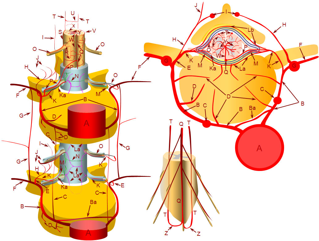
A – aorta; B – segmental artery; Ba – intersegmental arterial anastomosis; C – prevertebral anastomotic network; D – direct vertebral body feeding arteries; E – dorsal spinal artery; F – intercostal/muscular artery; G – pretransverse anastomotic network; H – dorsal division of the dorsal spinal artery; I – post-transverse anastomotic network; J – muscular branches of the post-transverse anastomotic network; K – ventral division of the dorsal spinal artery; Ka – radicular artery; La – ventral epidural arcade; Lb – dorsal epidural arcade; M – nerve root sleeve dural branch of the ventral division dorsal spinal artery; N – dural branch of the ventral division dorsal spinal artery; O – radiculopial artery; P – radiculomedullary artery; Q – anterior spinal artery; R – mesh-like pial arterial network; S, T – posterior spinal artery; U, V – pial arterial network (a.k.a. vasocorona) anastomoses between anterior and posterior spinal arterial systems, W – sulco-commissural artery, X – rami perforantes of the peripheral (centripetal) system, Y – central (centrifugal) system of sulcal arteries, originating from pial network of the cord; altogether, the pial network and rami perforantes (R+Y) are called the vasocorona or corona vasorum; Z – rami cruciantes (a.k.a. crux vasculosa, a.k.a. rami anastomotici arcuati)
Back to Basket and Balance
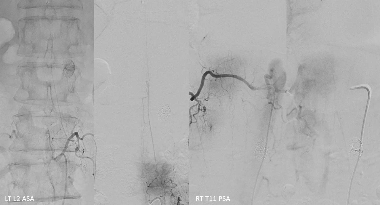
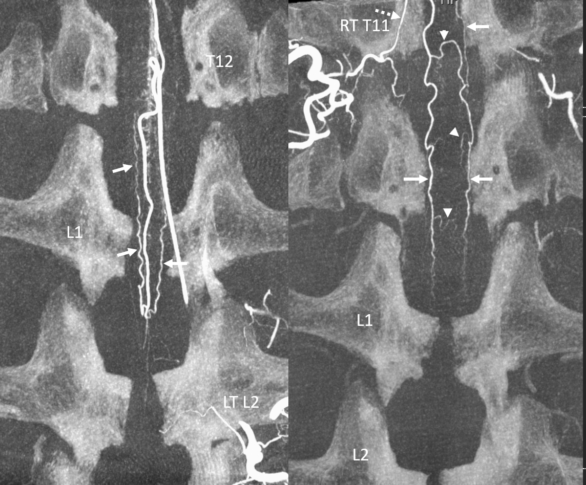
Full spinal angio should give the overall sense of how all is put together. A low origin L2 Adamkiewicz (two left images) is in balance with a well-developed posterior spinal network, with arterial and venous phases (two left images). Dashed white — radiculopial. Arrowhead — vasocorona. White arrows — PSAs. Venous phase — dorsal spinal vein (black arrows) and radicular vein (black dashed arrow)
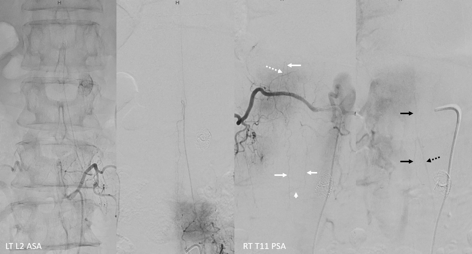
Without arrows
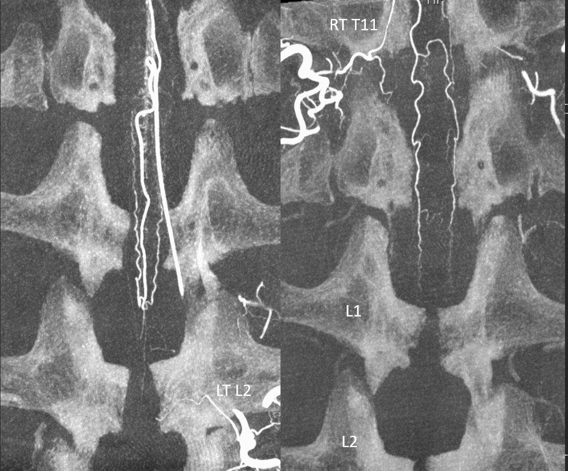
The same on CBCT — see the “basket” supply by ASA and balance with RT T11 injection. Obviously, CBCT is much more informative.
Without arrows
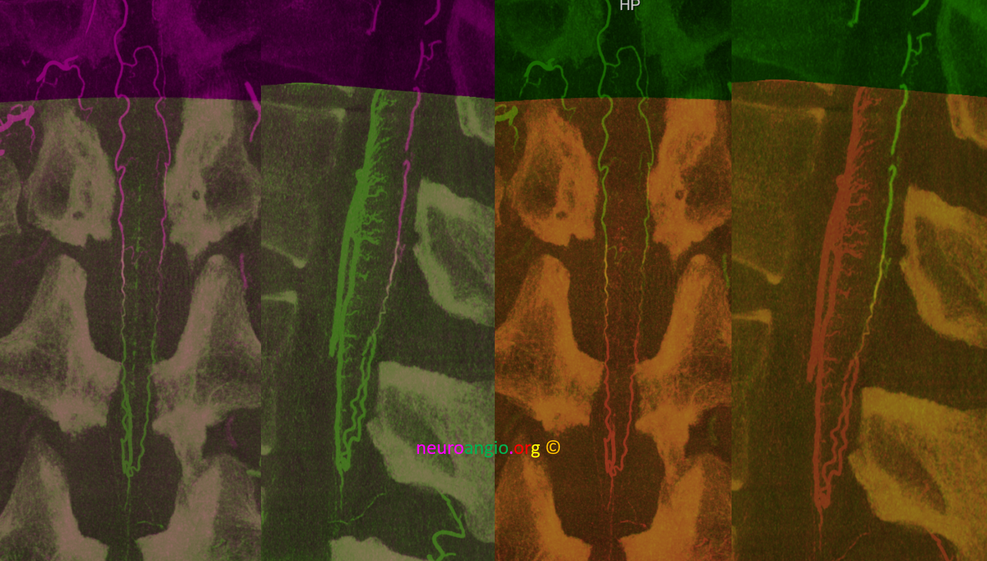
Fusion of two CBCT datasets is visually stunning, but understanding comes from raw data. Same data in two different color schemes. The “watershed” PSA region is where colors mix. It is important not to overthink this — a stronger injection from one pedicle may artificially shift the watershed for example. Would the PSA connections be enough to supply the cord if right T11 radiculopial artery was occluded? Probably yes in this case. What about the reverse — probably not.
Stereo pairs
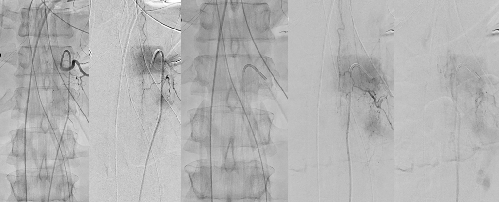
Below is another example in suspected spinal stroke. Patient had a recent myelogram, which gives unique images. ASA — two leftmost images. PSA is rest. The PSA is prominent, and gives nice venous phase images (rightmost).
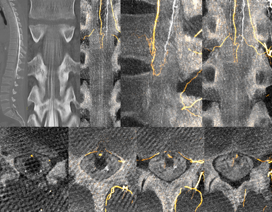
CBCT fusion — why is quality of images inferior to prior CBCT? Patient body habitus.
“Radiculomedullopial” Artery
Not uncommonly, both anterior and posterior spinal arteries can be supplied from the same segmental artery (via different nerve root arteries — which is clinically important — see case of spinal dural fistula embolization here). Again, anterior spinal should be dead center, unless shifted by some mass effect, and posterior spinal to the side. Compare the qualitatively different “hairpin turns” at transition from radiculomedullary / radiculopial to ASA/PSA — PSA is tighter. Also see how large the PSA system is here, extending far inferior
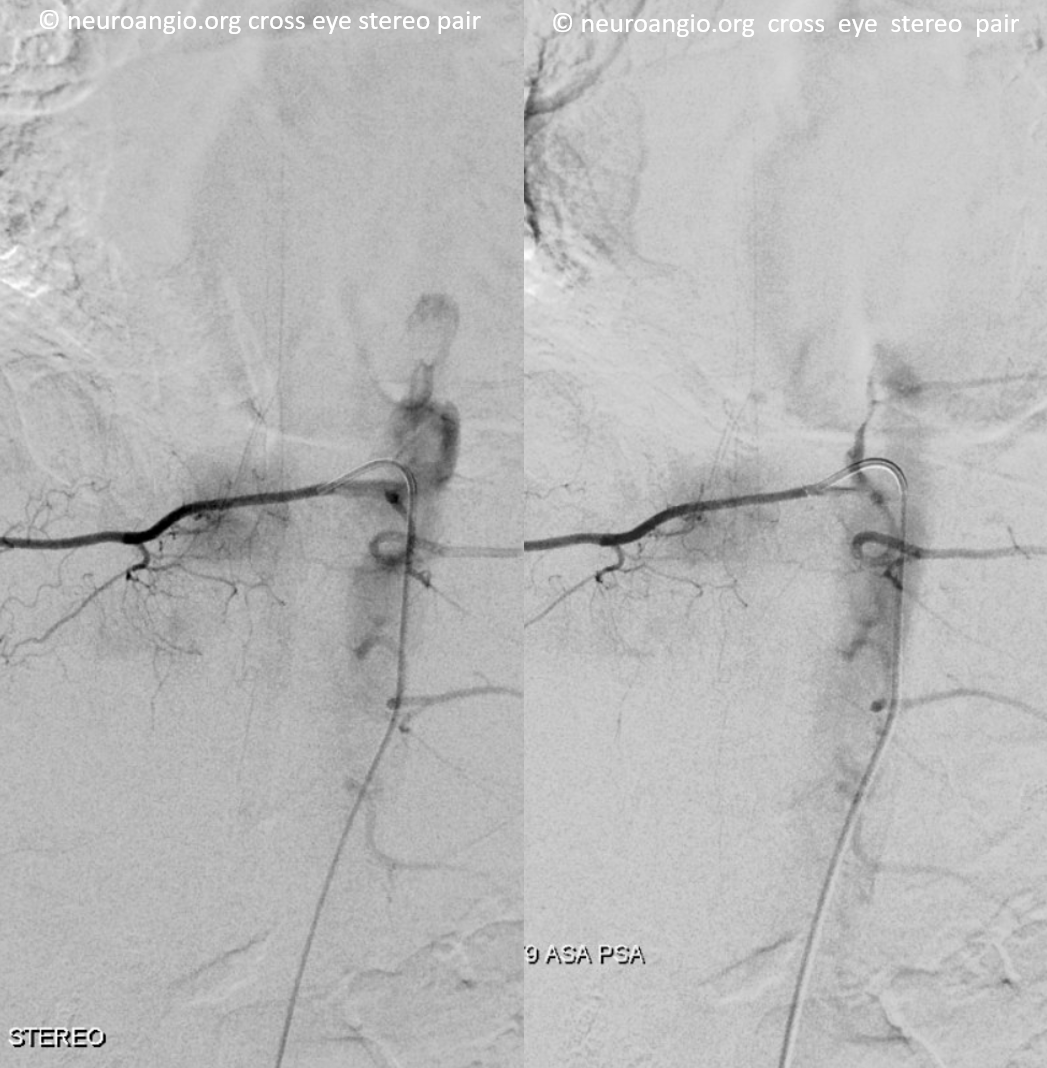
Unsubtracted image, same case
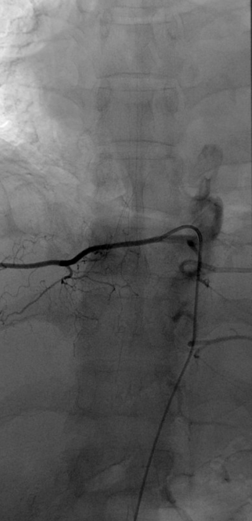
CBCT MIP of same case — note tighter and more posterior hairpin turn of much larger PSA, as compared with the ASA at this thoracic level.
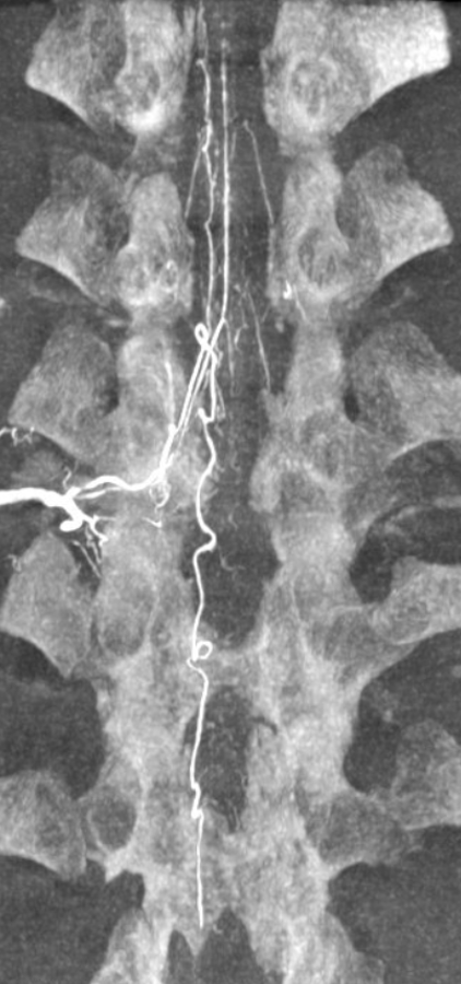
Beautiful volume rendered cross-eye stereo pair
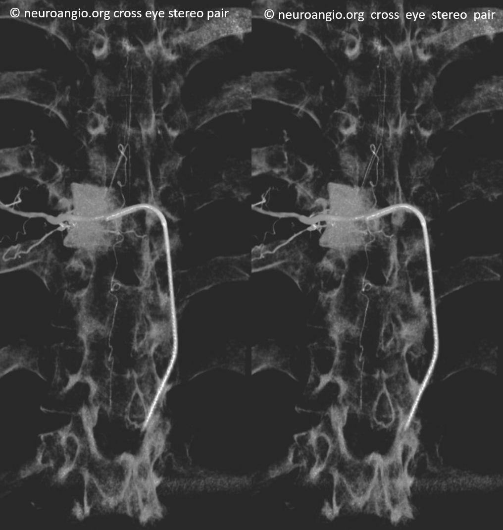
Below is another instance. Note well-separated dorsal (dashed arrows) and ventral (solid arrows) radicular arteries, supplying the PSA and ASA, respectively.
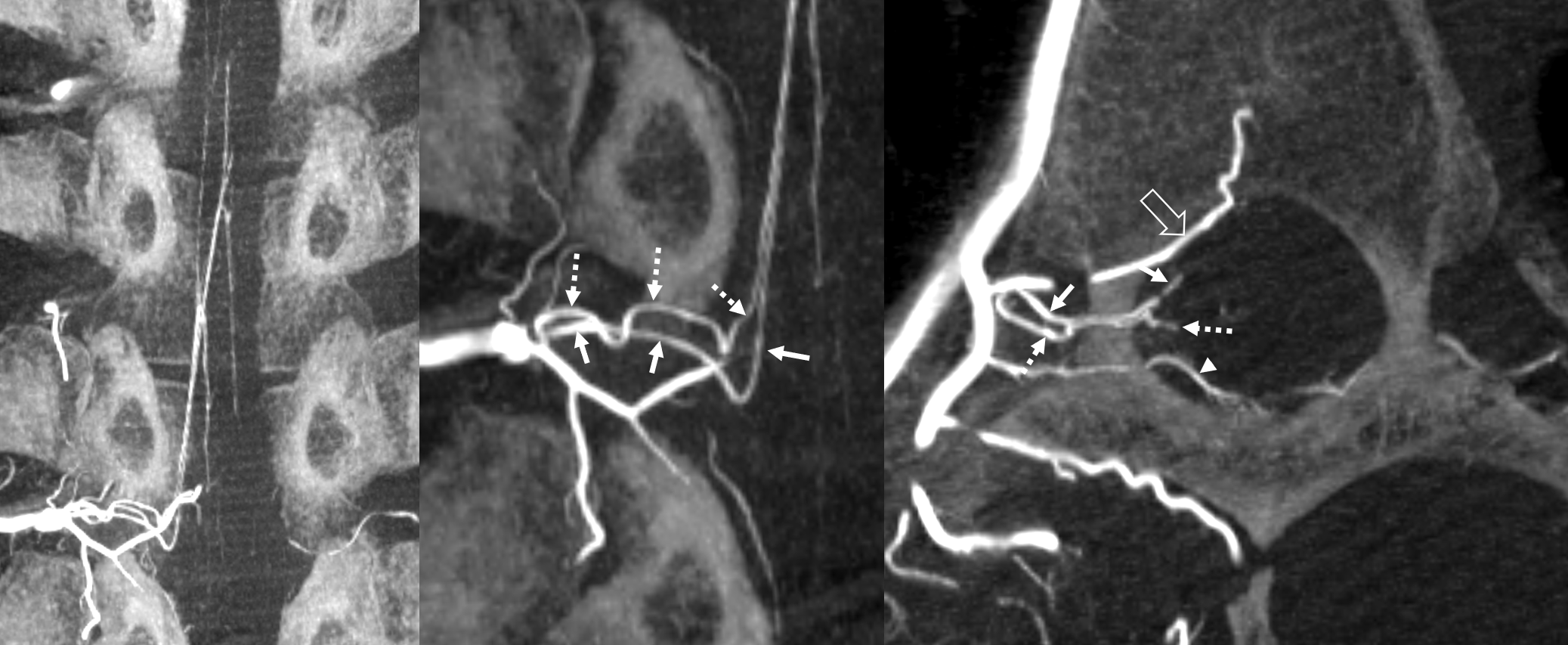
Another example from supreme intercostal segmental artery — 2D DSA PSA is faint
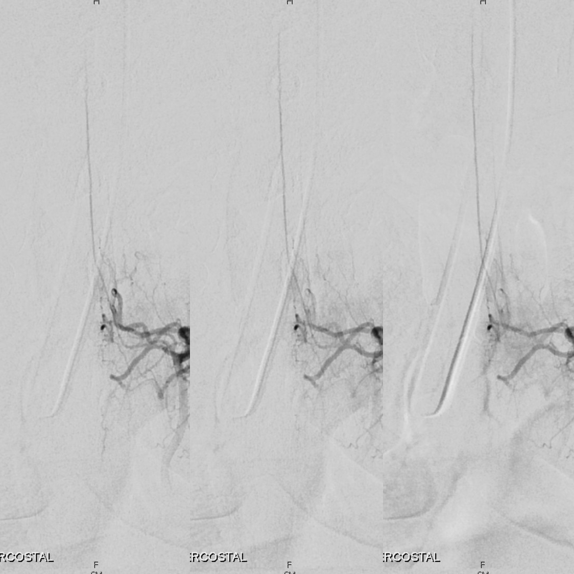
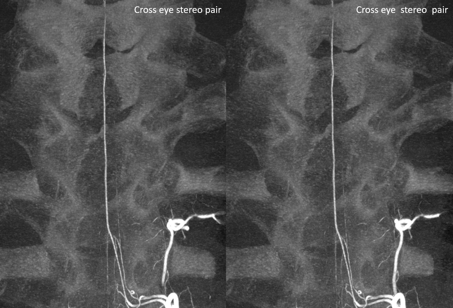
CBCT of same case. In all of these cases, see distinctly separate dorsal and ventral radicular arteries supplying respective PSA and ASA systems. Thoracic PSAs can be particularly large. This is important, as damage to them can produce major neurologic deficits, and spinal dural fistulas are more commonly associated with the dorsal nerve root, placing the PSA at particular risk during embolization. The thoracic cord is thinner than cervical and lumbar enlargements, and more damage can be done. Our practice is to routinely obtain CBCT at level of fistula to decide on embolization.
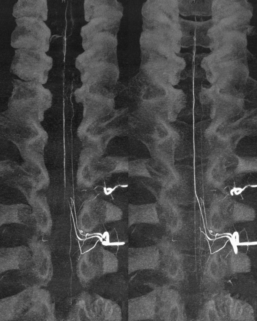
Cross-eye stereos
And GIF
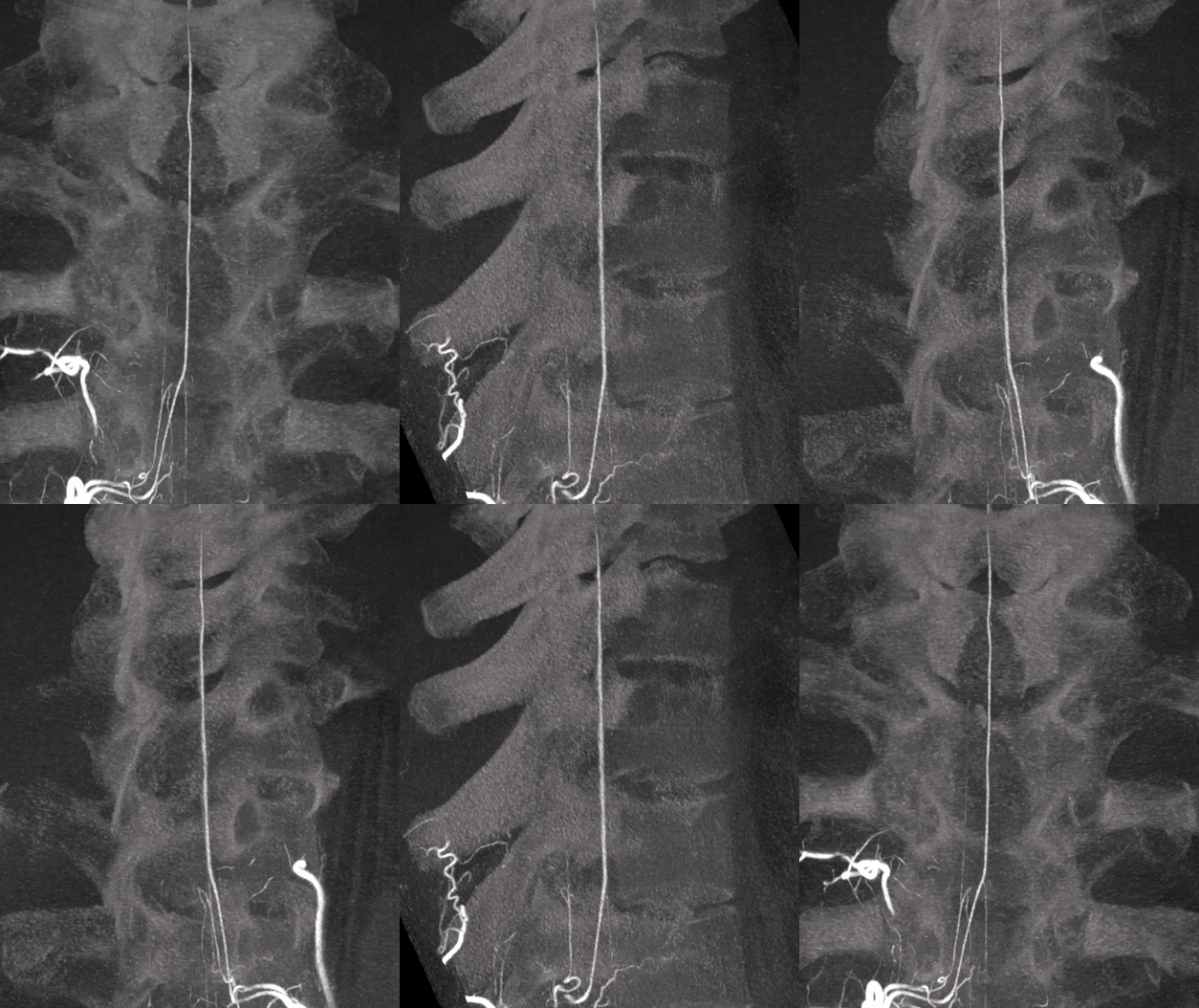
Unusual situations are possible. Below is an injection of what might look to be a very large Adamkiewicz, with the anterior spinal system contributing to supply of both PSAs. In fact, this is the reverse — large radiculopial / PSA system supplies lower ASA via particularly large vasocorona connection. Note renal cell tumor met at this level, precluding embolization.
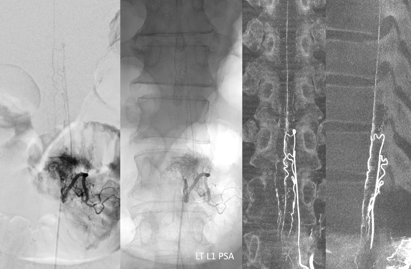
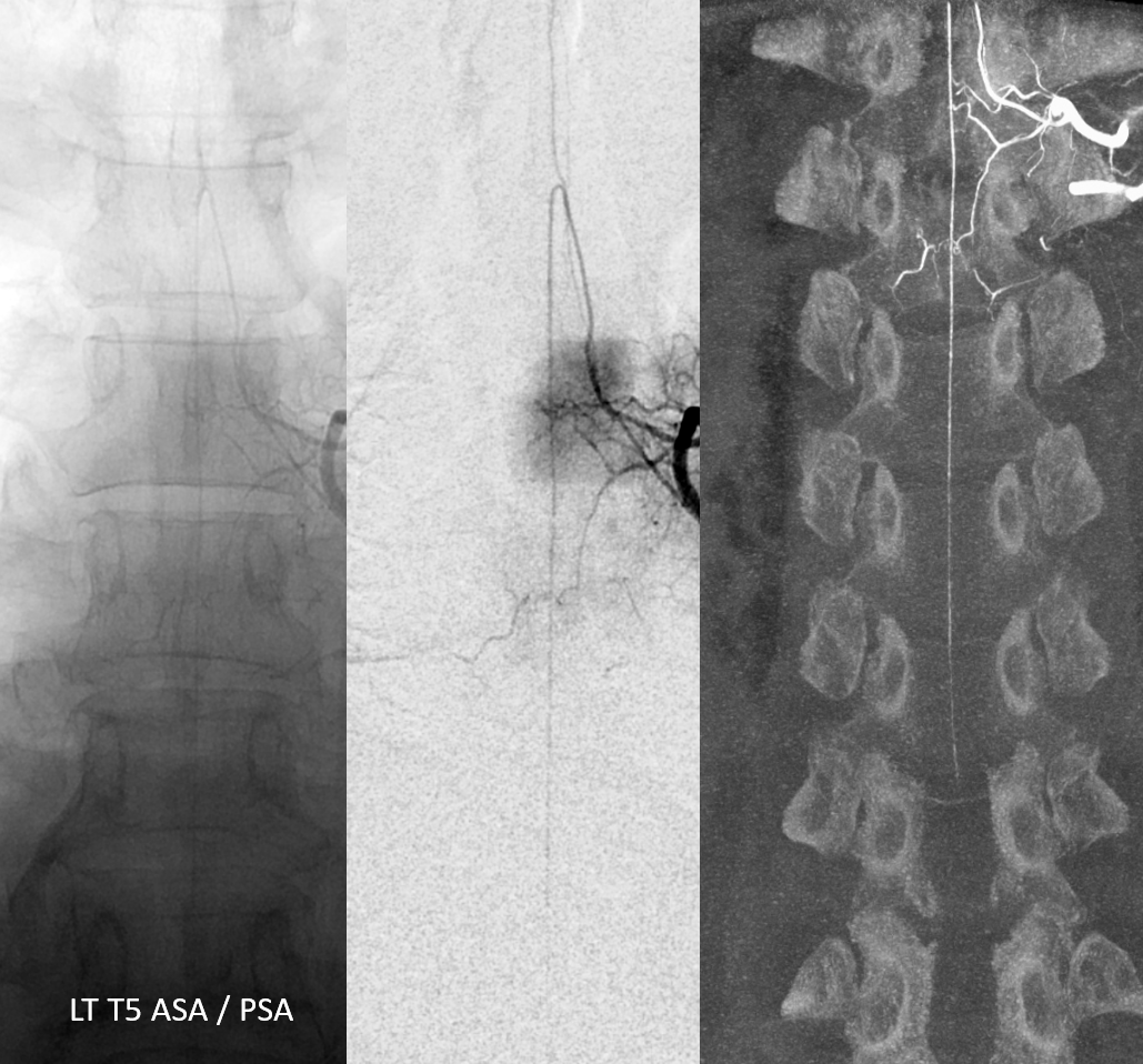
The upper ASA origin is from T5 — in fact another “radiculomedulopial” ASA/PSA level
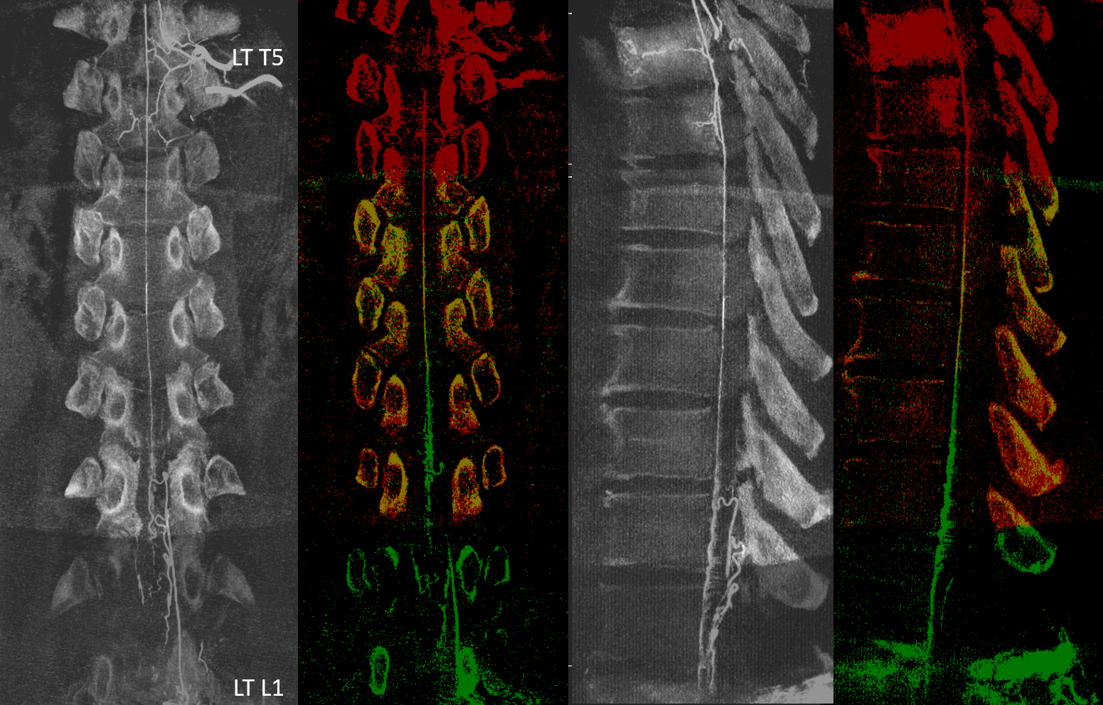
Fusion then is possible — the “anterior spinal watershed” yellow color mix
Preop angiogram of Meningioma with cord compression. Fusion of ASA (T5) and PSA (T4) shows clear PSA and ASA presence on CBCT, whereas both are quite faint, including faint blush of the meningioma (black arrow)
Cervical Segment Posterior / Lateral Spinal Arteries
Confusion exists here because in the cervical cord, the name is “lateral spinal” artery. Diagrams are developed to theorize what is posterior and what is lateral. It is best not to overthink it, and consider the whole thing as a posterior spinal network, whatever the name. Strictly speaking, the boundary are the dorsal nerve rootlets — posterior/dorsal to these is posterior spinal, and anterior / ventral to them is lateral spinal. Guess what — the vessels meander and traverse between them back and forth across different levels. Just keep the big picture in mind. For reference:
Because the cervicomedullary junction is so important from interventional perspective, it deserves its own “Lateral Spinal Artery” page for more details.
Spinal arteries are easier to see in the neck, generally, because necks are thinner than associated bodies — particularly well seen in this lateral (dashed white — PSA, dashed black — sulcocommissural line, black arrowheads — radicular veins)
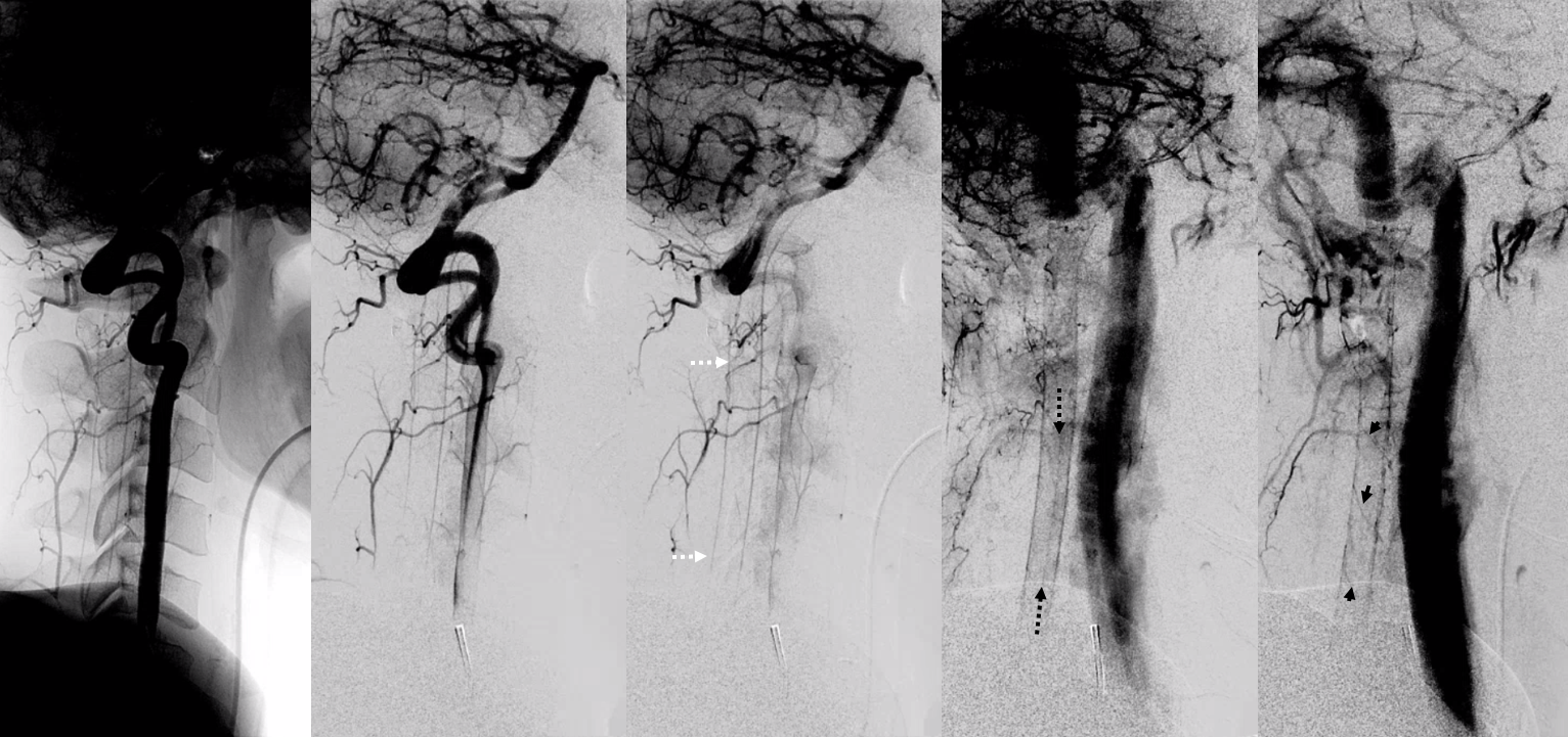
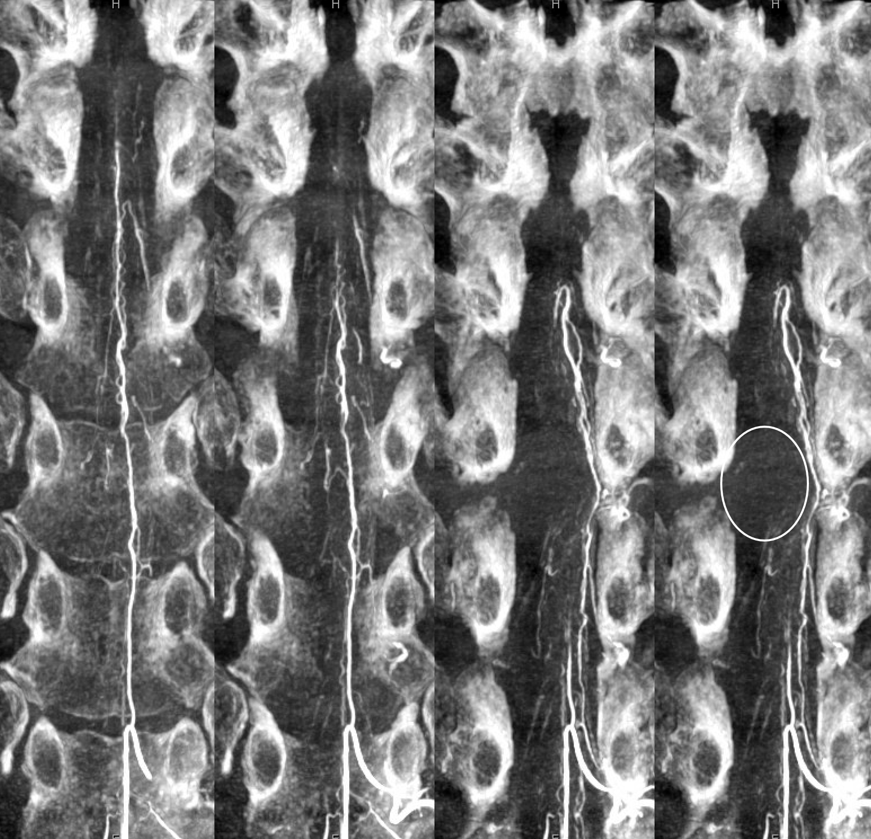
CBCT if best of course. Lots of detail here on both ASA and LSA/PSA. The posterior / lateral spinal is inside solid white oval. For the splitters, the lateral spinal is smaller and more ventral. For the lumpers like us, its all posterior spinal system. Middle image — vasocorona connecting to lateral spinal artery. Dashed white oval — same — ASA supplying bilateral lateral cord. Dashed black oval — from the back — posterior spinal more prominent, lateral spinal smaller and shorter.

Without labels

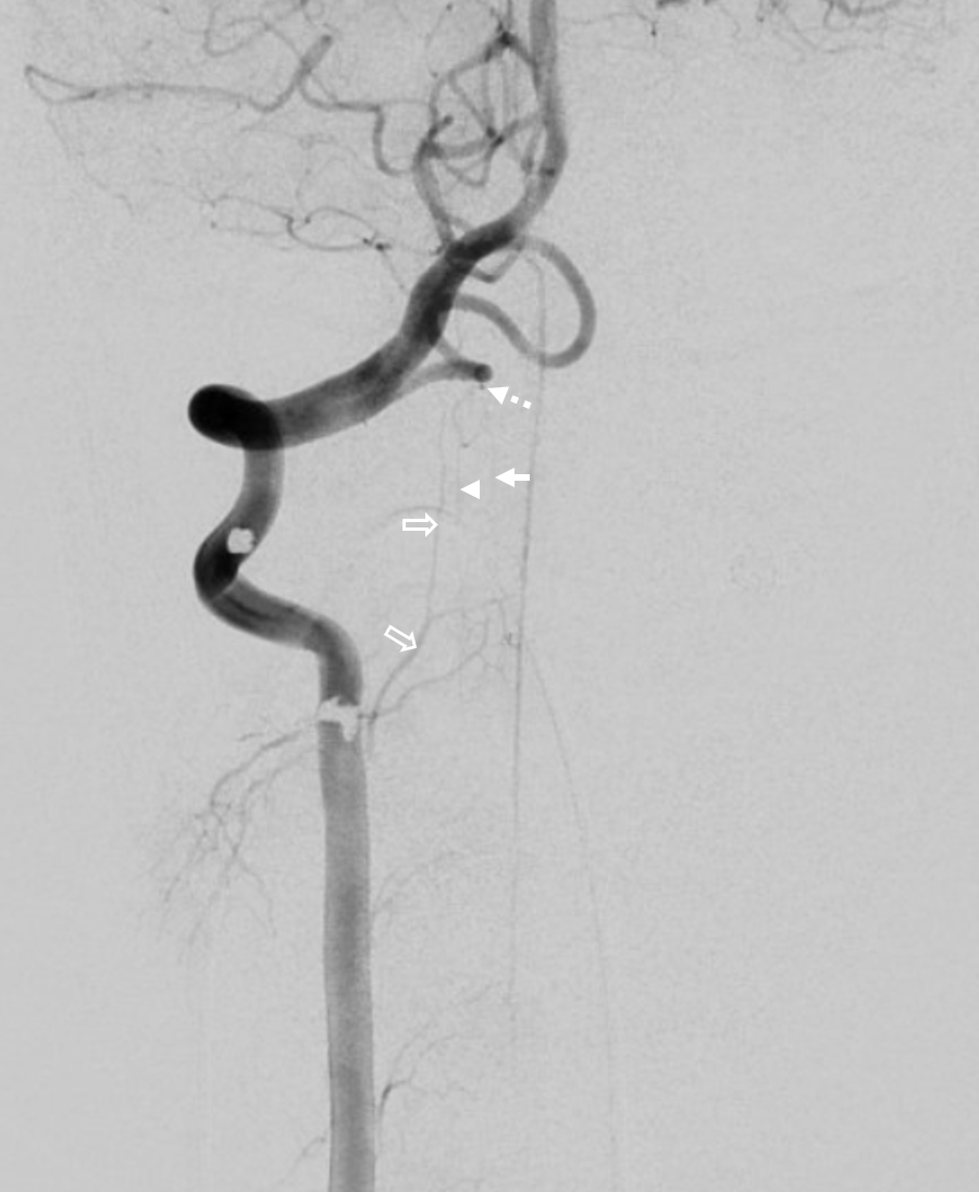
Another 2D-DSA anterior/lateral/posterior threesome, for the anatomically dorky… The artery ventral to the ASA is what?
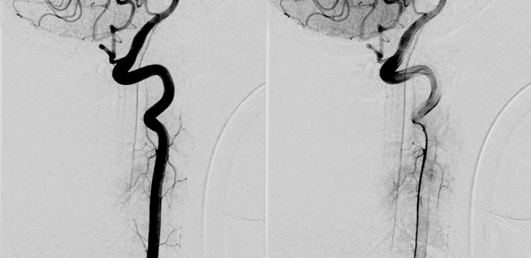
Frontal view of same case: arrow – PSA, arrowhead — LSA, dashed arrow — PICA origin of the whole thing, open arrows — you tell me…
CBCT axial MIP video of same
Application — Cases, Embolizations
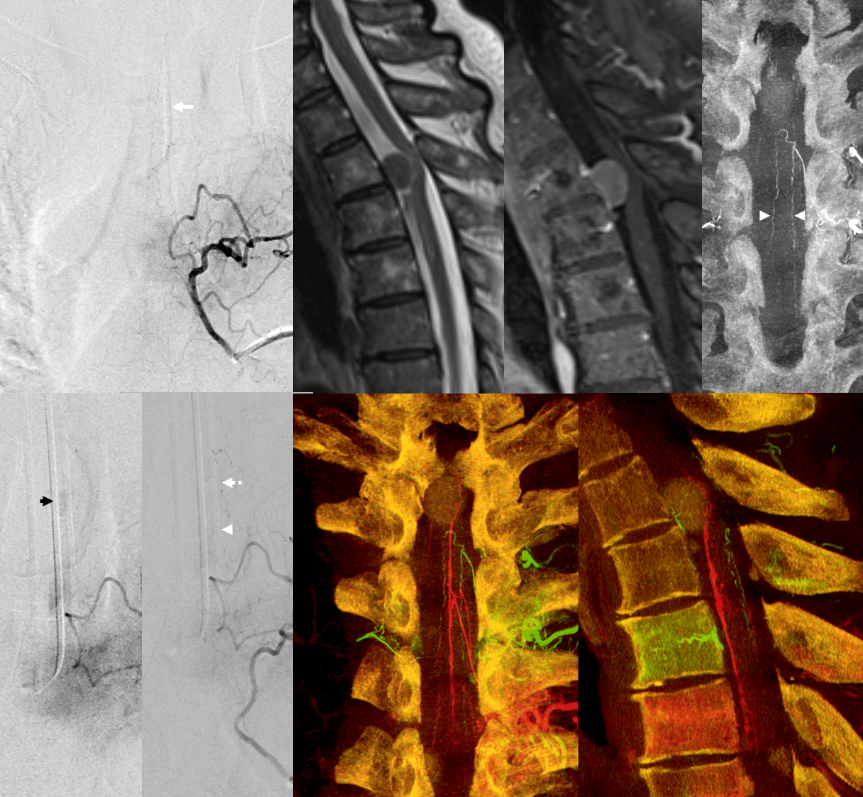
The outsized attention paid to the Anterior Spinal Artery may come at expense of the PSAs. Infarcts of the PSA are clinically important and often come with disturbing pain symptoms that can be extremely difficult to manage. Looking for PSA, not just ASA, in vicinity of spinal dural fistulas before embo is mandatory for example.
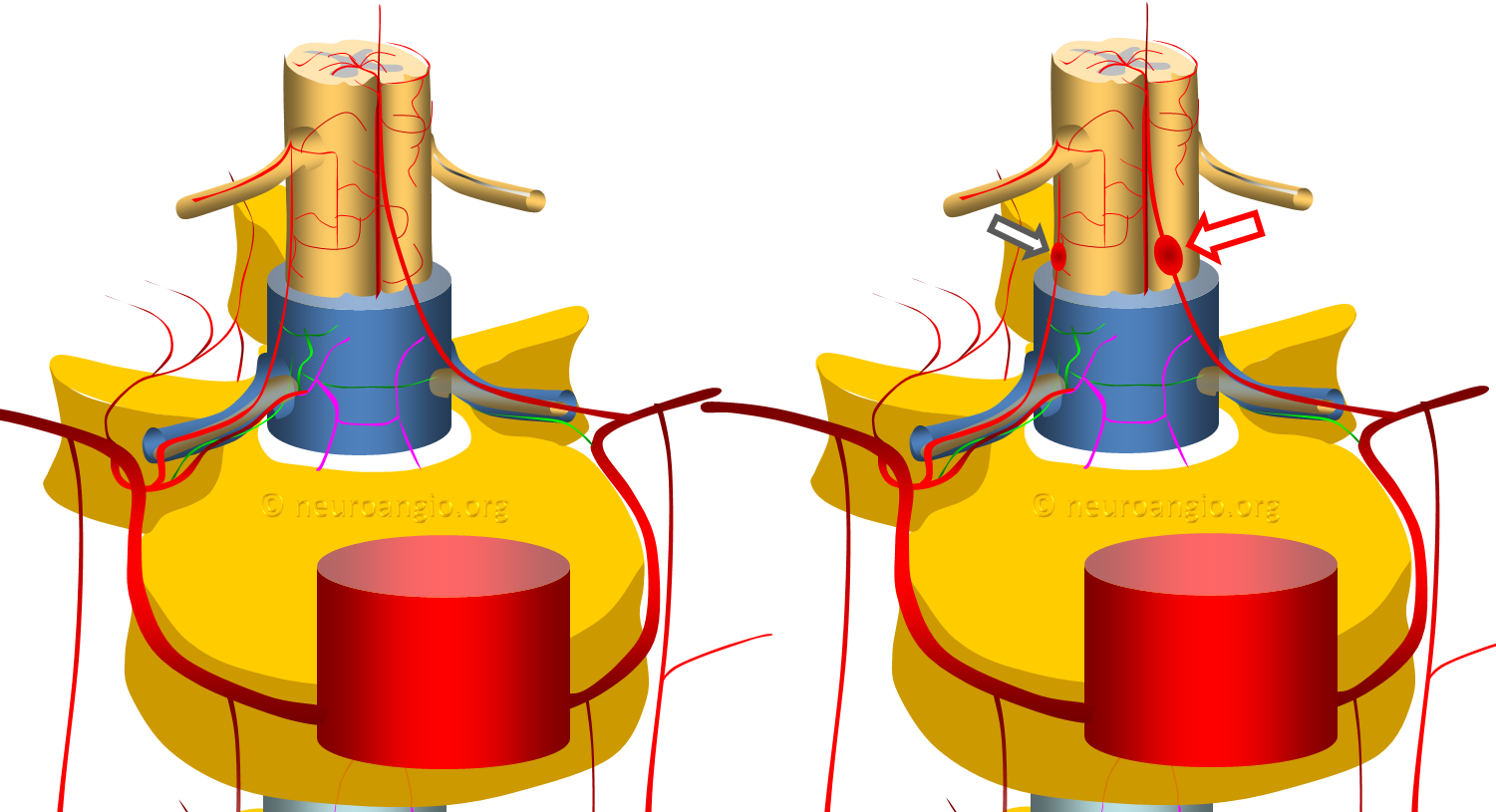
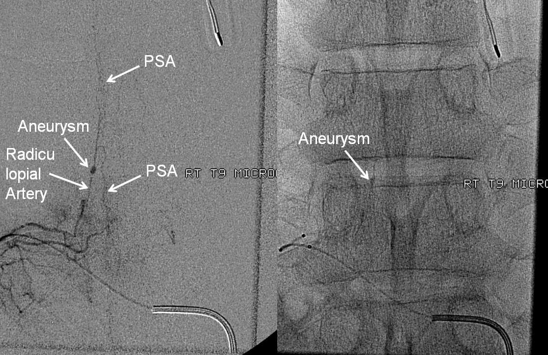
Rare and often underdiagnosed — presents with hemorrhage. Dedicated page here. It is a dissection (dissecting pseudoaneurysm) of the radiculomedullary or radiculopial artery as it enters the dura. Below is a typical, older image, with many more on the dedicated page.
Intramedullary Tumor
Hemangioblastoma — dominant ASA supply, some contribution from PSAs. Full case here

Intradural Extramedullary Tumor
Full case here — good CBCTs, etc
Spinal Dural Fistula
Importance of identifying the PSA before embolization. Full case here.
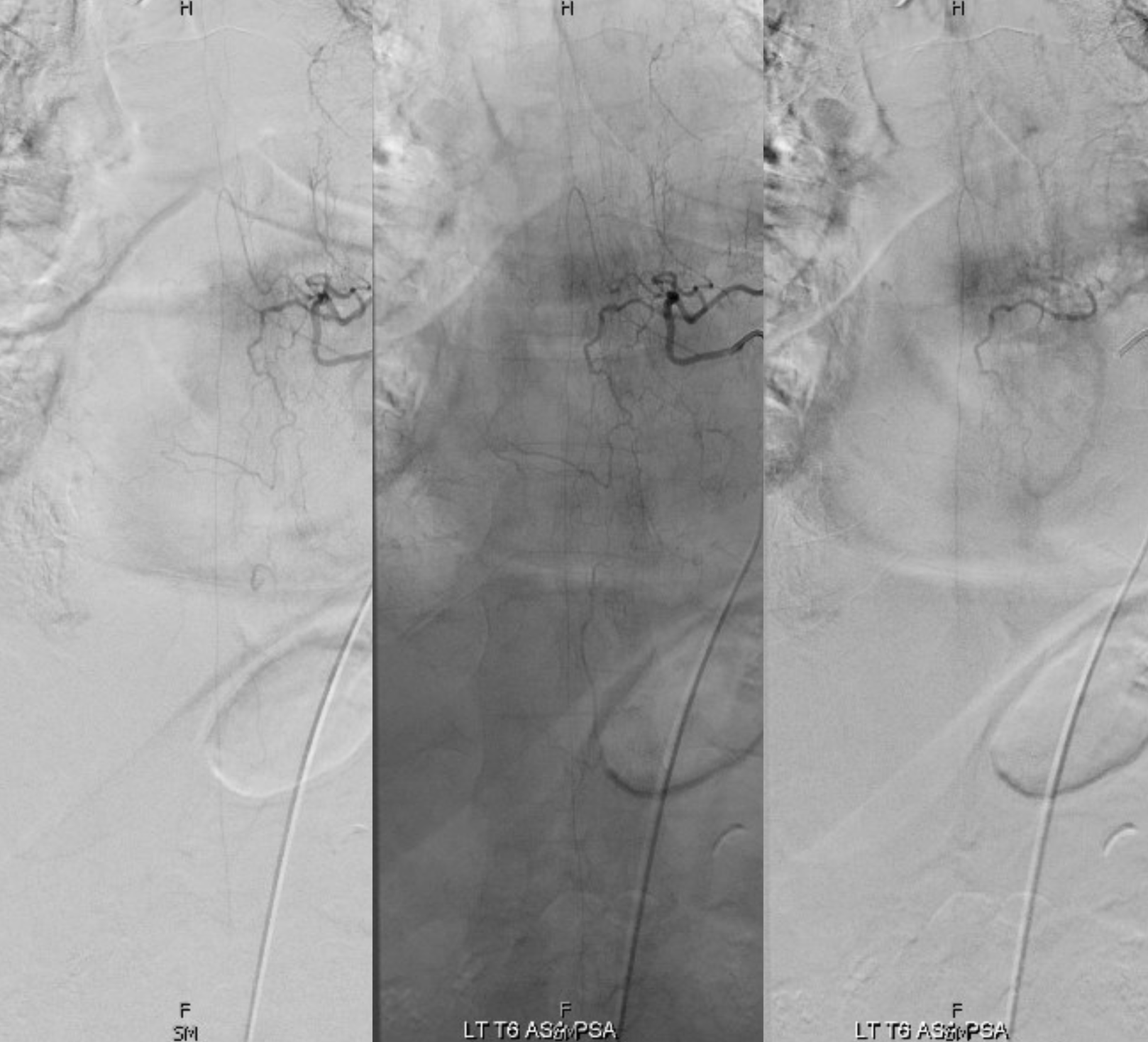
Case 2 — RT T6 fistula, left T6 radiculomedullopial supply embolization
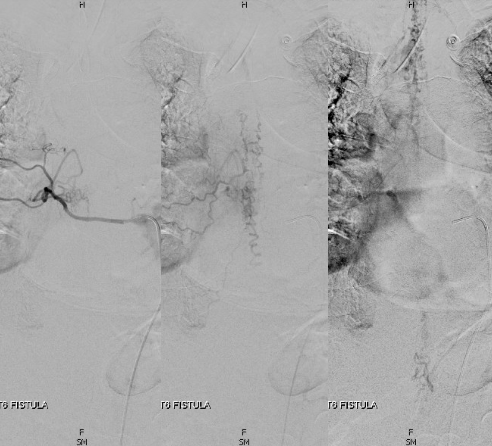
Superselective Fistula embolization at SAME level as ASA — very rare case
Embo is possible because ASA is supplied by ventral radicular branch, and fistula by dorsal one. Full case here
Lots more cases in Case Library section