Comprehensive look at ASA embryology, imaging, physiology, and pathology.
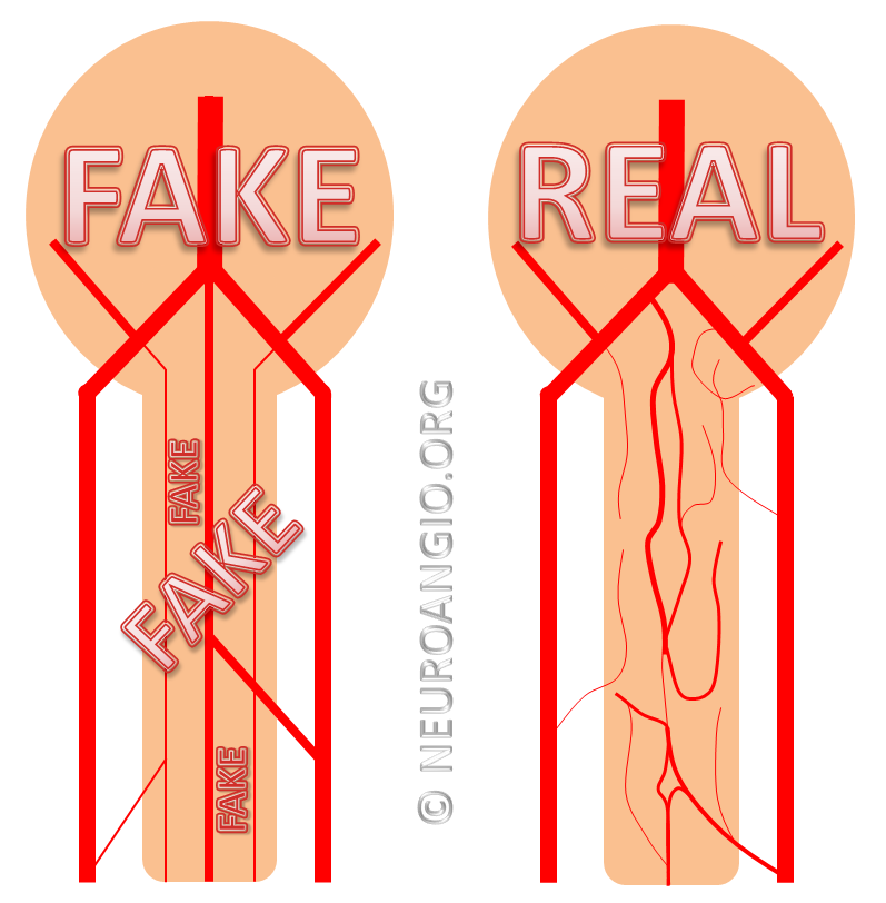
The ASA is a mostly continuous system extending along the ventral cord from craniocervical junction to conus medullaris, traditionally responsible for the bulk of cord blood supply. A big part of this page is to show exceptions to these generally useful rules, why exceptions matter, and therefore the need to determine case-specific vascular disposition.
Embryology
Quite important in understanding ASA, and in context the larger spinal arterial system, so summary is below. A more complete discussion is found in the dedicated section of neurovascular embryology. It is also functionally grave error to separate anterior spinal system from the posterior and lateral spinal ones. Although discussed on their respective pages, many cases are purposefully duplicated/triplicated, as all are part of the whole.
The basic arrangement of the spinal system consists of a metameric grid of transversely oriented intersegmental vessels, connected by various longitudinal channels. This simple bit of knowledge goes a long way in understanding spinal anatomy. Millions of years of speciation have taken place upon a basic building block of the organism — the metameric segment. Just like the fly and the worm, the human body consists of metameric segments, with ecto-, meso-, and endodermal elements. Each vertebral body, its ribs, muscle, nerves, and dermatome, correspond to one level or segment. It is perhaps easiest to appreciate this concept at the thoracic level, where each rib, vertebral body, and other elements constitute the prototypical segment. In the early human embryo, the neural tube is first supplied by simple diffusion. When its limits are reached, a primitive vascular system consisting of paired dorsal and ventral aortae (longitudinal vessels) and transversely oriented segmental arteries come into play to vascularize the developing tissue of the embryo.
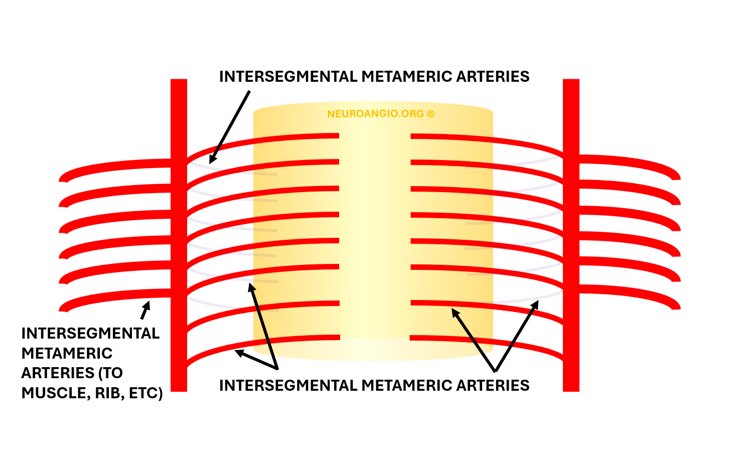
As the volume of spinal progressively enlarges, new longitudinal connections form between the transverse segmental arteries, most likely to facilitate distribution of blood within the vascular system. This pattern is seen throughout the body, but is somewhat easier to recognize in the vertebrospinal arterial system, where it gives rise to adult anterior and posterior spinal arteries, and numerous extradural longitudinal segmental connections (vertebral, deep cervical, ascending cervical, prevertebral, post-vertebral, lateral sacral, median sacral arteries) which are discussed in their respective pages, and most notably the Spinal Arterial Anatomy page.
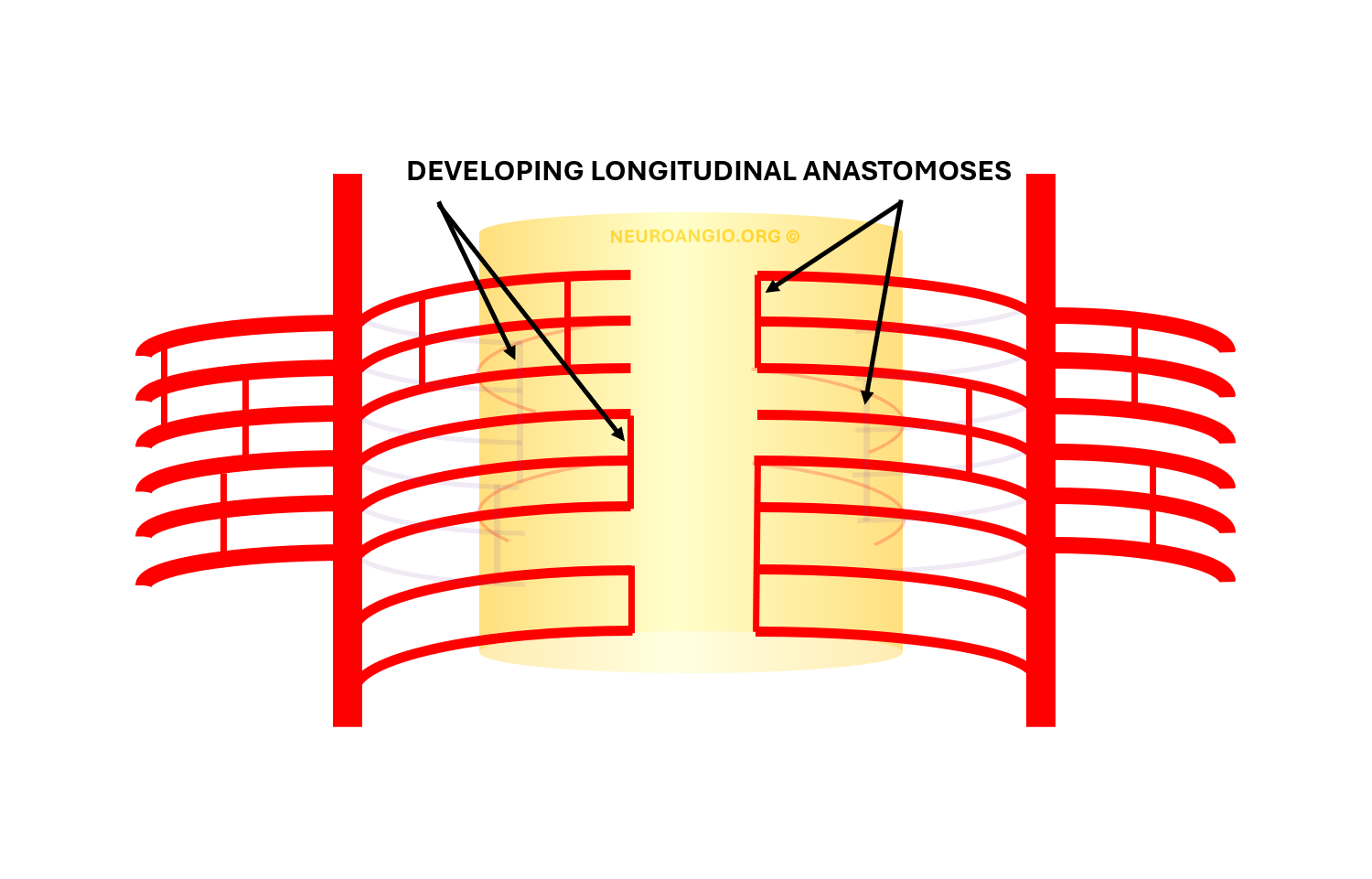
A grid-like network may be present at some stage, probably with a lot of “variations”. Additional longitudinal “secondary” vessels develop as above — future vertebral, deep cervical, pertransverse anastomotic etc arteries (not labeled). When seen like this, the ASA is a “secondary” vessel, developing as a result of hemodynamic needs — as are all longitudinal ones.
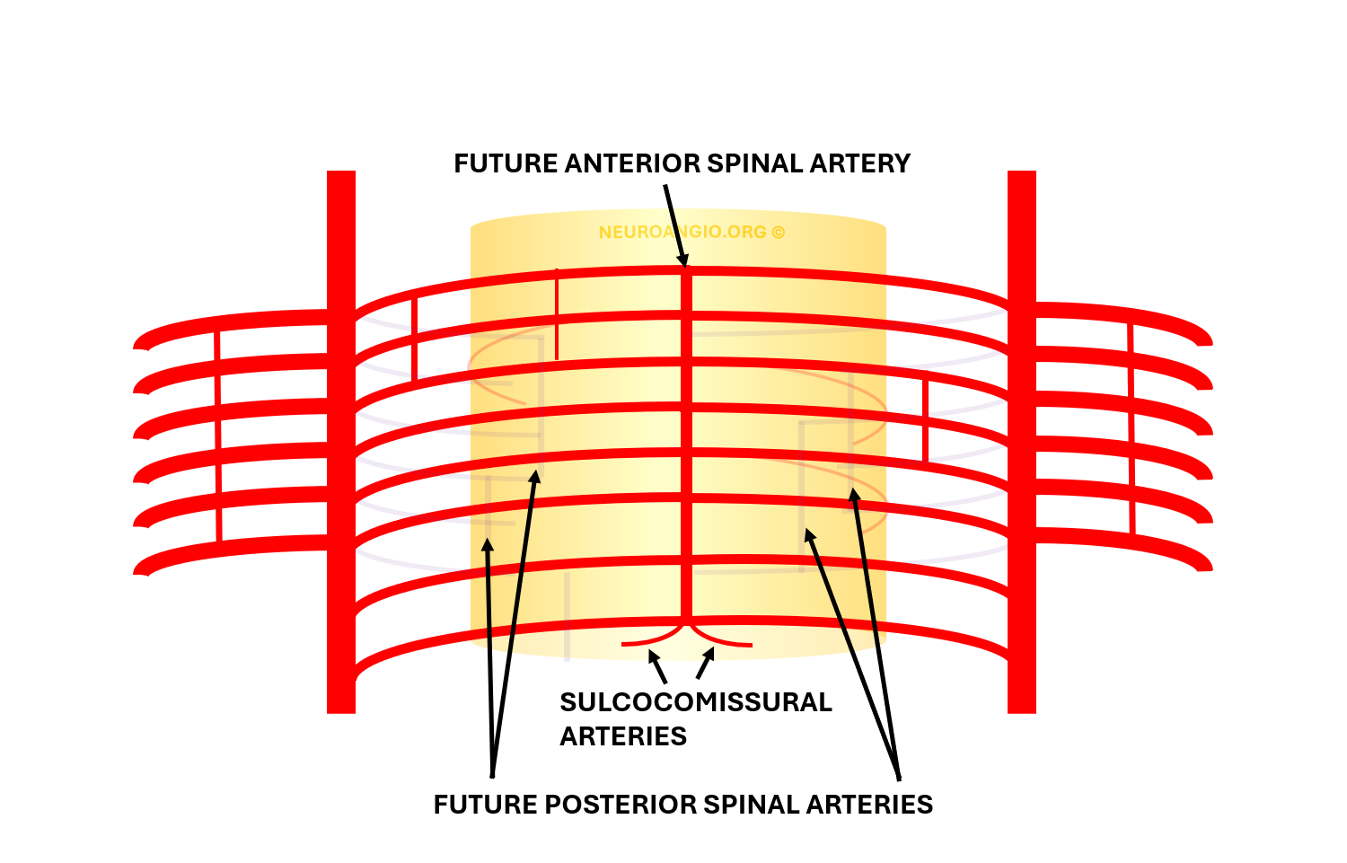
Gradual establishment of several dominant longitudinal vessels leads to recognizable “mature” anatomy. The other intersegmental vessels, often said to “regress”, probably do not actually do so. They more likely remain in “rudimentary” size, below angiographic resolution, yet capable of enlargement when required (see examples below).
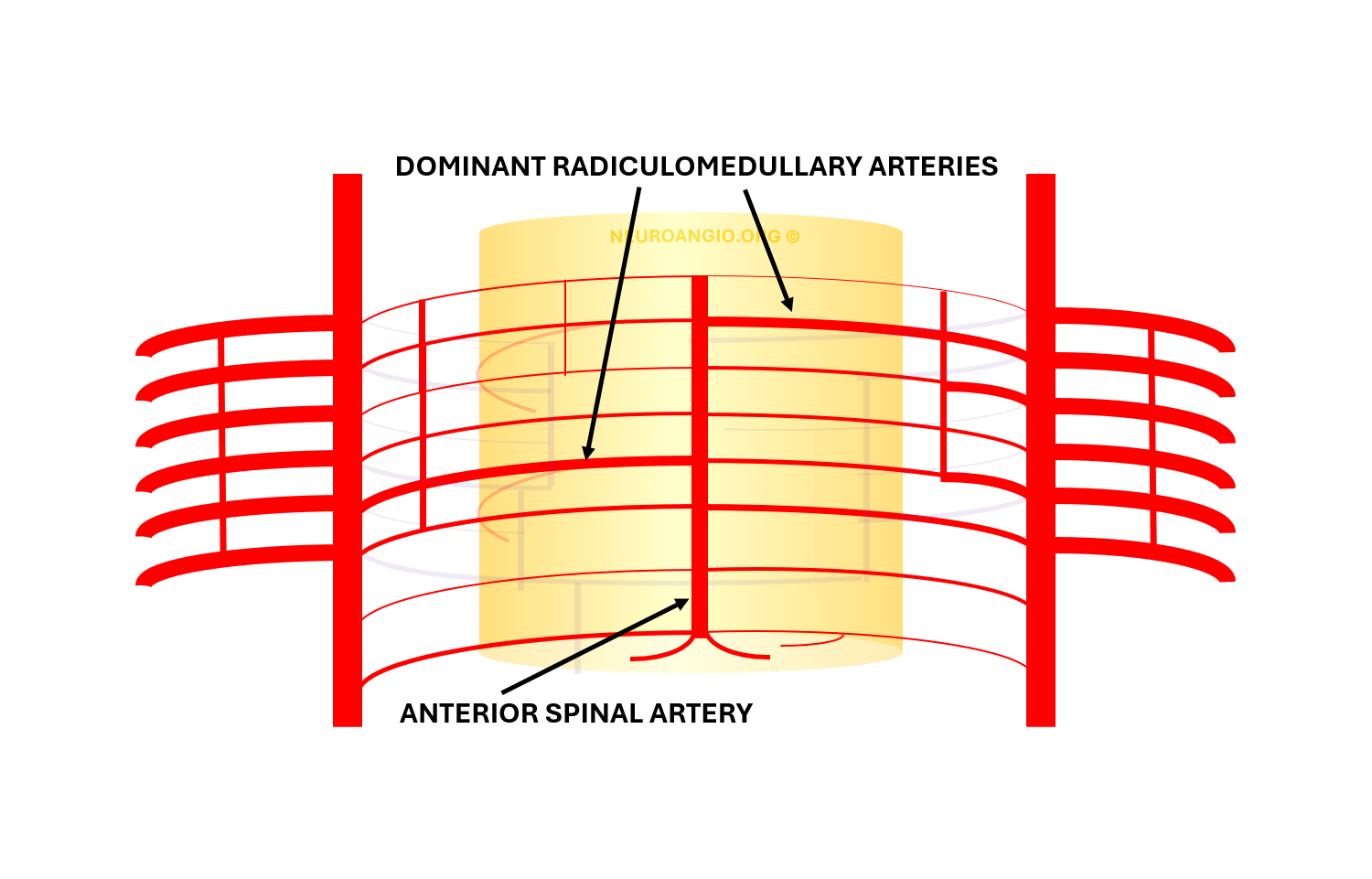
This process, in terms of the spinal cord, gives rise to the familiar adult appearance of the anterior spinal artery and its remaining radiculomedullary feeders, while most intersegmental arteries previously connected to it in early fetal life are limited to supply of the nerve root and adjacent tissues in the adult — and called radicular or, more properly, radiculodural arteries.
The same process applies to the radiculopial arteries supplying the posterior spinal system.
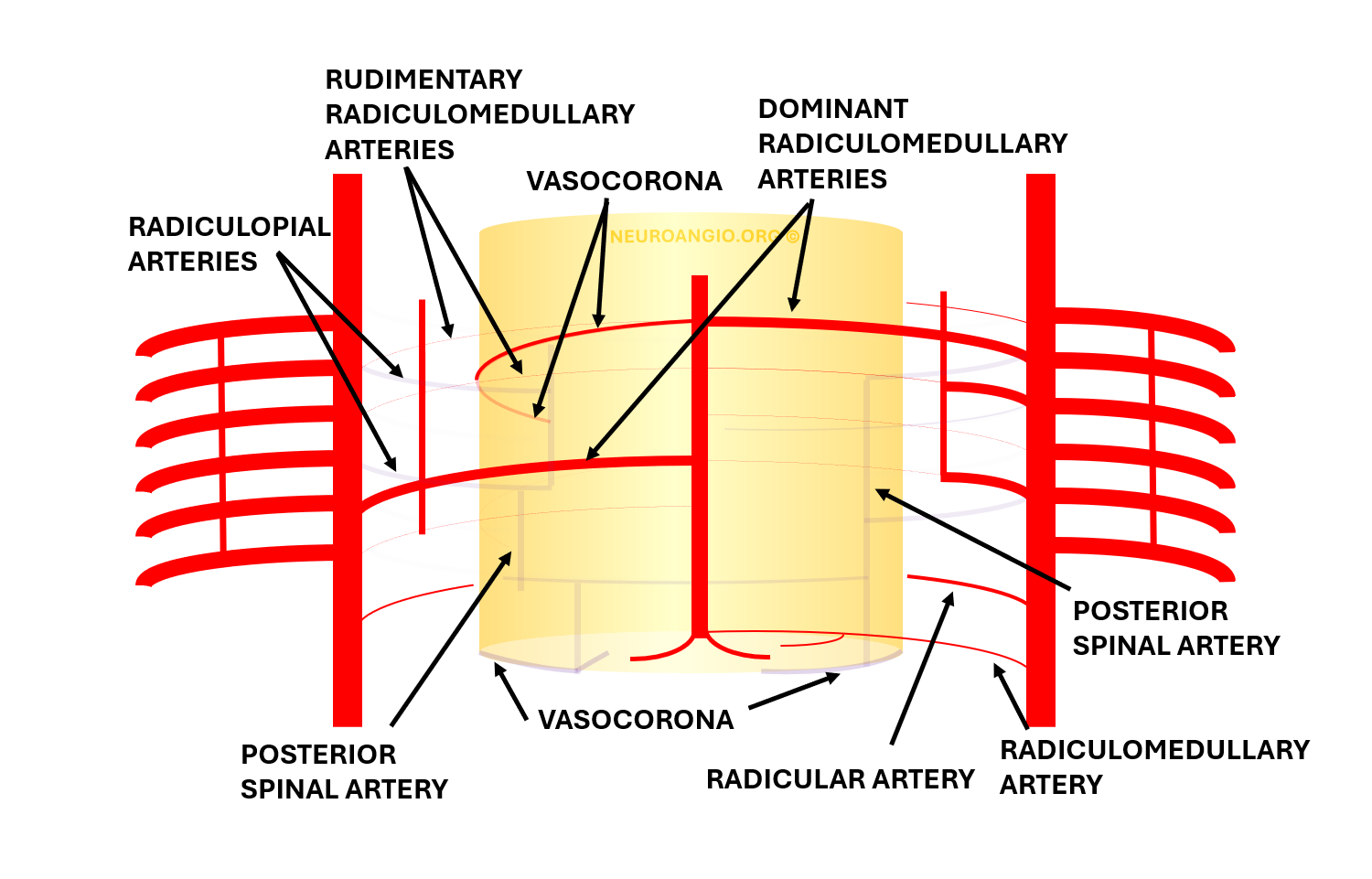
The above concepts help one understand the variability in spinal cord supply. For example, two co-dominant radiculomedullary arteries (as above) may be present, or one larger single vessel — called the Adamkiewicz (unlabeled, below)
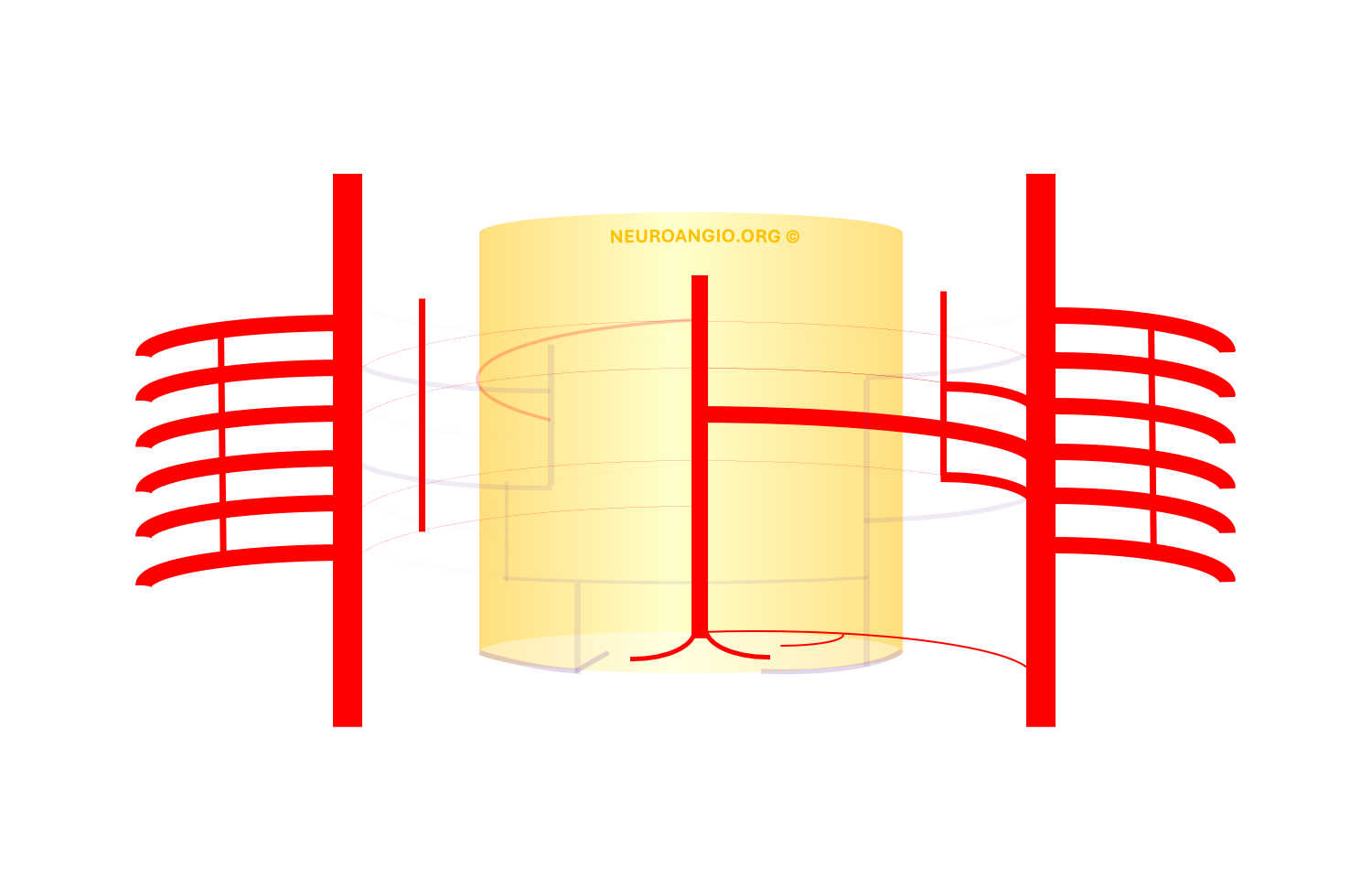
Vasocorona — surface anastomoses between anterior and posterior spinal arteries, may on occasion be the dominant source of supply to the anterior spinal artery, via the posterior spinal system, as below
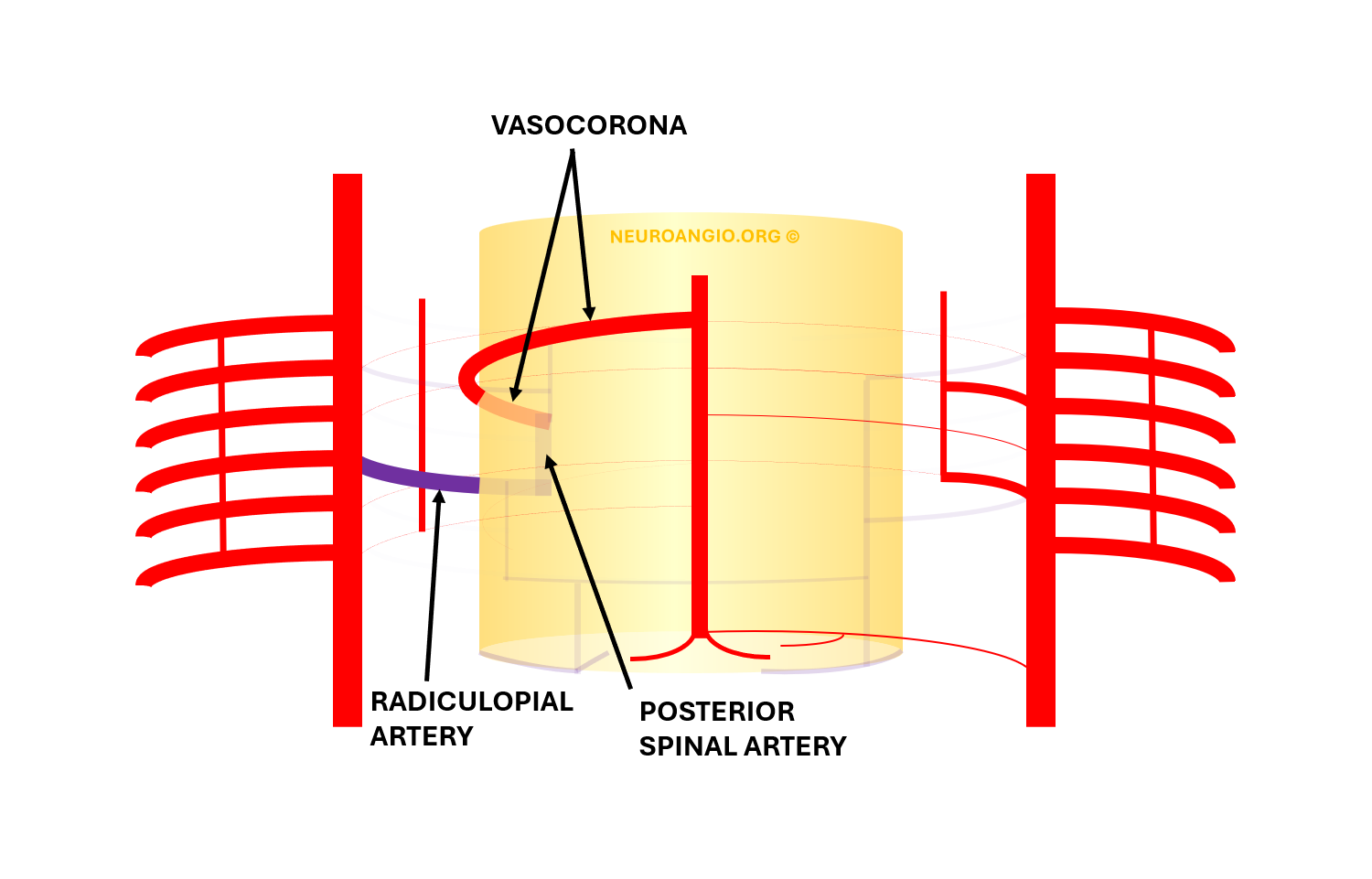
At the conus level, the posterior spinal system is usually prominent, due to the larger amount cord tissue. Well-developed anastomoses between anterior and posterior systems at that level go by various names — basket, rami cruciantes, etc. Not infrequently, the anterior spinal artery is predominantly supplied by one or more Posterior Spinals via “rami cruciantes” (which are just named vasocorona — same thing — arteries.
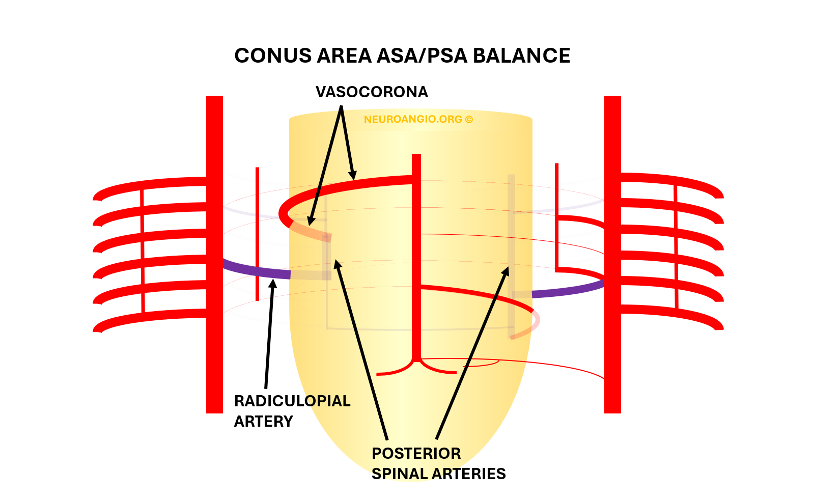
The more classic disposition is ASA supply of both PSAs via the same rami cruciantes…
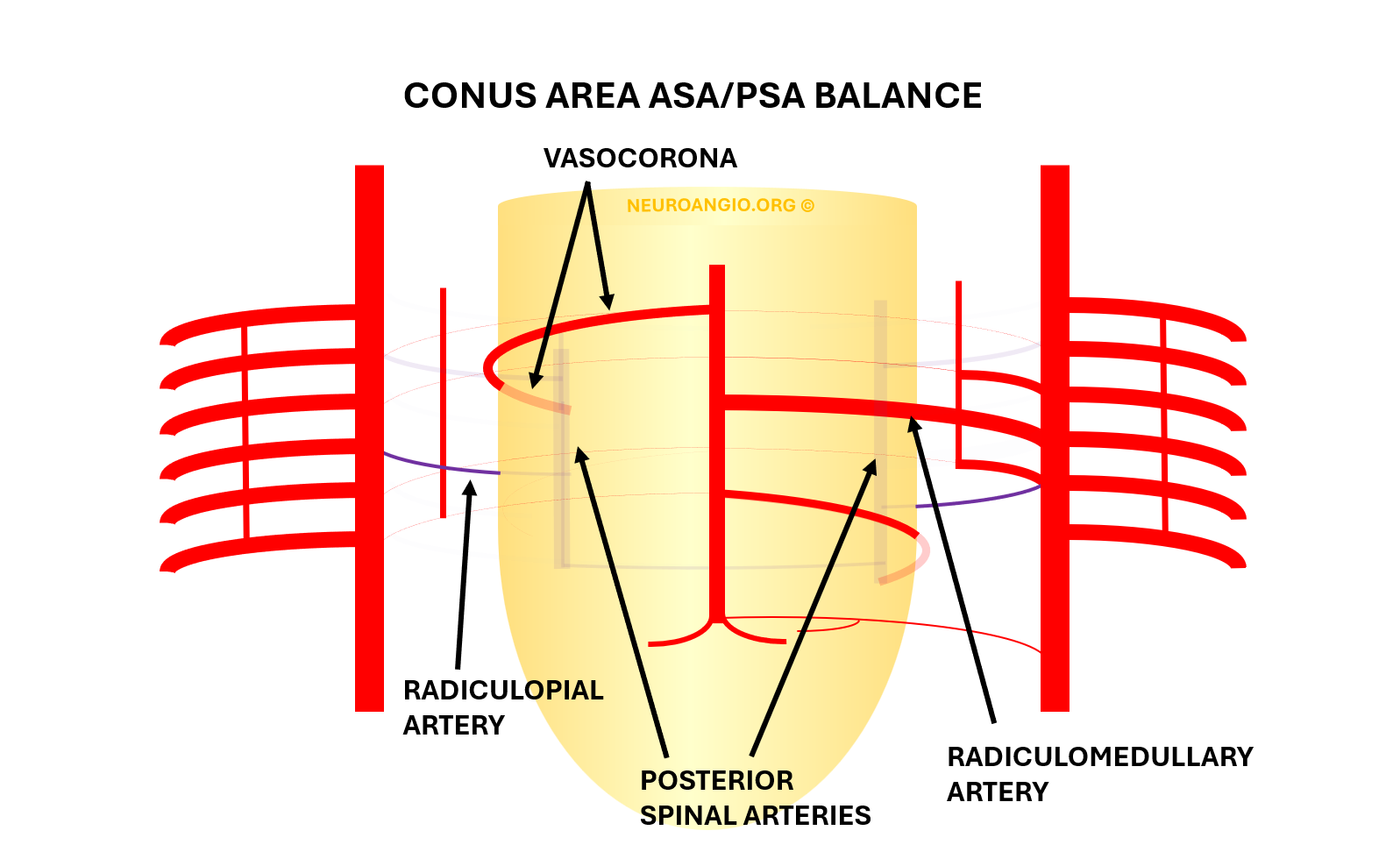
In the cervical spine, ASA is often duplicated or discontiguous, and its tributaries frequently originate from deep cervical and anterior cervical longitudinal arterial networks, as well as from the vertebral artery. Posterior and Lateral Spinal Arteries are correspondingly more prominent.
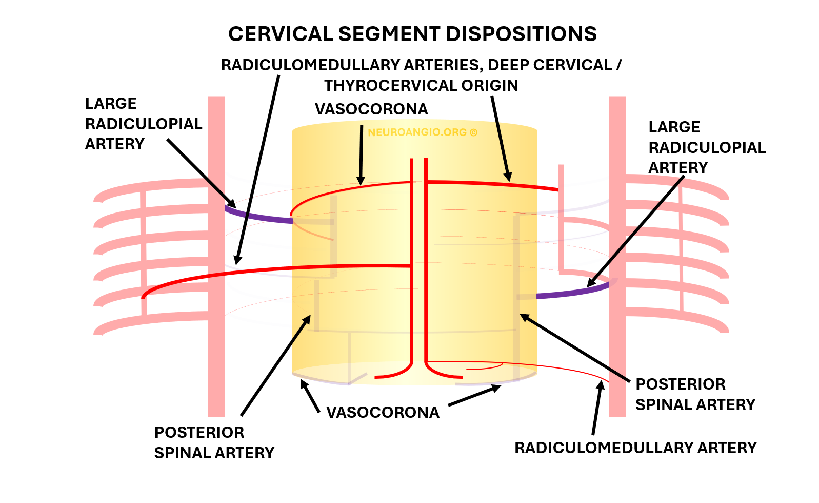
The same pattern of development takes place in the extra-axial, paravertebral space, where longitudinal connections between segmental arteries form a multitude of adult vessels, such as the vertebral, pre-vertebral, pre-transverse, deep cervical, lateral spinal, and other arteries, comprehensively addressed in the Spinal Arterial Anatomy Section
Visualization
2D-DSA and Cone Beam CT are still the best. Noninvasive imaging generally lacks resolution, even for 3T MRI. Photon Counting Detector CT is still early in spinal experience, i’m sure it will find dominant ASA contributors.
Technique of 2D-DSA is key — General Anesthesia is best, pharmacologic paralysis, positioning patient and imaging so that spinous processes are midline at level being images, all important. Elevating arms helps for CBCT of lower thoracic and conus regions. Details below
Schematic
One of our better drawings
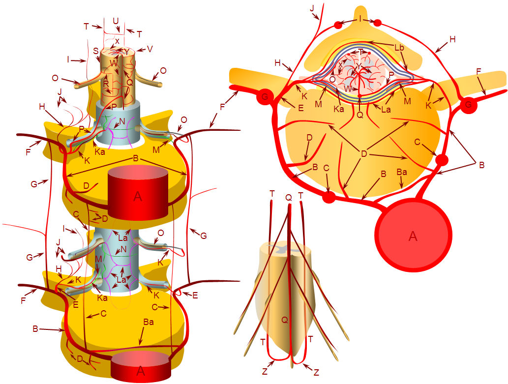
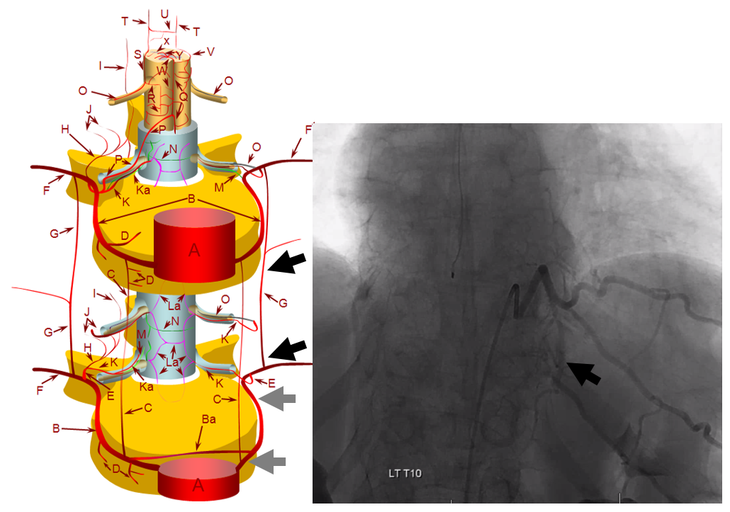
A – aorta; B – segmental artery; Ba – intersegmental arterial anastomosis; C – prevertebral anastomotic network; D – direct vertebral body feeding arteries; E – dorsal spinal artery; F – intercostal/muscular artery; G – pretransverse anastomotic network; H – dorsal division of the dorsal spinal artery; I – post-transverse anastomotic network; J – muscular branches of the post-transverse anastomotic network; K – ventral division of the dorsal spinal artery; Ka – radicular artery; La – ventral epidural arcade; Lb – dorsal epidural arcade; M – nerve root sleeve dural branch of the ventral division dorsal spinal artery; N – dural branch of the ventral division dorsal spinal artery; O – radiculopial artery; P – radiculomedullary artery; Q – anterior spinal artery; R – mesh-like pial arterial network; S, T – posterior spinal artery; U, V – pial arterial network (a.k.a. vasocorona) anastomoses between anterior and posterior spinal arterial systems, W – sulco-commissural artery, X – rami perforantes of the peripheral (centripetal) system, Y – central (centrifugal) system of sulcal arteries, originating from pial network of the cord; altogether, the pial network and rami perforantes (R+Y) are called the vasocorona or corona vasorum; Z – rami cruciantes (a.k.a. crux vasculosa, a.k.a. rami anastomotici arcuati)
Lets consider the artery generally from cervical to conus
Cervical ASA
Generally easier to visualize on angio due to smaller amount of bystander tissue — necks are generally smaller than chests or abdomens. In up to one third of subjects, the cervical ASA is not well seen, and posterior / lateral spinals can be quite large. The radiculomedullary and radiculopial artery origins are more variable than thoracic or lumbar segments — vertebral, ascending cervical, and deep cervical origins (especially at lower levels) are common.
Examples
Typical dominant ASA (Q). The letters correspond to figure above. Note central position projecting over the spinous processes in the frontal view. Lateral stereo views have no bone landmarks — which makes it harder — in particular one should not confuse the anterior spinal with the odontoid arcade, which is more ventral.
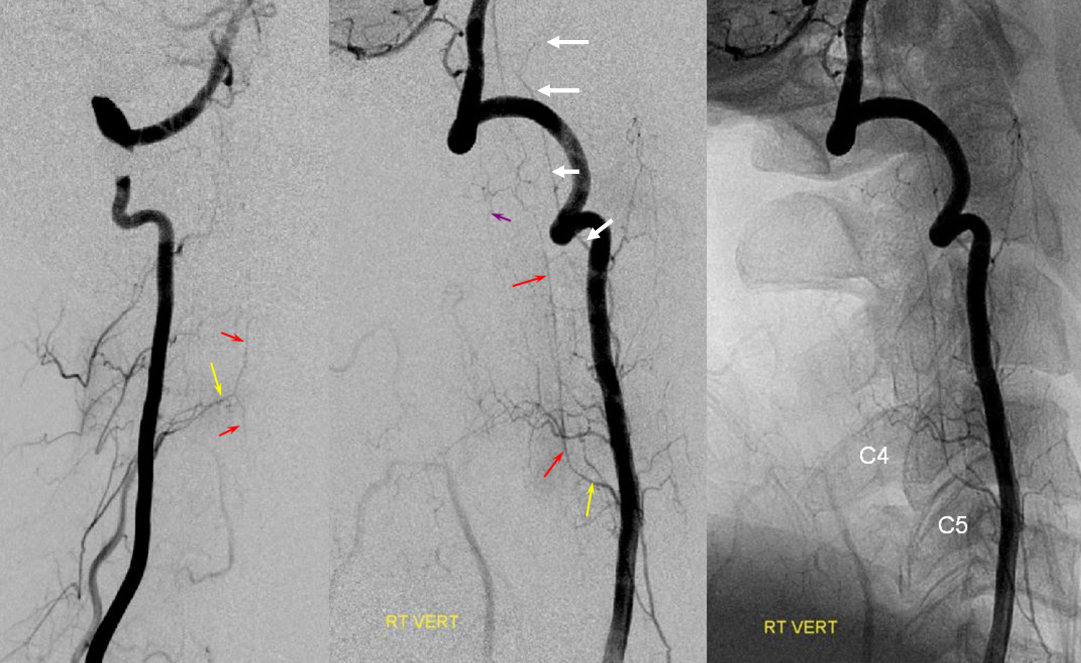
Old example with radiculomedullary (yellow) supply of the ASA (red). At C2 and C1 levels, the odontoid arcade (white arrows) is noted
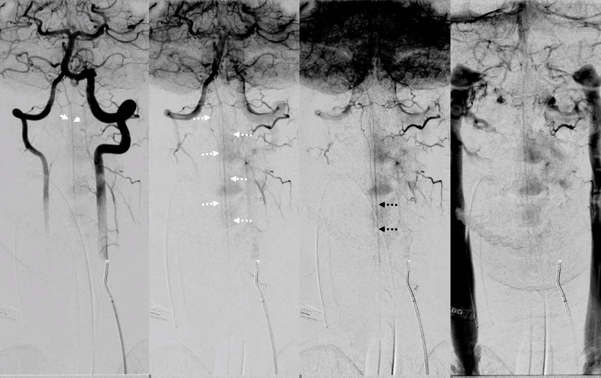
Look at this! Ridiculously “one-sided” origins of all three major systems — the anterior spinal and both posterior spinal arteries are supremely well-seen — originating from distal right vert (asa) and both PICAs (posterior spinals). This is most unusual. Frontal views show duplicated upper cervical asa (white arrowheads), long contiguous posterior/lateral spinal arteries (dashed white arrows), and faintly seen sulco-comissural arteries (black dashed arrows)
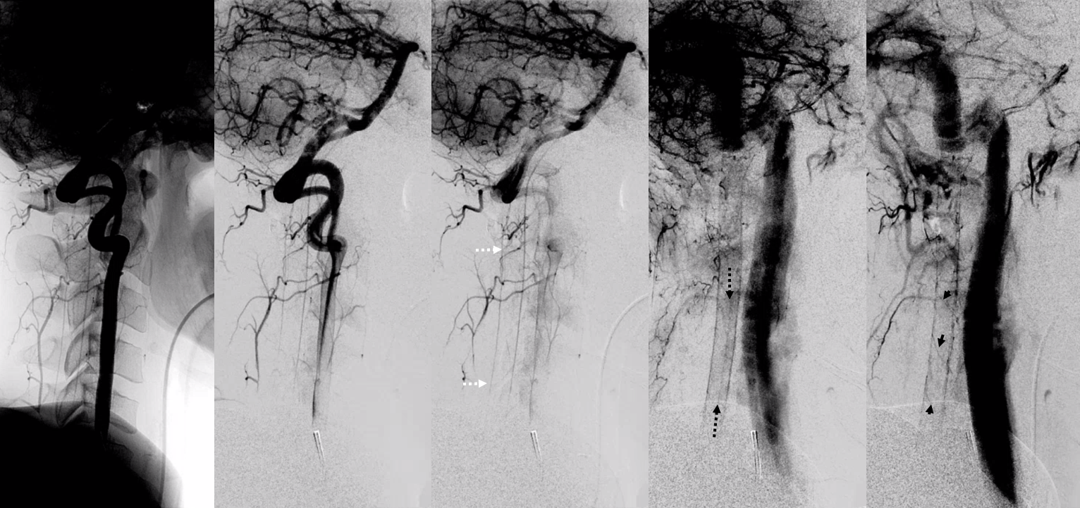
Lateral views show extremely well the posterior spinal axis (white dashed arrows), the position of spinal cord in the osseous spinal canal (leftmost image), a beautifully seen faint line of sulco-comissural vessels terminating in the depth of the ventral median sulcus (black dashed arrows), and circumferential / transmedullary spinal veins (black arrowheads)
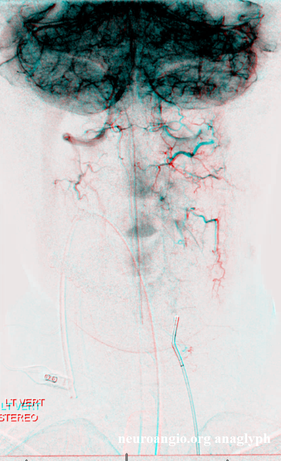
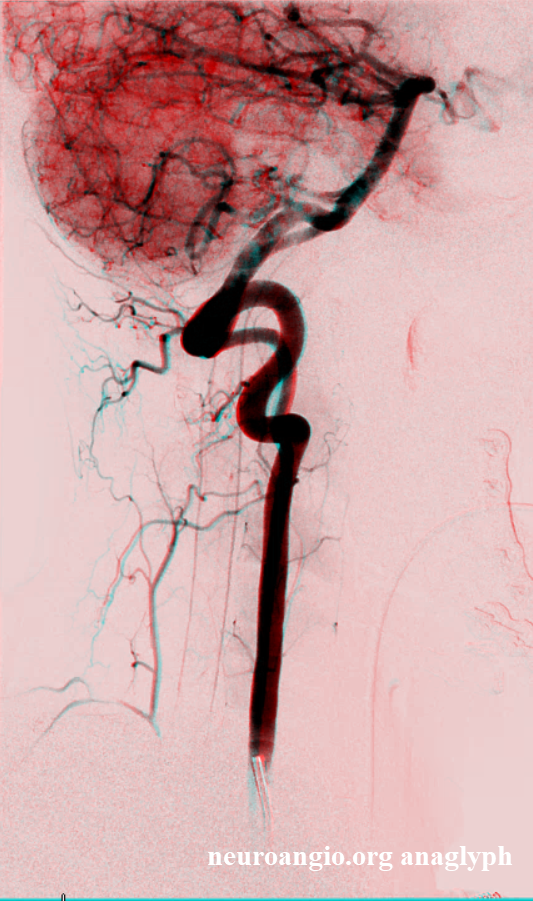
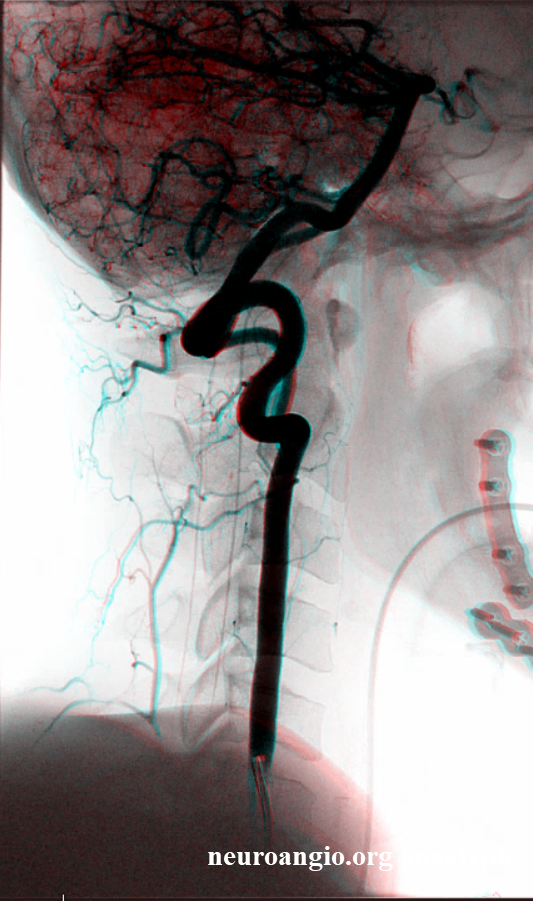
Another view of the cervical cord, this one also displaying the posterior spinal (brown) axis and the vasocorona vessels (yellow) connect to the anterior spinal axis (red), supplied by two co-dominant radiculomedullary arteries.
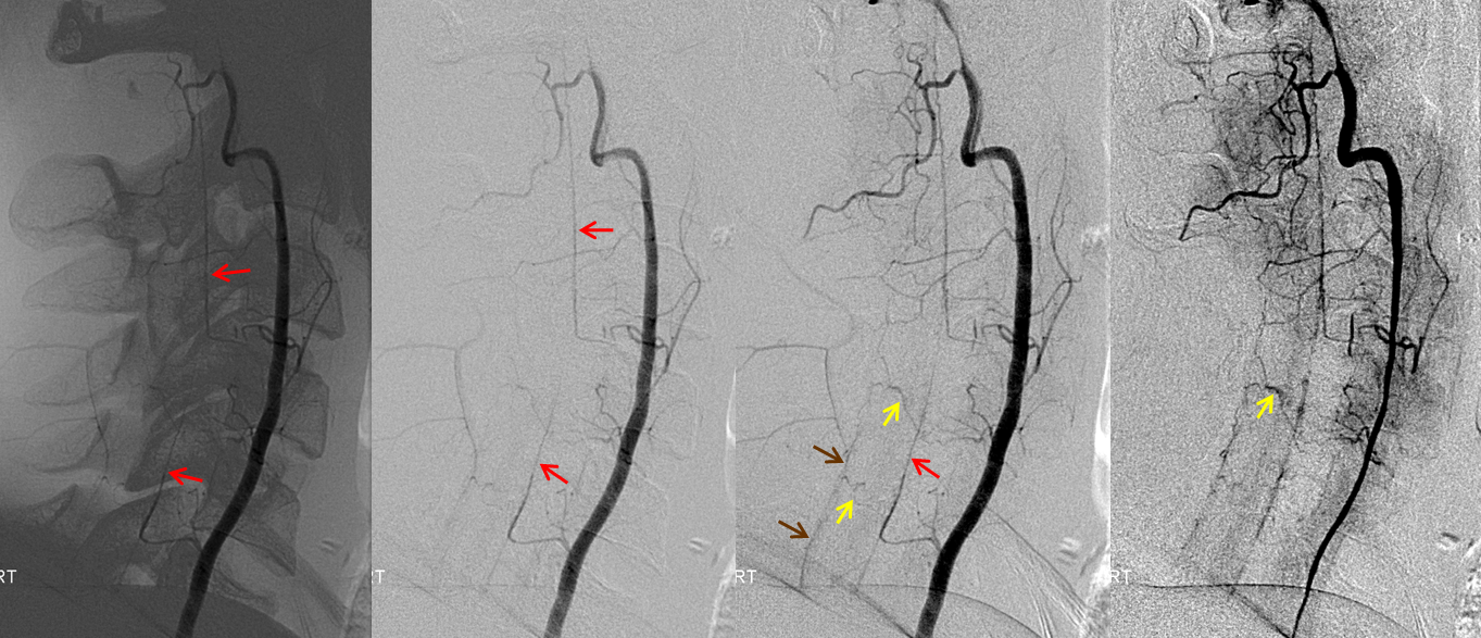
It is exceptional, and probably not so advantageous, to have highly dominant cervical ASAs, like the one below

Laterals. Unfortunately this was before our HRCBCT era…


The more typical, not so well seen ASA system, but it is there if you look carefully

Cone Beam CT and Intrinsic Cord Arteries
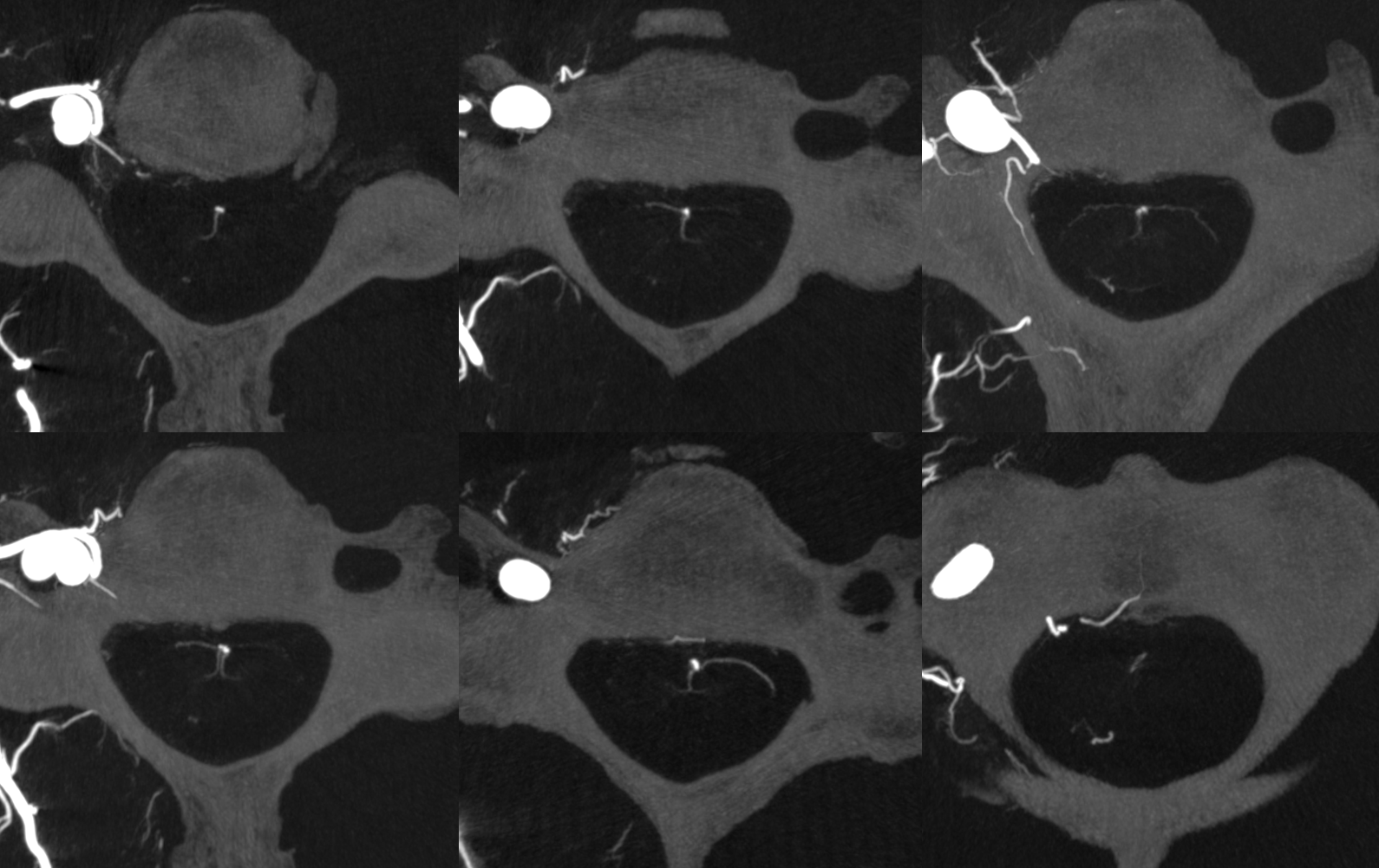
The state of art imaging is High Resolution Cone Beam CT Angiography (HR-CBCTA, used interchangeably with CBCT, HRCBCT, etc). How to obtain such images — see comprehensive section. Same case as above. Amongst wealth of addition info are sulco-comissural arteries and vasocorona connecting anterior, lateral, and posterior systems
The posterior/lateral spinals are inside solid white oval. The lateral spinal is smaller and more ventral. Middle image — vasocorona connecting to lateral spinal artery. Dashed white oval — same — ASA supplying bilateral lateral cord. Dashed black oval — from the back — posterior spinal more prominent, lateral spinal smaller and shorter.

No labels. Note the well-seen and unlabeled sulco-comissural arteries running in the ventral median sulcus

Now take a look at the axials. Each sulco-comissural artery supplies its own half of the cord. Only at the cervicomedullary junction do those perforators typical feed both paramedian segments (hence the dreaded syndrome of their occlusion). Typically, and probably for reasons listed above in the embryology section, the sulco-comissural supply is lateralized, as indeed it is indeed for brainstem and brain homologs as well. The lower left image is probably the best, and resolution of at least 100 microns is required.
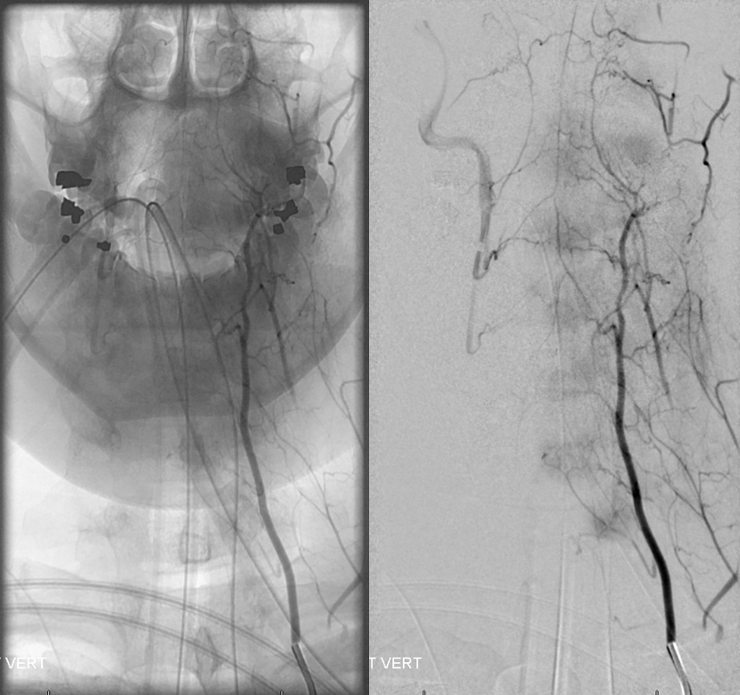
Rich collateral network of vertebral system
The connections between cervical anterior spinal system and the myriad other vessels in the neck are plenty — bilateral vertebral, occipital, anterior cervical, deep cervical, ascending pharyngeal — many ways to get in trouble during embolizations. Most of these connections are relevant in terms of vertebral artery anastomoses (see vertebral artery page). Here is a superb illustration of these connections in a patient with a hypoplastic vert, where the diagnostic catheter is occlusive at the origin and therefore allows for reflux into all kinds of adjacent vessels — all images courtesy of Dr. Eytan Raz
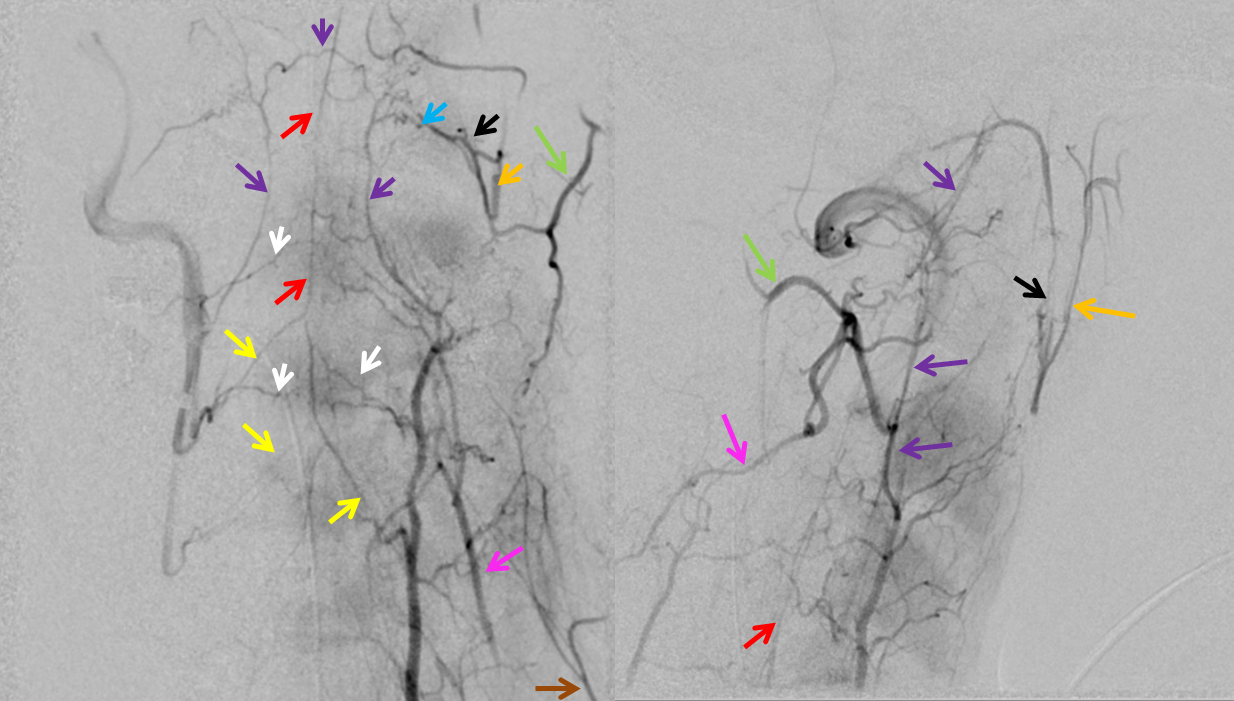
Same with labels and lateral. Red – ASA, white – radiculomedullary, yellow — epidural arcade, purple — odontoid arcade, blue – muscular branch ascending pharyngeal; black – neuromeningeal trunk, ascending pharyngeal; orange — pharyngeal trunk, ascending pharyngeal; green — occipital; pink — deep cervical; brown – ascending cervical
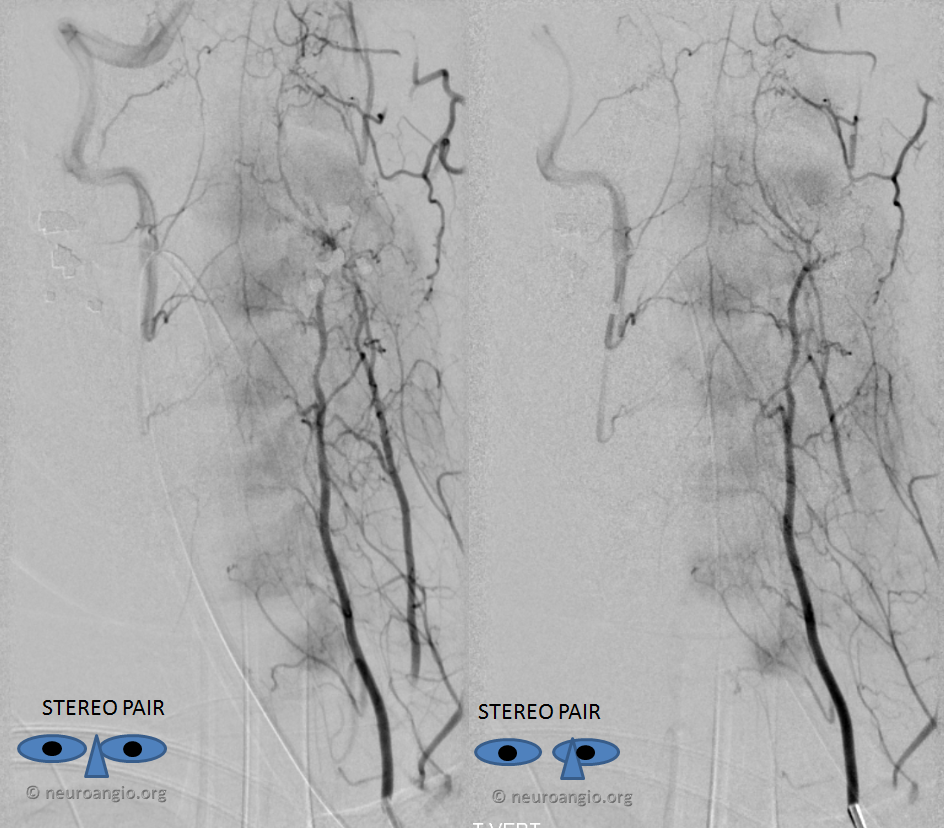
Now, for one of the most amazing stereos on the website
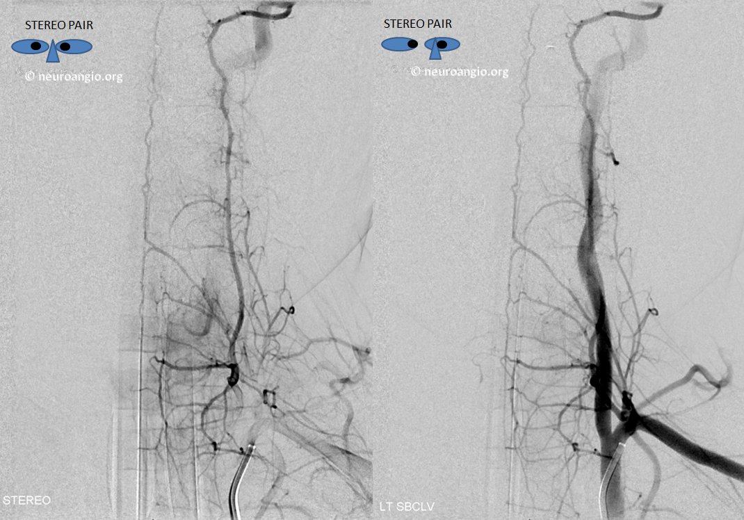
The Cervical ASA is often “duplicated” or incompletely fused (see embryology section). In such cases, each parasagittal vessel supplies its respective side of cord
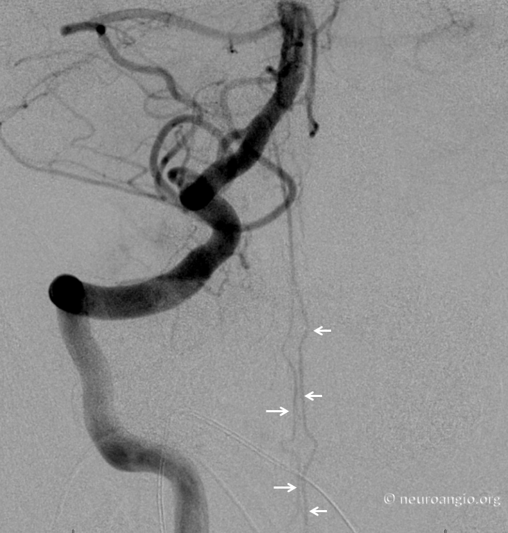
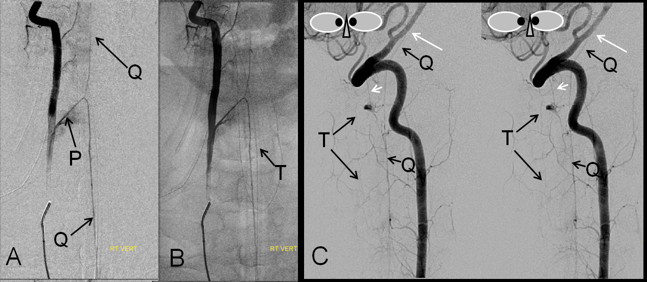
Deep Cervical origin of the radiculomedullary artery — second most common after the vert. At our institution, all cases of posterior fossa subarachnoid hemorrhage with no intracranial cause REQUIRE identification of the anterior spinal artery, as in ~10% of cases (in-house experience) the pathology turns out to be in the cervical spine. Subselective catheterization of thyrocervical and costocervical trunks is mandatory to map out the upper cord, and supreme intercostals maybe also (see below)
Anterior spinal artery (Q) origin from deep cervical artery, P= radiculomedullary artery; notice collateral opacification of the vertebral artery (long white arrow) via the C2 intersegmental artery (short white arrow).
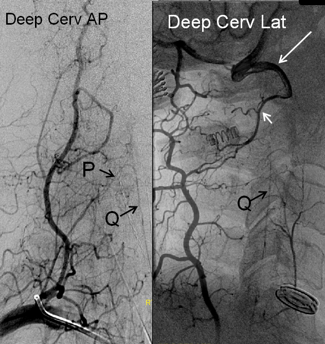
Typical appearance of another case, with radiculomedullary (black) supply of the ASA (white)
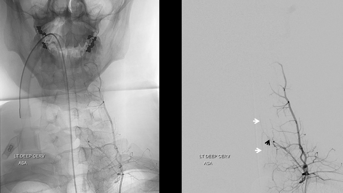
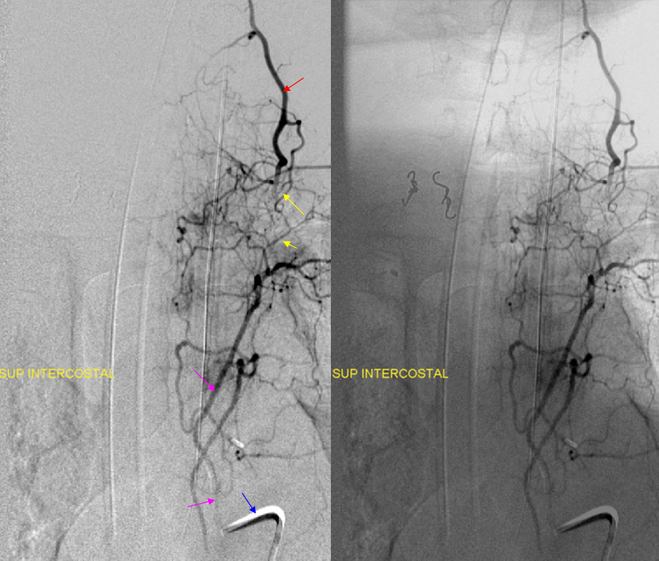
Another deep cervical origin — any longitudinal system can give origin to the radiculomedullary artery — in this case the radiculomedullary artery (orange) originates from the deep cervical branch (red). Notice also injection of supreme intercostal artery (pink, lower two images) with extensive deep cervical artery anastomoses (yellow) through which the anterior spinal artery can be inadvertently embolized. The catheter, barely engaged in the supreme intercostal, is labeled in blue — visualizing the deep cervical network via paravertebral anastomoses.
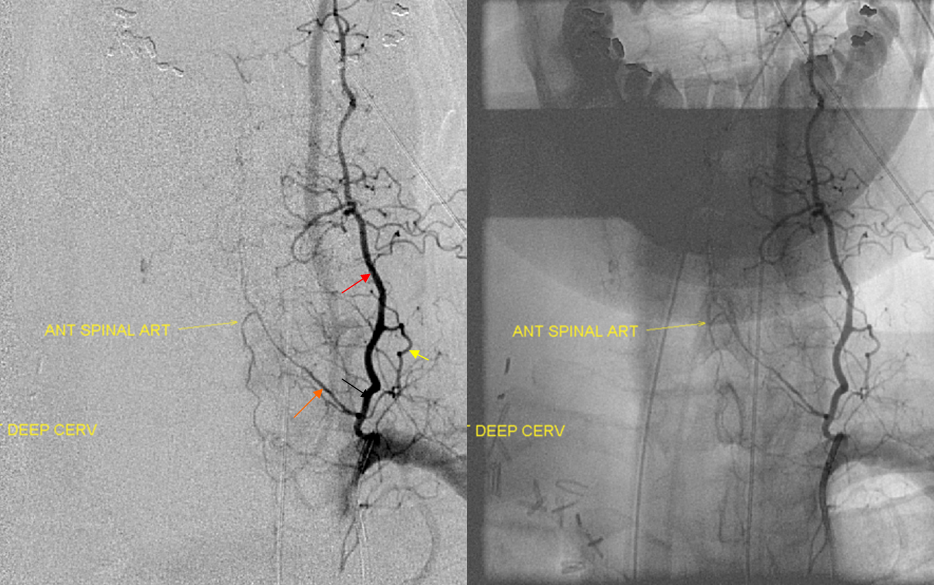
Stereo views of deep cervical injection, associated with more extreme example of anterior spinal artery “duplication”
THORACIC REGION
The cervical/thoracic boundary with respect to the cord is illogical, as is thoracic/lumbar one for that matter. Having ribs at some levels and not at others has nothing to do with any specific distinctions at cord level. More important are changes in cord volume at thoracic and lumbar enlargement — more volume needs more blood, its that simple. Thus, seemingly more prominent posterior/lateral spinal systems at cervical level, and posterior spinals / rami cruciantes at the conus (see below). The ASA continues without regard to the ribs, but it is also wrong to think that because the thoracic cord is thinner, the posterior spinals are less important. Indeed, at every level of cord, there is a balance, and in some individuals the posterior spinals can be larger and anterior smaller, or vice versa.
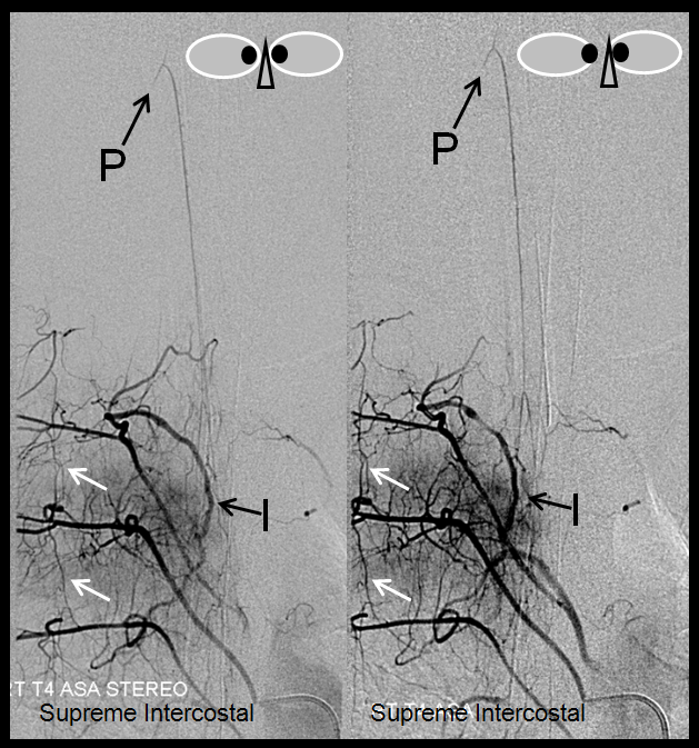
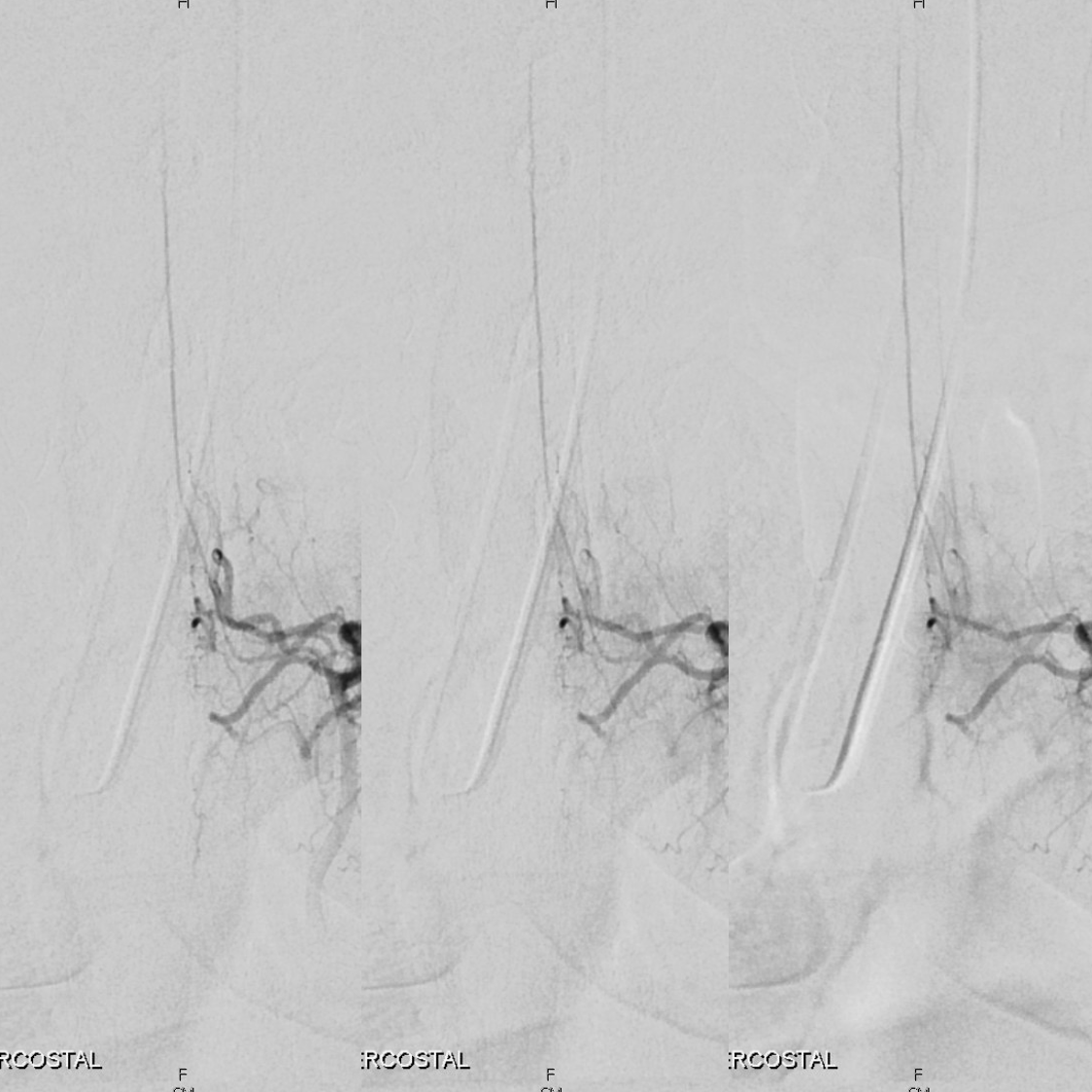
Supreme Intercostal Origin of Cervical Spinal Artery — seen often as well, and important to know. The supreme intercostal supplies at least T1-T3 or more levels, so the odds of seeing a quasi-random from embryology perspective supply to either ASA or PSA is at least 3 times as high as from any individual level. Upper thoracic arteries can be difficult to catheterize, especially in patients with capacious dilated atherosclerotic aortas. We use a 4F or 5F RDC (which can be too small for the upper thoracic spine); if that does not work, one can try an appropriately-sized Cobra, or perhaps a Simmons 1. Sometimes, hand-shaping an RDC to produce a bigger curve (so as to push against the contralateral aortic wall) is more helpful than another catheter. In this case, the supreme intercostal was visualized via the T4 segmenal injection through a prominent paraspinal anastomosis (I)
Stereo pair, supreme intercostal artery origin of the anterior spinal artery (same legends as above), visualized via T4 injection through a prominent post-transverse anastomosis (I). Notice transient contrast reflux into a cervical radiculomedullary branch (P); another longitudinal anastomosis (white arrow) between adjacent T3, T4, and T5 segmental arteries
Supreme intercostal artery (red/P origin from the vertebral artery — another example of homology between various longitudinal anastomoses. Notice multiple intercostal arteries (yellow)
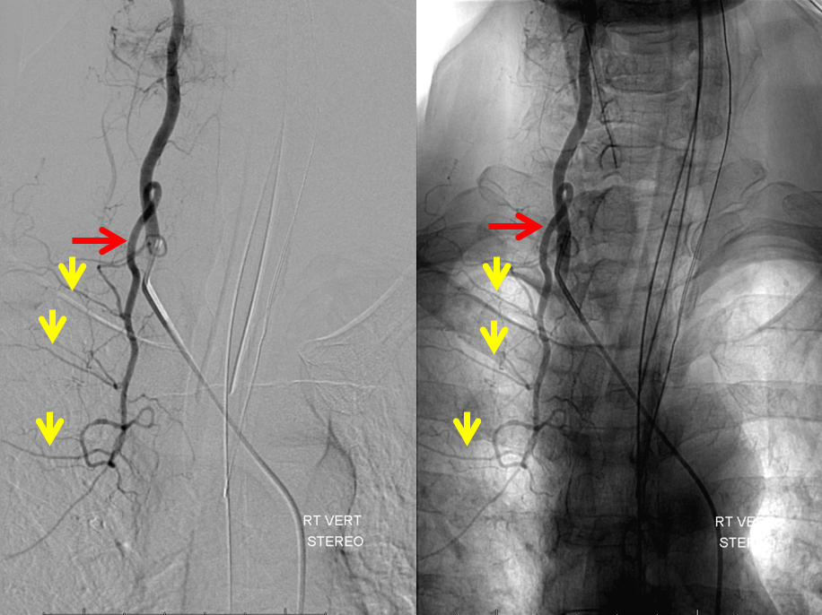
Supreme Intercostal and Collaterals
A “wedge” or flow control position with a catheter is a very important concept. Injecting from wedge control allows one to build up supraphysiologic pressures inside the system being injected — reversing flow in some arteries, etc. It can of course be dangerous with possible rupture of vessels etc. Wedge position and other ways to manipulate pressure-flow dynamics are important to master.
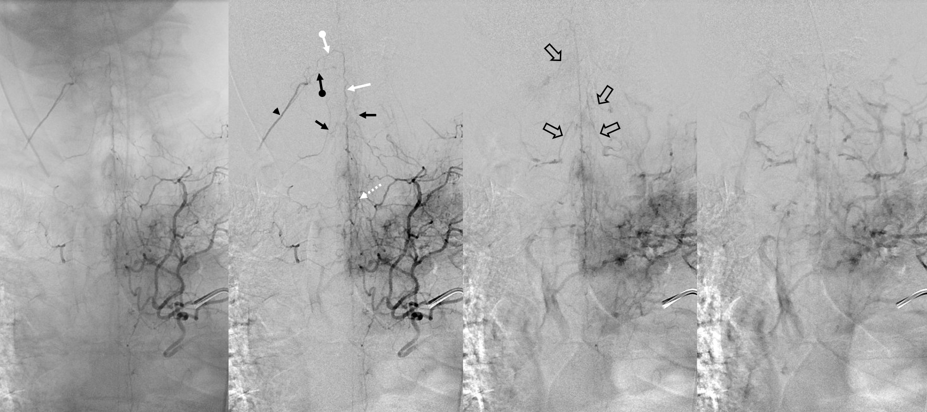
Radiculomedullary artery (dashed white arrow) supplies the asa system (white arrow). Retrogradely opacified is a right deep cervical level radiculomedullary artery (white ball arrow) as well as radiculopial artery (black ball arrow), with visualization of the common trunk radiculomedullopial artery (black arrowhead). Also seen are two other radiculopial arteries (black arrows), likely opacified retrogradely via vasocorona. The obvious conclusion here is that the system is well-collateralized. Occlusion of either radiculomedullary artery seen here is likely to be asymptomatic, unless there is embolus into the asa.
On the venous side, multiple radicular veins (open black arrows) are visible.
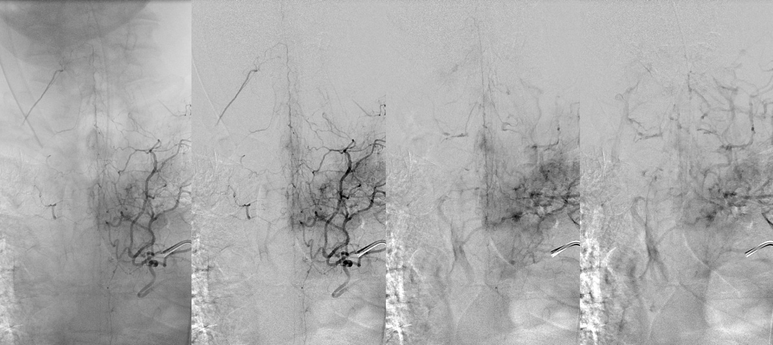
Without arrows
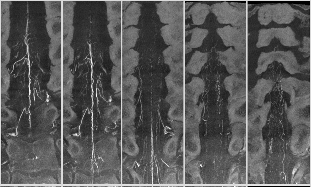
CBCT of the same case — can you recognize some of the vessels labeled above? Hint — its a mixed phase injection, so both arteries and veins are visible — if it were easy, anyone could do it…
Anterior and Posterior Spinal Thoracic Network
As seen above, the posterior spinal arterial system is very important. There are many radiculopial and radiculomedullary arteries beyond our 2D-DSA resolution, and beyond HRCBCT as well. Not infrequently, PSA system can be quite prominent, as in this example
Here is a supreme intercostal 2D-DSA – there is a large obvious ASA. Can you see the PSA?
HR-CBCT of same patient — see why Cone Beam CT is important?
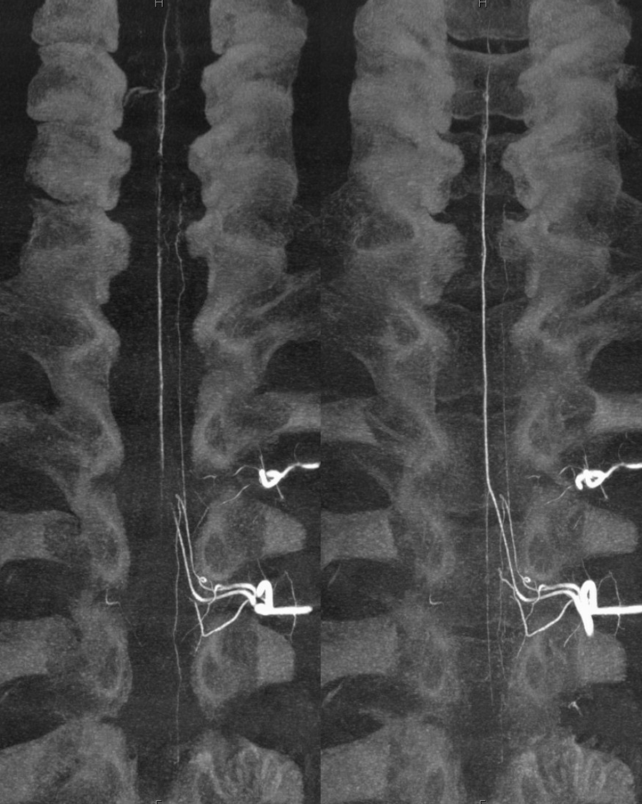
Another example
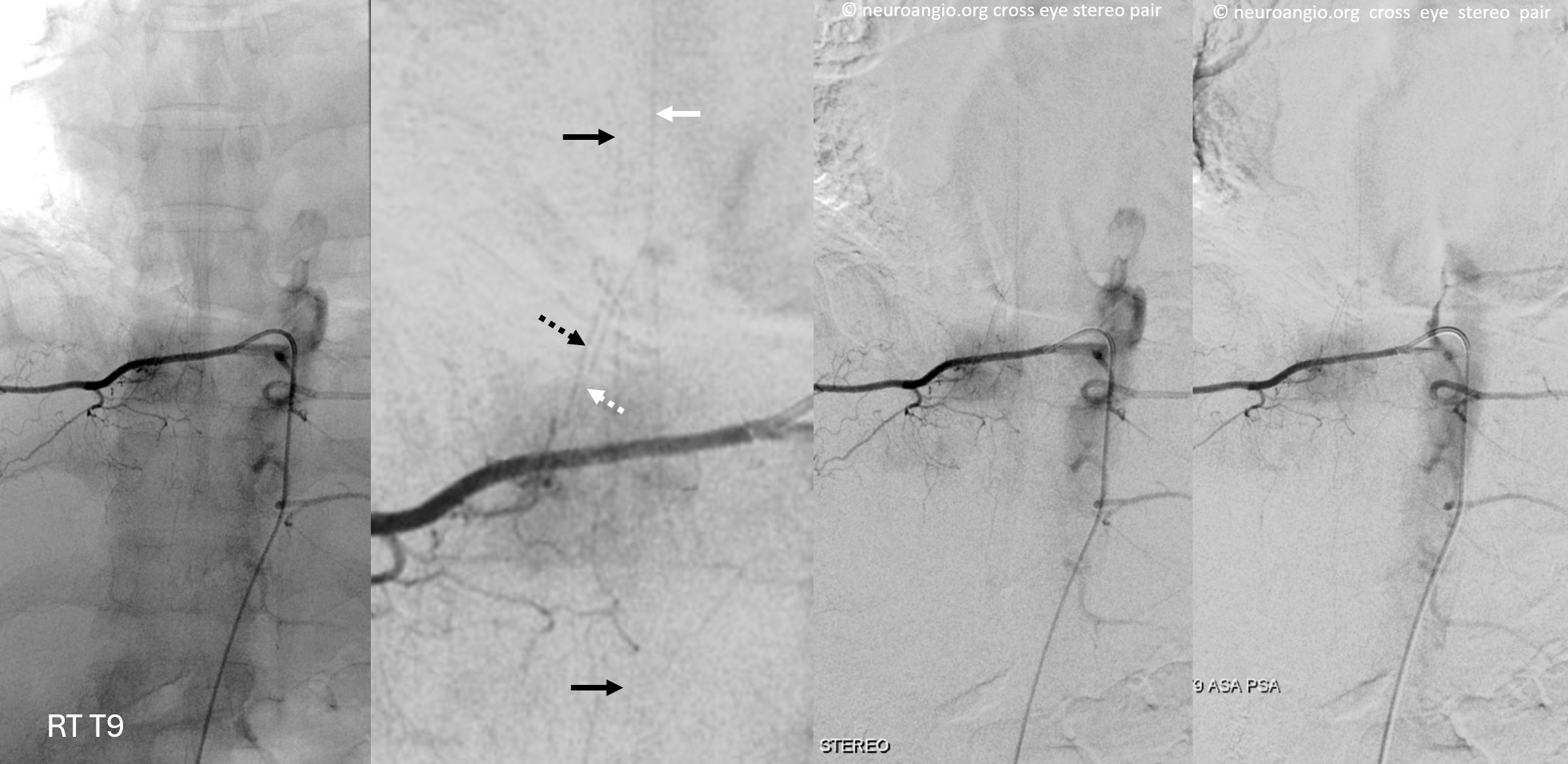
Right T9 injection — radiculomedullopial artery, with radiculomedullary (dashed black) and radiculopial (dashed white) contributions to respective posterior (black) and anterior (white) spinal arteries. Two images on right are stereo pairs. Proper midline spinous process alignment (leftmost image) shows the posterior spinal artery slightly off midline.
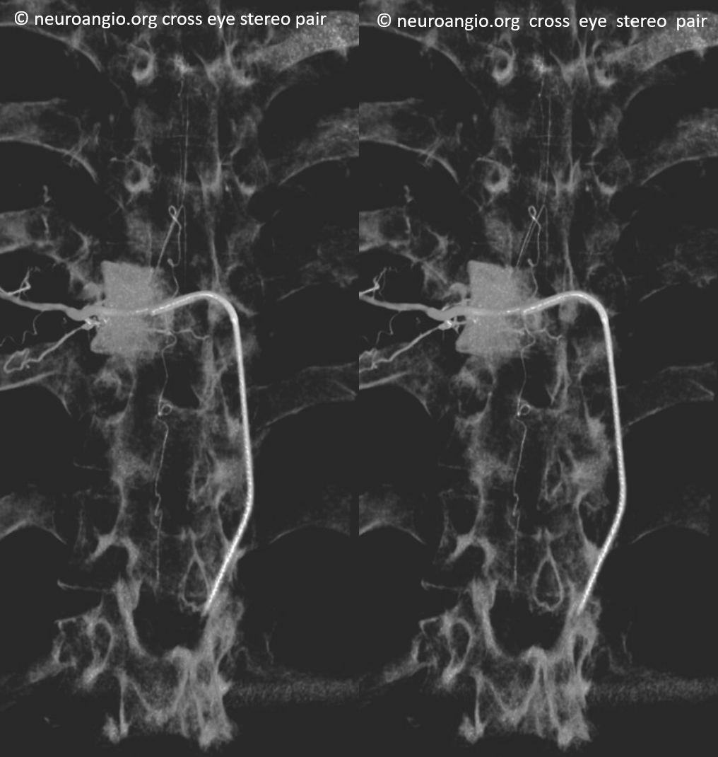
The posterior spinal is much larger than it looks on 2D-DSA — see HRCBCT stereo pair of the same patient below
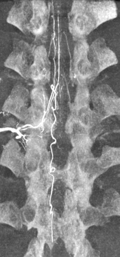
Note several additional retrogradely visualized radiculopial arteries (unlabeled)
Conus Region — Artery of Adamkiewicz and Thoracolumbar Junction
The most consistent and earliest recognized supply to the cord is the artery of Adamkiewicz. It technically refers to the largest radiculomedullary artery, supplying the largest by volume conus medullaris section. In practice, many textbooks and individuals refer by Adamkiewicz both the radiculomedullary artery and its ASA continuation, however this is incorrect, and important to note given that many contributions to the ASA are possible.
From the embryology discussion, its clear that any radiculomedullary, including Adamkiewicz, can arise from any segmental artery level. In roughly 75% of cases, the Adamkiewicz is found on the left side between T9 and L2. In practice, this wide range is not very helpful — just to say that its there somewhere near the terminal cord. More important is the predominant left-sided origin.
Below is a very typical Adamkiewicz — can also see posterior spinal arteries — the “basket”, visualized via rami cruciantes. Here ASA is dominant (see schematic below)
Schematic of same
The same case in HRCBCT — with minimal venous admixture. Note typical appearance of abundant sulco-comissural arteries
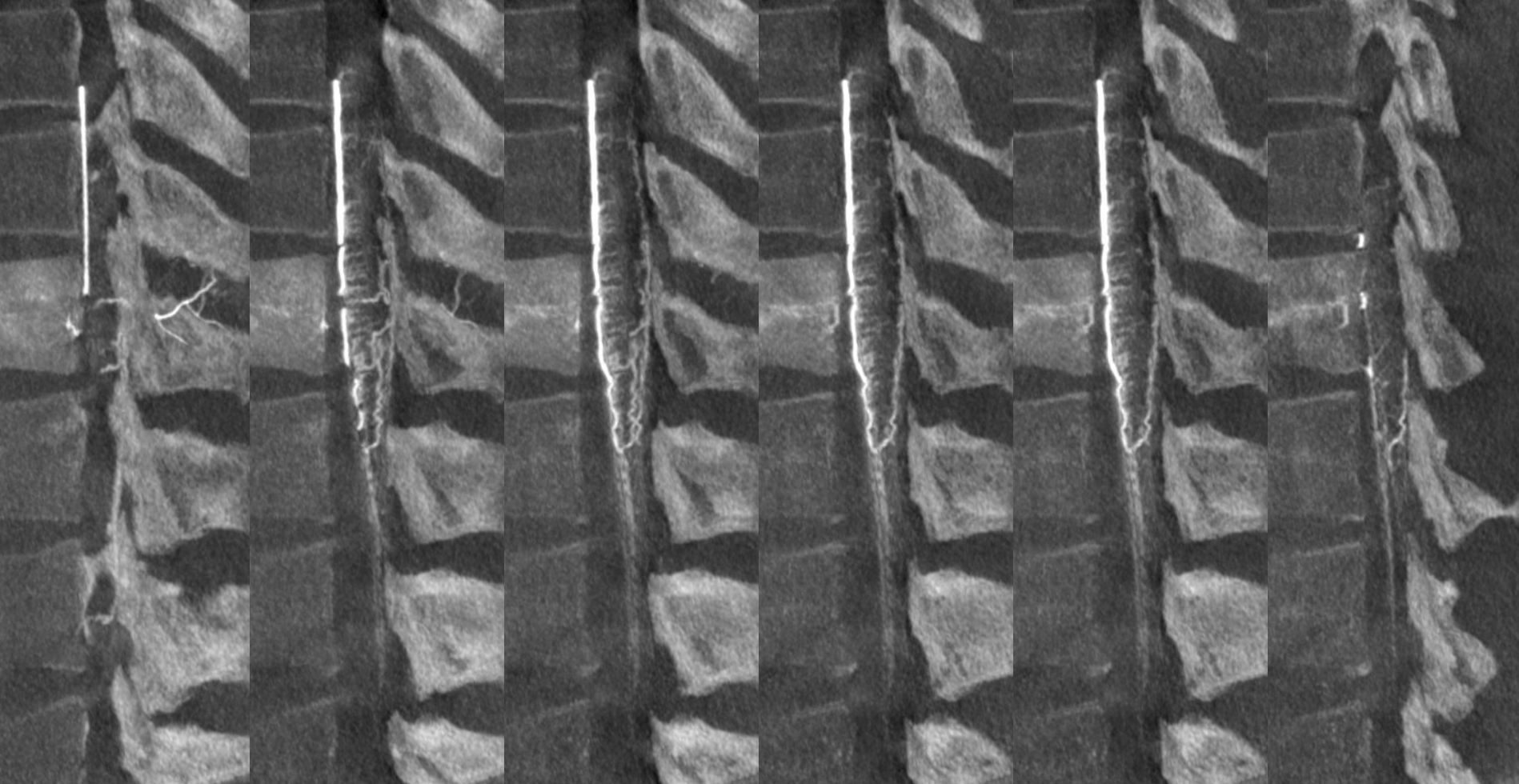
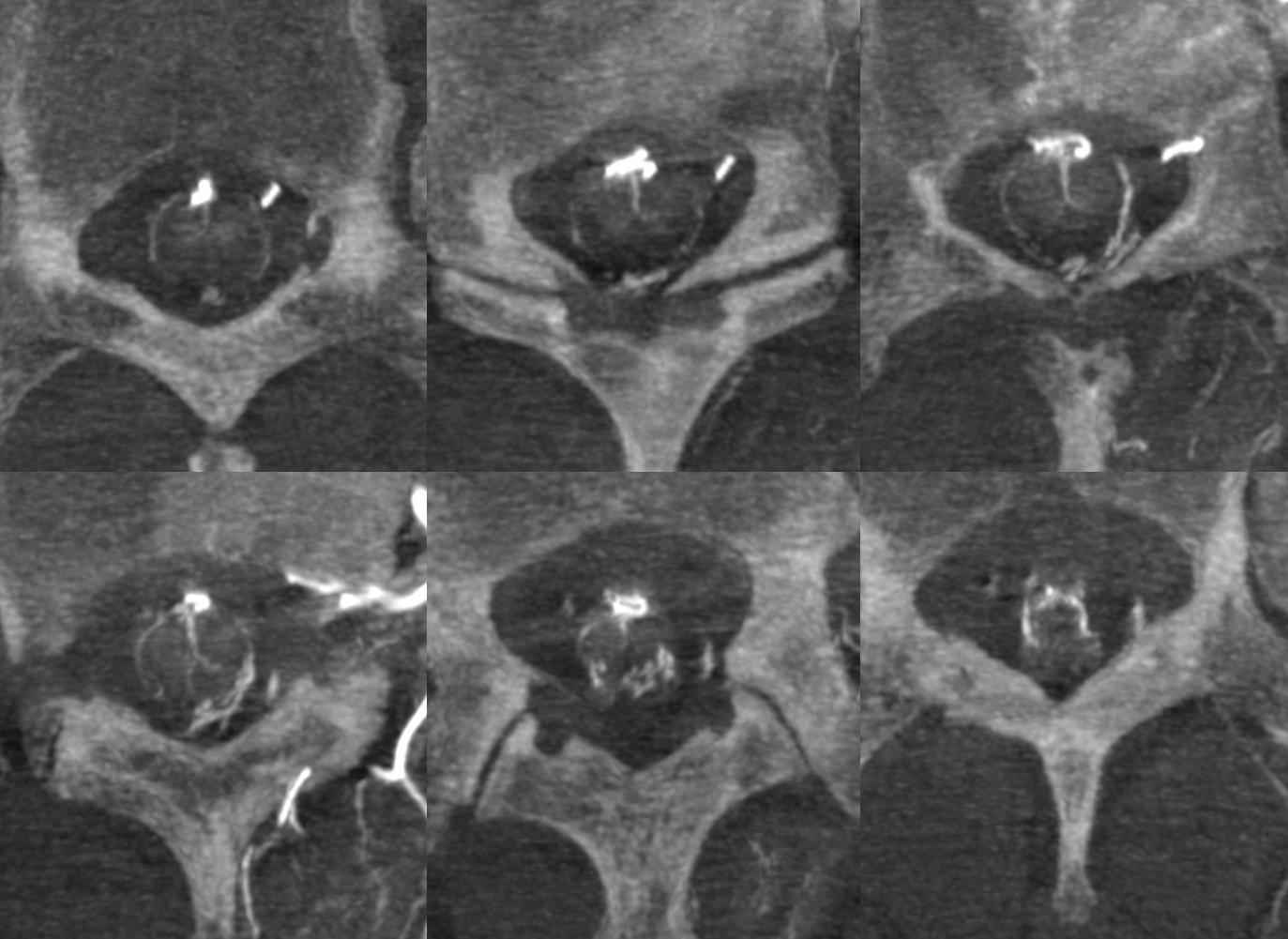
Video
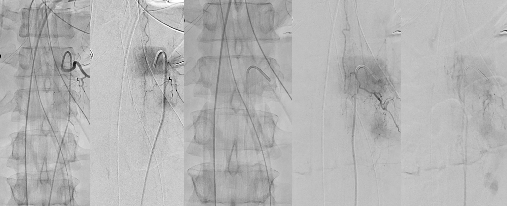
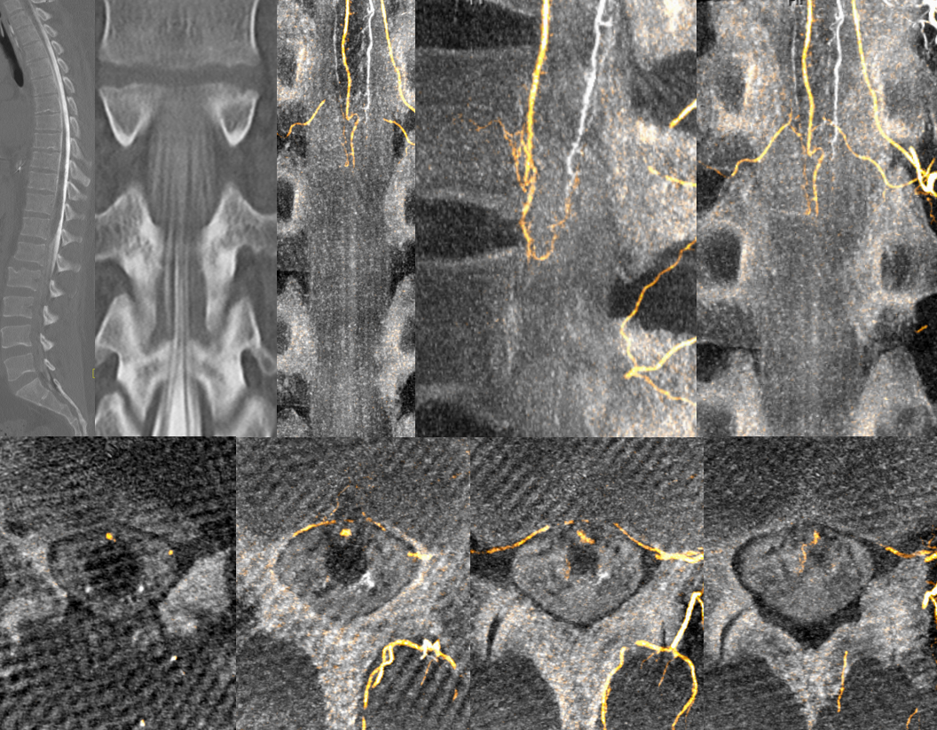
A unique case of suspected spinal artery stroke, where angio was immediately preceded by a myelogram… Injections of anterior (two leftmost images) and posterior spinal contributors proved normal appearance of the basket
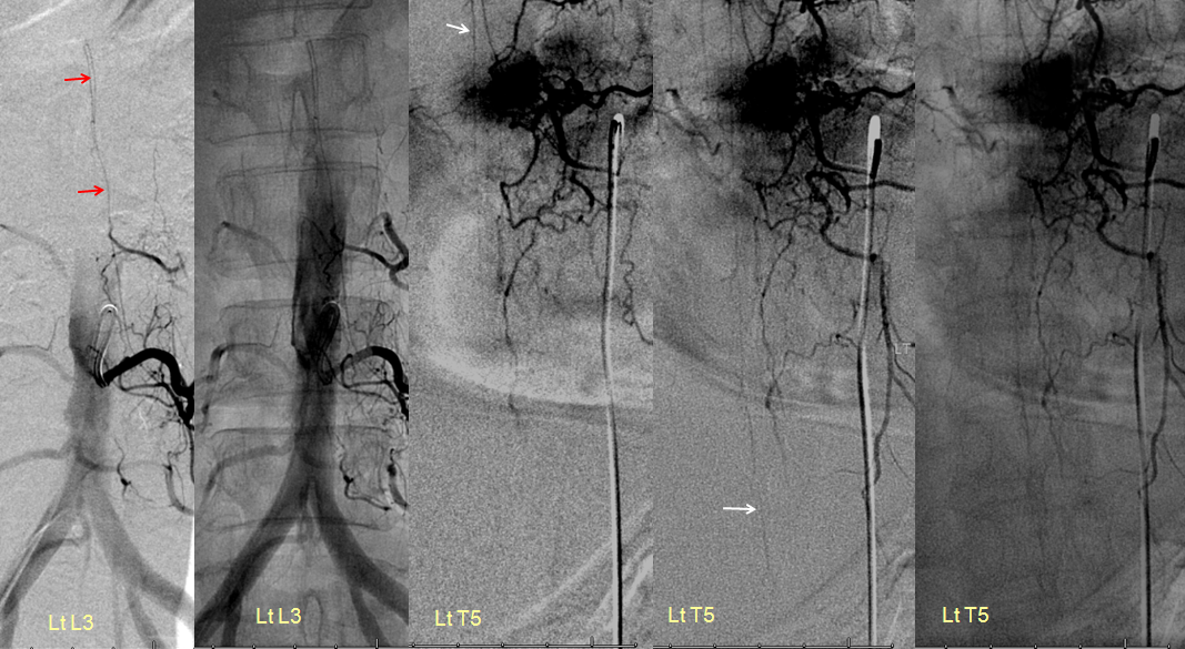
Here, balance as in everything. Higher origin Adamkiewicz (T5) ASA (white), associated with prominent conus posterior spinal (red) supply from L3
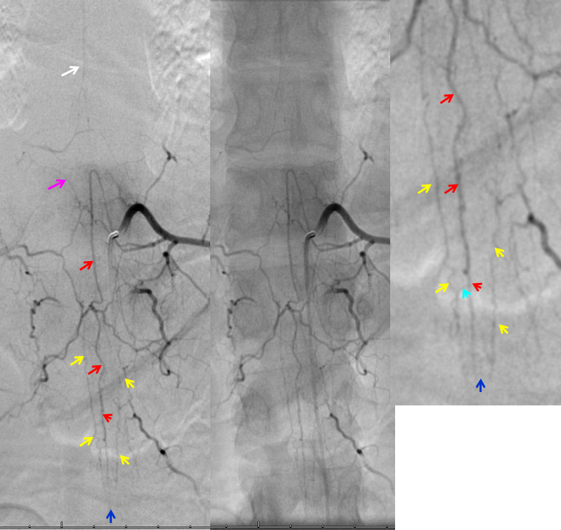
In this young patient, the conus region “arterial basket” is particularly well displayed. The anterior spinal artery (red) opacifies, via coronary arteries at the bottom of the conus (dark and light blue arrows) the posterior spinal systems (yellow arrows). Notice, in an image to the left, how a more superior segment of the anterior spinal artery (white) is visualized through the retrocorporeal arcade (pink arrow).
Stereo views below. Arterial “basket” consisting of the anterior spinal artery (black) and posterior/lateral spinal arteries (white) can often be visualized from an appropriate radiculomedullary artery injection (brown for radiculomedullary and red for radiculopial). In this case, the posterior spinal network is coming from right L1, and radiculomedullary artery (brown) to the ASA from the left L1 (opacified via the retrocorporeal arcade)
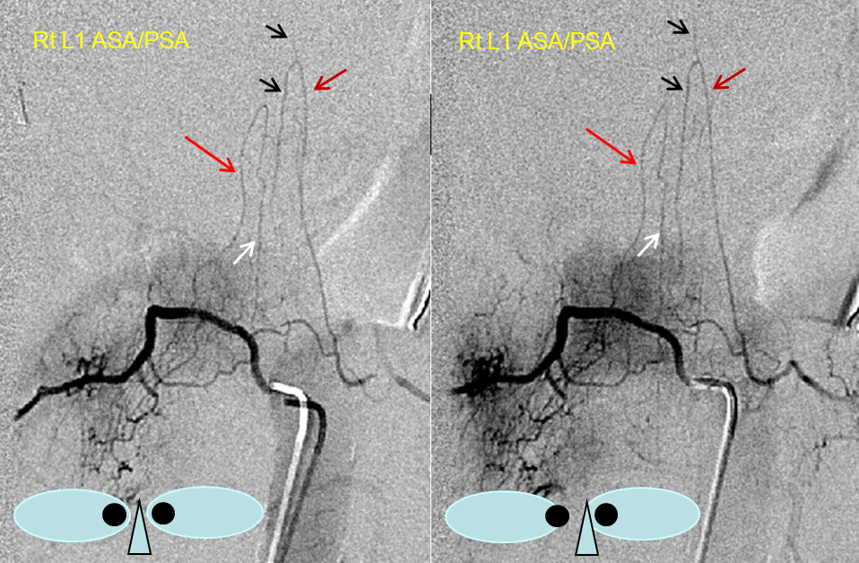
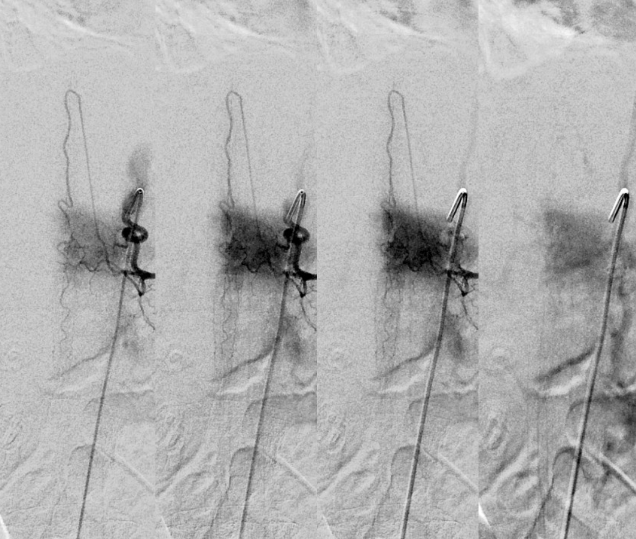
Standard DSA shows large Adamkiewicz from left T9, however it does not go to conus. Venous phase image on right shows good filling of spinal veins.
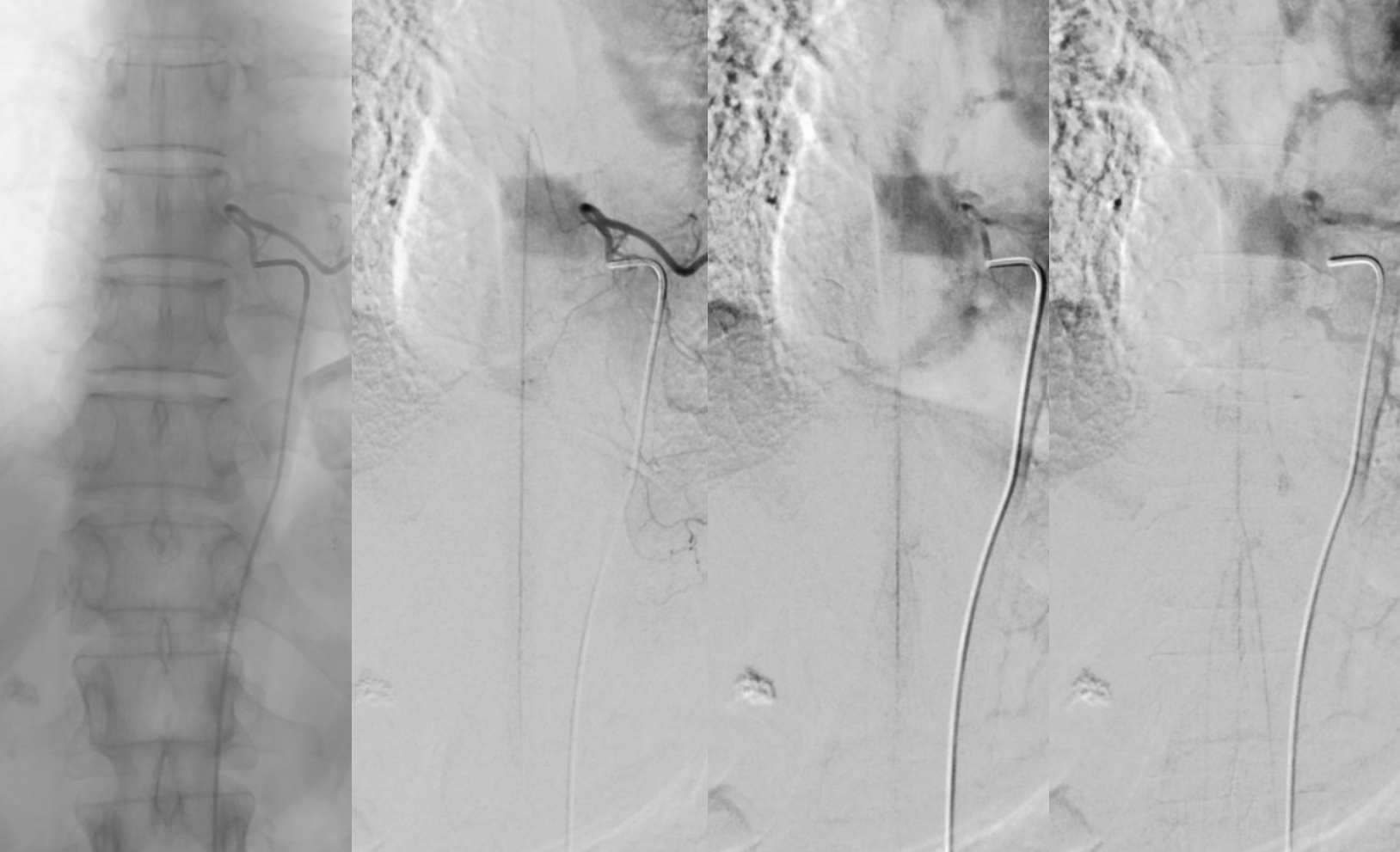
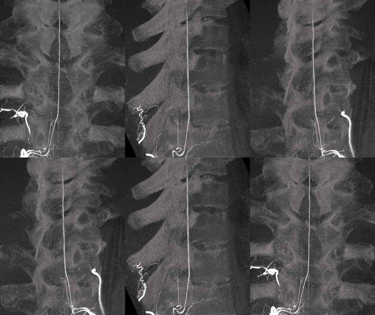
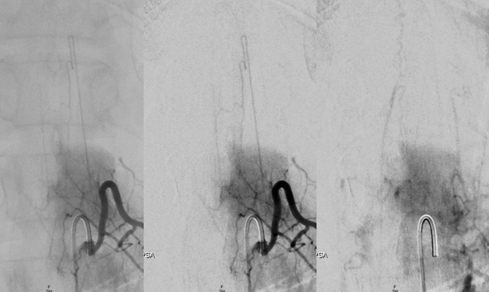
The conus is supplied by a large posterior spinal from left L1 (the origins of posterior spinals from thoracolumbar junction area are much more consistent than origin of Adamkiewicz). The transverse “vasocorona” vessels on the posterior cord connect to the large right posterior spinal as well. Image on right shows well the venous system — testament to large area of supply by the posterior spinal system. More info on Posterior Spinal Artery and Spinal Venous Anatomy page
HRCBCT of same case — 7 second “DYNA” CT 22 cm FOV unbinned, 100% contrast 1cc/sec for 10 seconds with 3 second delay.
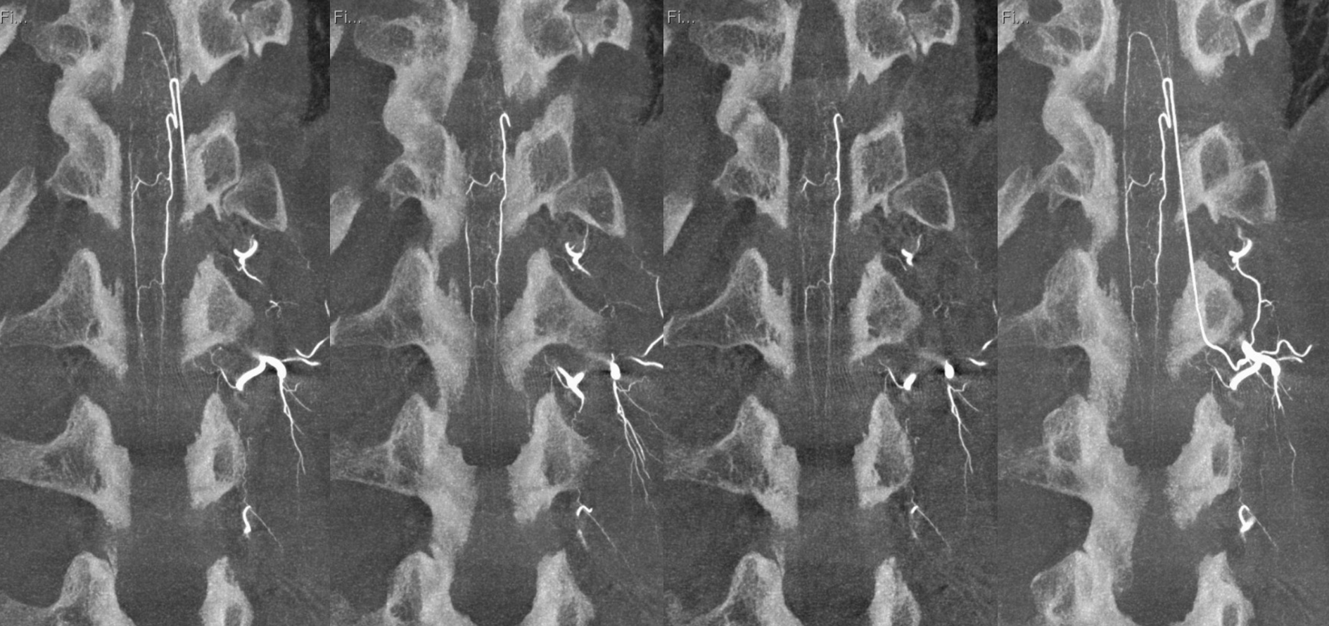
Stereo
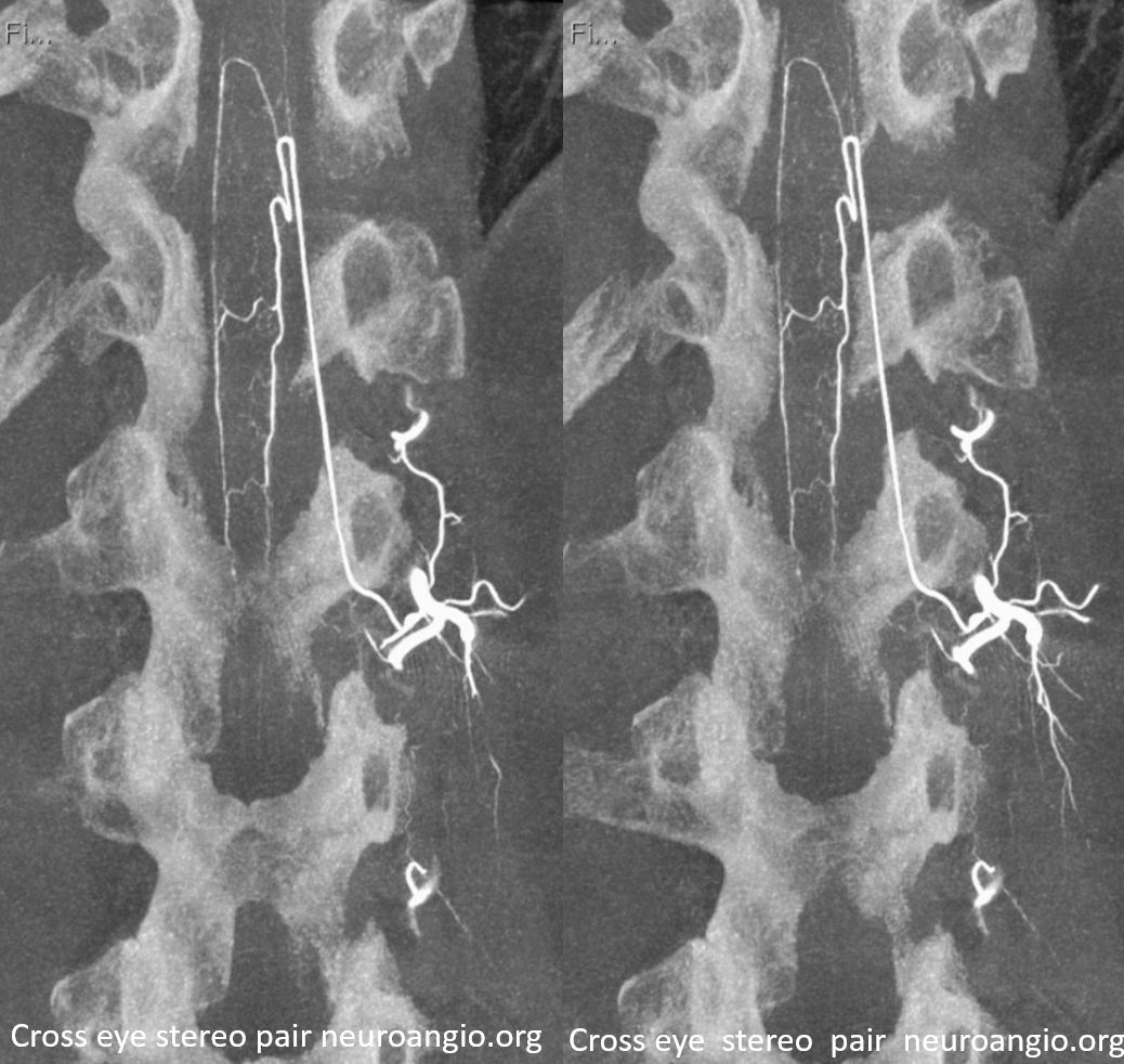
Where can ASA be hiding?
It is important to understand that in a neurologically normal patient the ASA has to be there, somewhere. if you are not finding it, look harder. Here is a nice illustration of “common origin” right T9, T10, and T11 intercostal arteries from the right T10 segmental artery
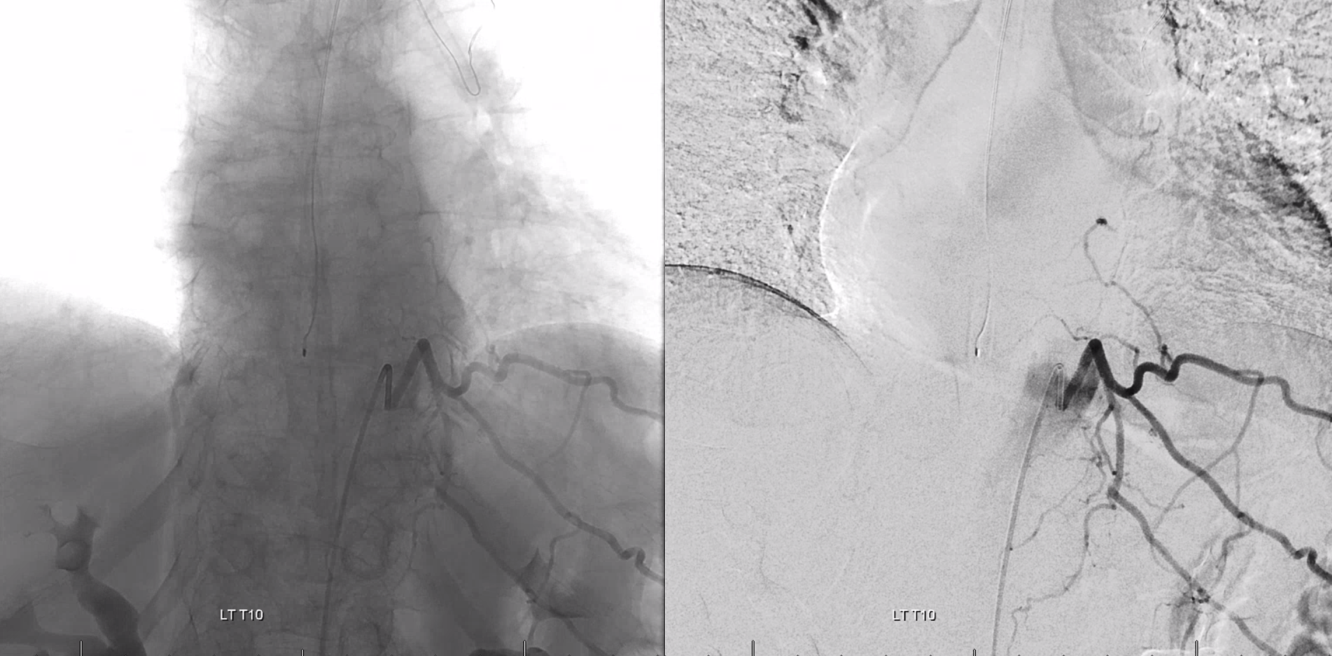
However, understanding of anatomy lead leads one to know that neither T9 nor T11 segmental arteries are completely missing. The anastomosis (G, black arrows below) is distal to origin of the radicular / radiculomedullary region branch (E). A small segmental artery is present, with its supply limited to the anterolateral vertebral body (gray arrows)
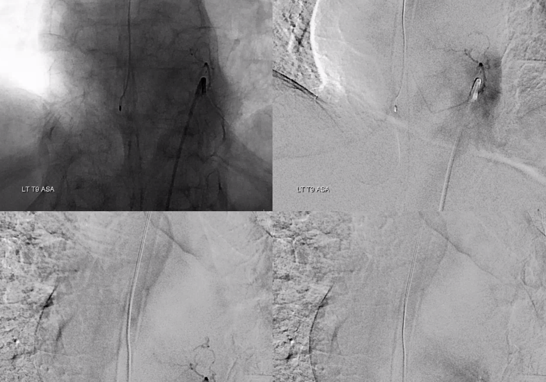
Indeed, after some effort exactly this kind of hypoplastic right T9 segmental artery is identified, supplying the vertebral body and giving rise to the radiculomedullary artery
Sulcocomissural Arteries and Intrinsic Arterial Cord Vasculature
Already seen in multiple examples above. Usually below any noninvasive as well as 2D-DSA resolution. Can be seen with General Anesthesia, pharmacologic paralysis, and apnea, and 100% contrast concentration injection end-on (frontal views) as dots superimposed on the anterior spinal artery, most commonly in thin habitus subjects where resolution is better. HRCBCT is best, of course. Clinical relevance is currently limited, except in cases of cord AVMs
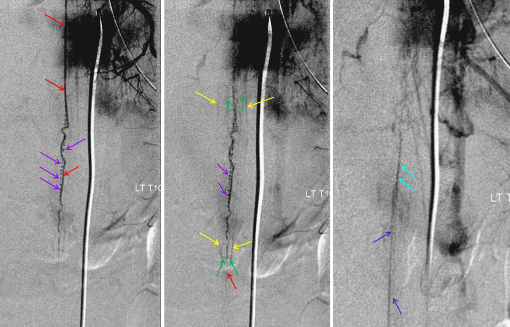
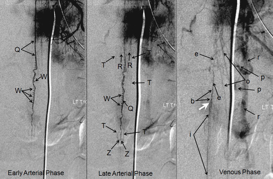
In this young, very thin patient, injection of the Adamkiewicz beautifully visualized multiple end-on sulco-comissural vessels, seen as dots (purple, W) next the anterior spinal artery (red, Q). Notice multiple coronal surface vessels (green, V) which opacify the posterior spinal channels (yellow, T), which are usually contiguous at the conus (rami cruciantes, or Z, labeled in green at the bottom). Venous phase image (right) shows a slanting direction radicular vein (dark blue), as is often the case, draining the basket. A hint of radiculomedullary veins is present also (light blue). Unfortunately, this image was obtained before our consistent HRCBCT acquisitions.
Same image with letter nomenclature corresponding to the master diagram above.
All of the Above – 6D Fusions — Anterior and Posterior Spinals, Sulcocomissurals
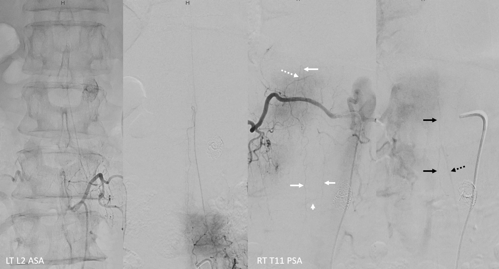
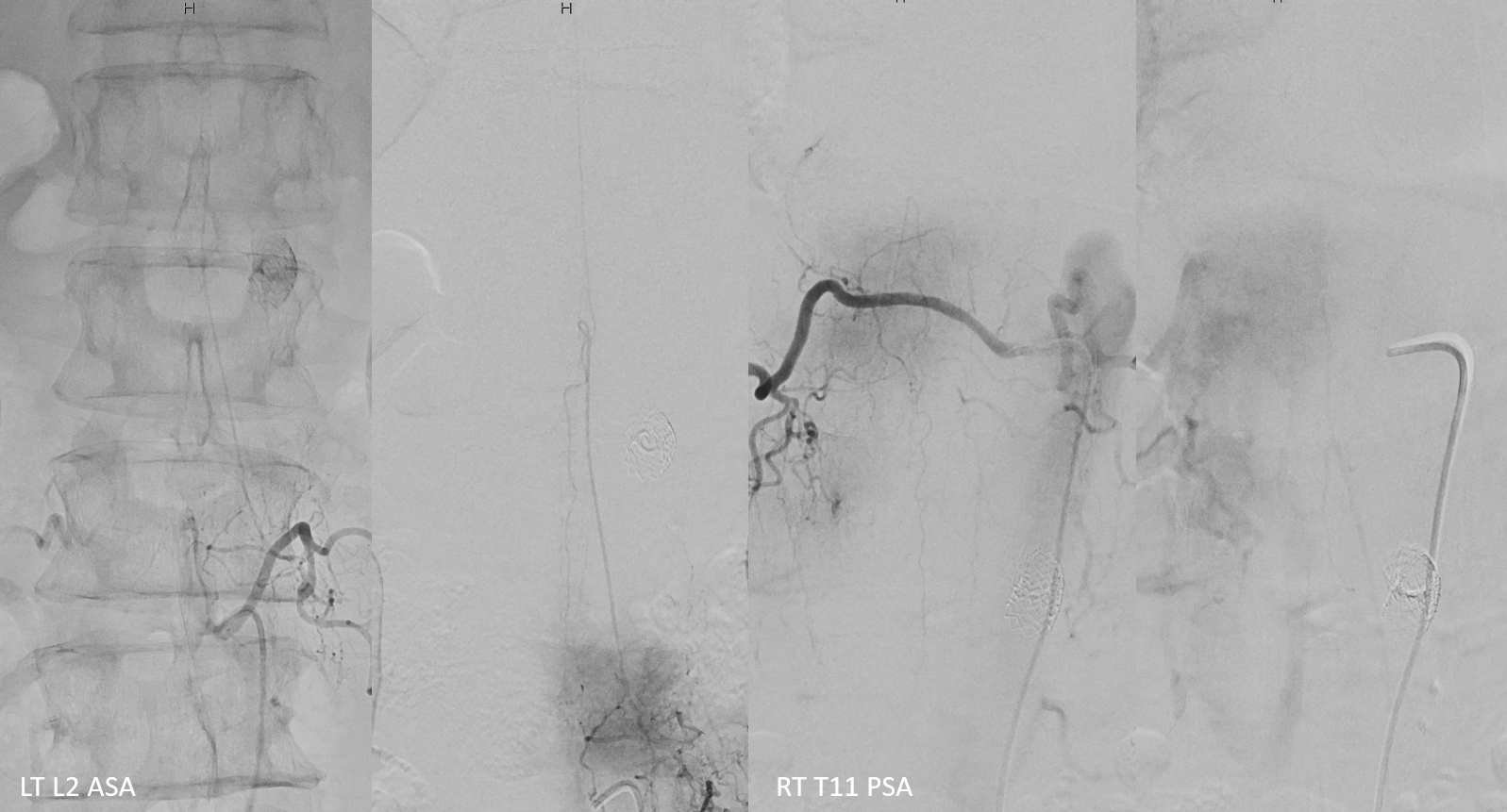
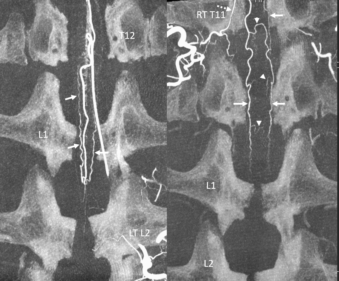
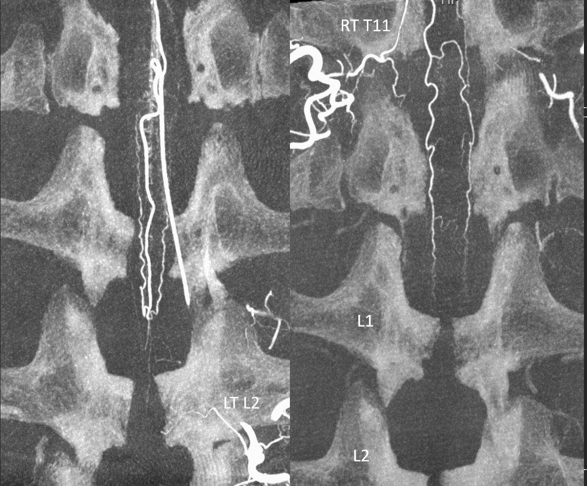
Full spinal angio should give the overall sense of how all is put together. A low origin L2 Adamkiewicz (two left images) is in balance with a well-developed posterior spinal network, with arterial and venous phases (two left images). Dashed white — radiculopial. Arrowhead — vasocorona. White arrows — PSAs. Venous phase — dorsal spinal vein (black arrows) and radicular vein (black dashed arrow)
without arrows
The same on HRCBCT — see the “basket” supply by ASA and balance with RT T11 injection. Obviously, HRCBCT is much more informative.
Without arrows
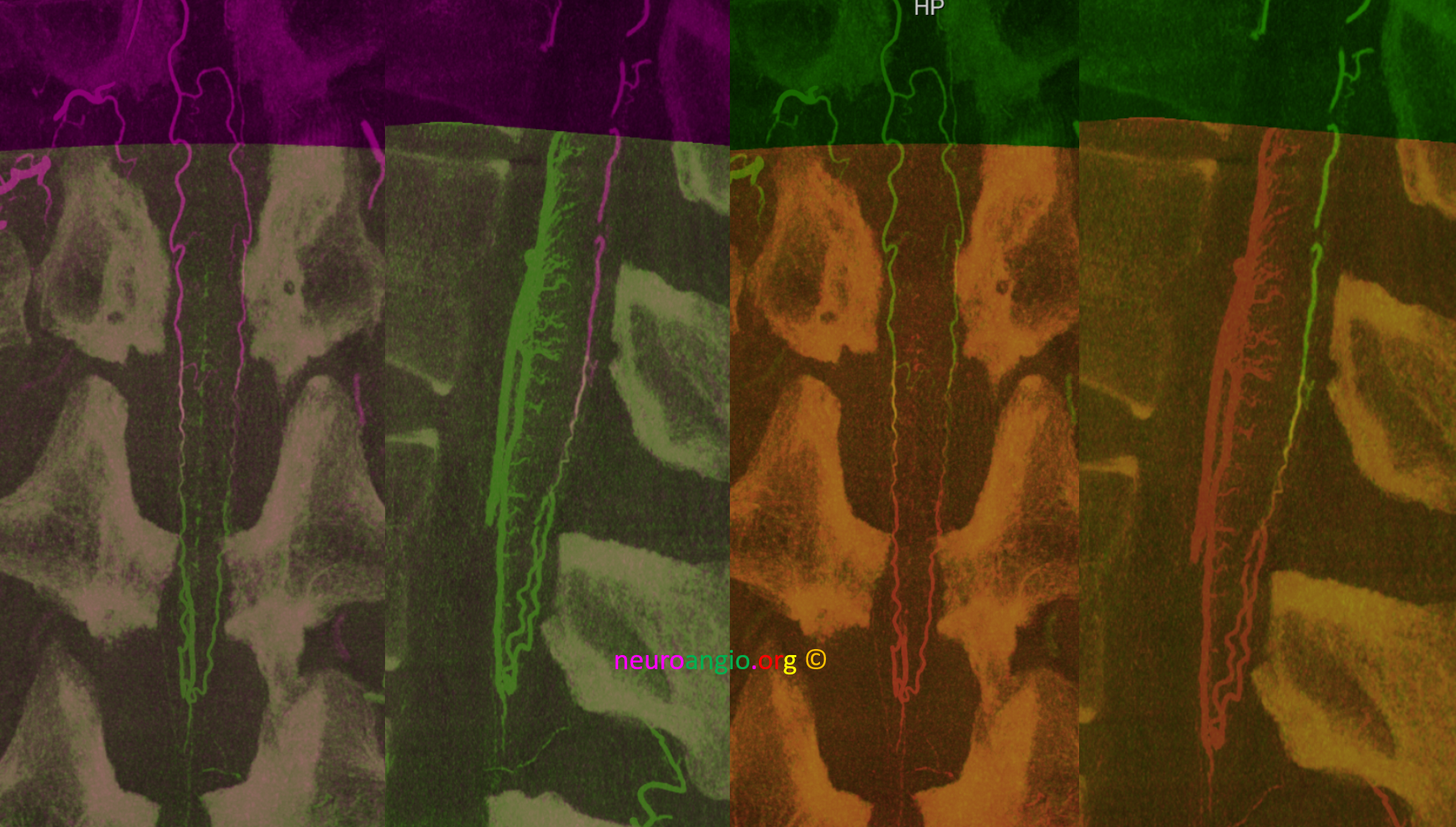
Fusion of two HRCBCT datasets is visually stunning, but understanding comes from raw data. Same data in two different color schemes. The “watershed” PSA region is where colors mix. It is important not to overthink this — a stronger injection from one pedicle may artificially shift the watershed for example. Would the PSA connections be enough to supply the cord if right T11 radiculopial artery was occluded? Probably yes in this case. What about the reverse — probably not.
Note high density of sulco-comissural arteries in areas of cord enlargement (cervical and conus regions)
PATHOLOGY
Spinal Cord Infarct: The anterior spinal artery is, overall, the largest and most consistent supplier of any section of cord — especially in cases where its failure is symptomatic. It is important to keep this in mind — cases where the ASA is either prominent or inadequately collateralized are more likely to present with, for example, classic ASA infarcts, compared to those where collaterals may be sufficient, and clinical symptoms either don’t exist or may be mild enough to be mistaken for other conditions. The above is a classic example of survivorship bias.
Spinal Dural Fistula
The most common spinal vascular shunt. The hallmark of dural fistula is cord venous congestion. In such cases, injecting the dominant ASA supply / Adamkiewicz consistently shows lack of venous phase, despite imaging for a long time (exact arteriovenous transit time in cord is hard to establish, somewhere between 4-8 seconds depending on cardiac output, blood pressure, and other factors. So, if you inject the Adamkiewicz, and don’t see spinal veins — that’s an indirect sign of spinal venous congestion.
Spinal dural fistula (see lots of cases in Case Library) is a prime example of spinal venous congestion pathology. Two factors determine how bad the clinical status is. One is competing inflow from the fistula. The other, just as important, is lack of venous outflow via radicular or bridging veins. The end result is cord venous congestion. How does that manifest angiographically? By not seeing cord veins in their expected venous phase following injection of cord arterial supply. Exact arteriovenous transit time in cord is hard to establish, somewhere between 4-8 seconds depending on cardiac output, blood pressure, and other factors. So, if you inject the Adamkiewicz, and don’t see spinal veins — that’s an indirect sign of spinal venous congestion.
Below is an example of Right TT origin Adamkiewicz injection. Center and right images show cord blush, but no radicular veins — congestion….
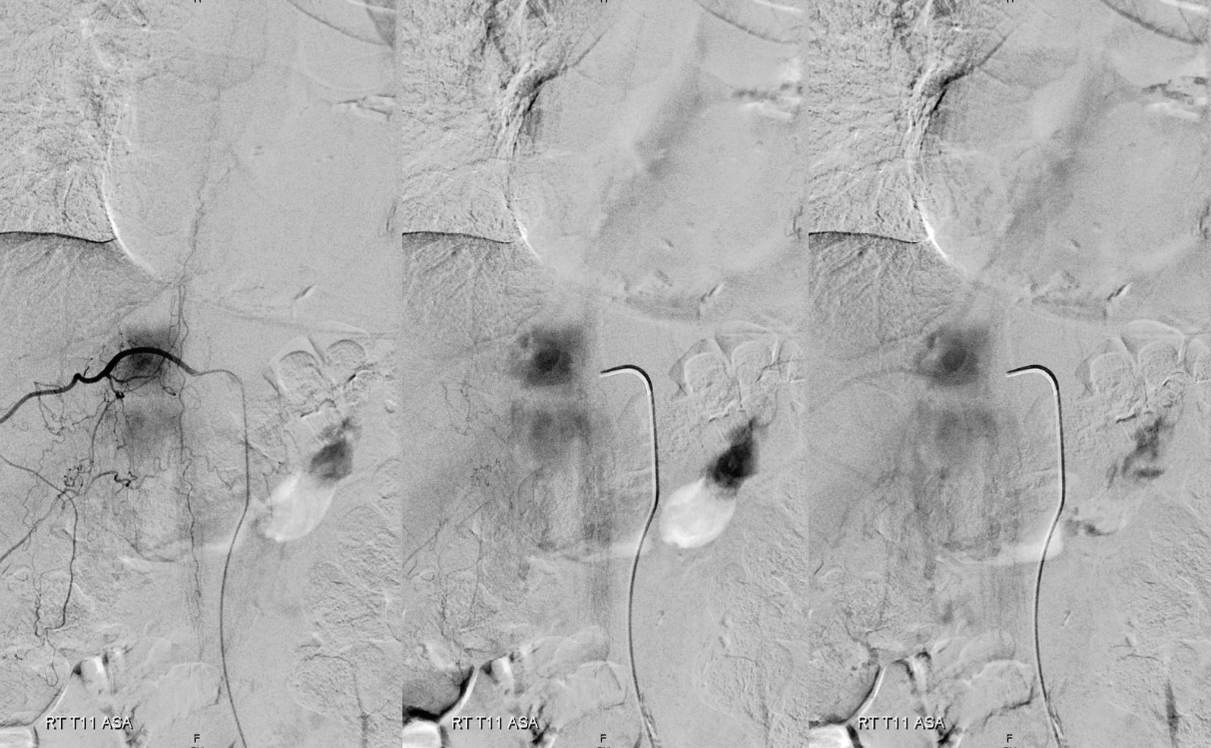
Fistula is at left T6. Note lack of radicular outflow, which is the main problem in highly symptomatic fistulas like this one
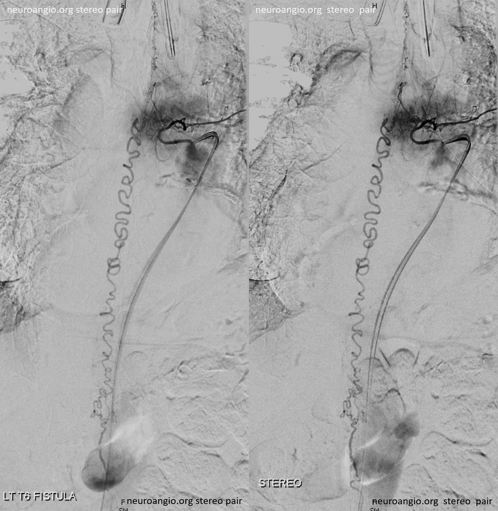
Following endovascular occlusion on the fistula (full case here), injection of the Adamkiewicz now visualizes the spinal veins previously parasitized by the fistula and now “returned” to the cord
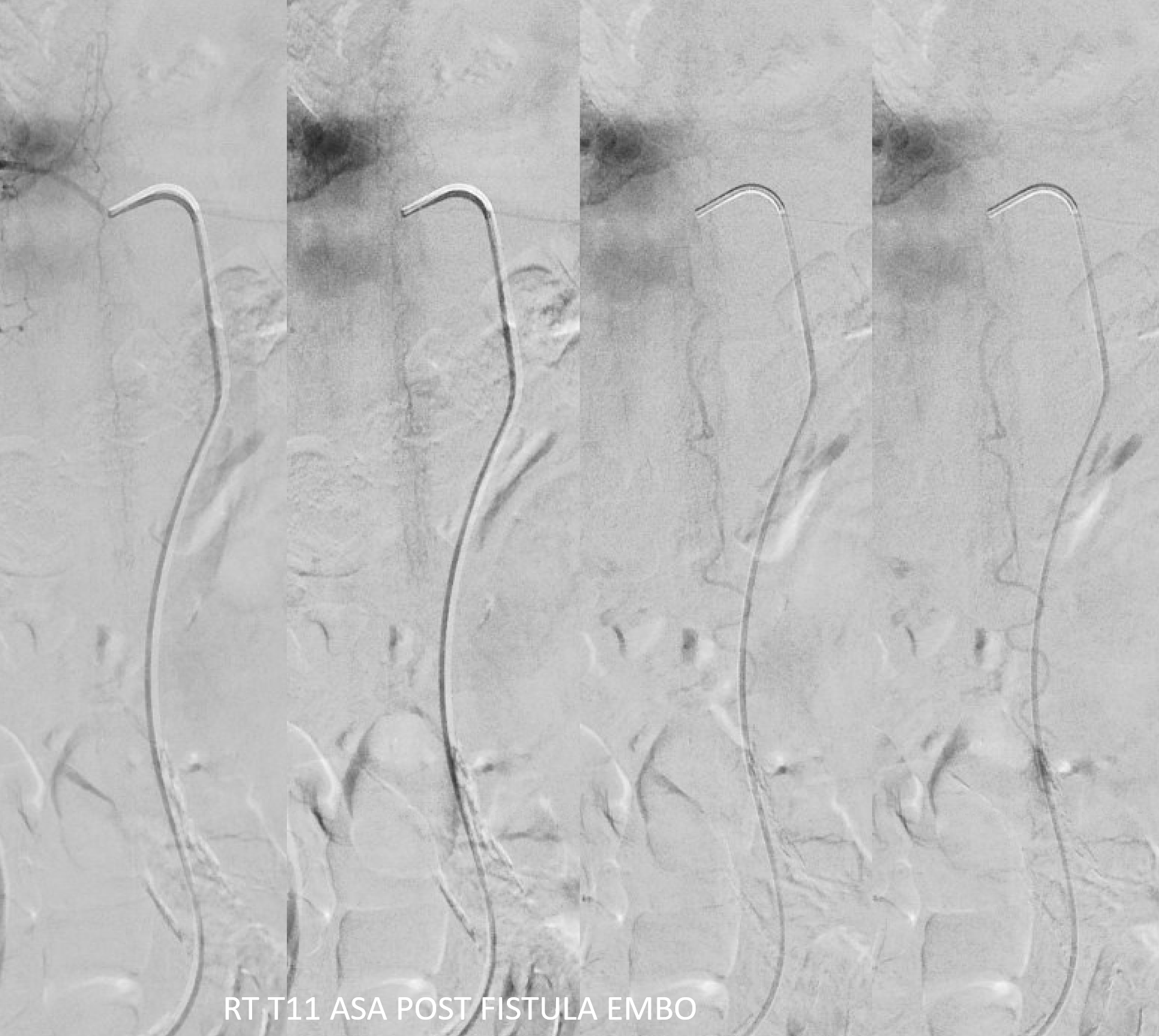
The venous system remains abnormal, with paucity of radicular drainage. This can contribute to limitations in recovery, along with any “permanent” congestion-related cord damage — a key under-recognized point. This, in a nutshell, is Dural Fistula 101 Primer.
Spinal AVM
By volume, majority of cord AVMs involve the ASA — indeed the choices of supply are either ASA, PSA, or both. Microcatheter injections and HRCBCT are key in understanding this very small and critical anatomy. Endovascular treatment is possible in select cases, and requires great familiarity with anatomy to maximize risk/benefit ratio. See cases on Case Archives page
Spinal Arterial Aneurysm
Aneurysms, likely dissecting, involve the radiculomedullary or radiculopial arteries, at the cite of their passage through the dura. The can be extremely symptomatic due to both rupture and / or occlusion of parent artery and cord ischemia. See many cases on Case Archives page
Had Enough? Of course not. There is so much more… email neuroangio@neuroangio.org if you have good cases to share or questions/comments.