
The potential for the ASA to reconstitute PICA territory has been described in the literature and illustrated on this site. For embryology discussion of this phenomenon see this article, this neuroangio.org page, or another neuroangio.org pages titled Neurovascular Evolution and Vascular Neuroembryology.
Case 1
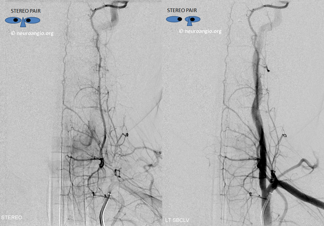
Here is another fascinating variant. Dissection of upper vertebral artery results in compromise of PICA flow. The collateral network which reconstitutes the PICA, without symptoms, is the anterior spinal system. Unique here is the duplicated nature of the ASA, which attests to the developmental nature of the ASA as a secondary vessel, bridging the primary metameric arteries supplying the developing cord.
Frontal view of right vert injection shows high cervical dissection (orange), the anterior spinal system (red, pink arrows) contributes to supply of the right PICA (black) via the C4 segment radiculomedullary artery (brown). Notice midline location on the frontal view and superimposition of the anterior spinal channels on the lateral view, confirming their anterior spinal nature. The odontoid arcade (white) also does the same, via its usual C3 segment radiculodural artery (purple)

Stereo of the same

Injection of the left vertebral artery reveals a healed, less severe superior vertebral dissection. In a mirror image fashion, the left C4 radiculomedullary artery (yellow) supplies the “left-sided” portion of the anterior spinal system (pink) which was also seen from the right vert injection. The odontoid arcade (blue) and its C3 radiculodural artery (green) are also well-seen.

Case 2
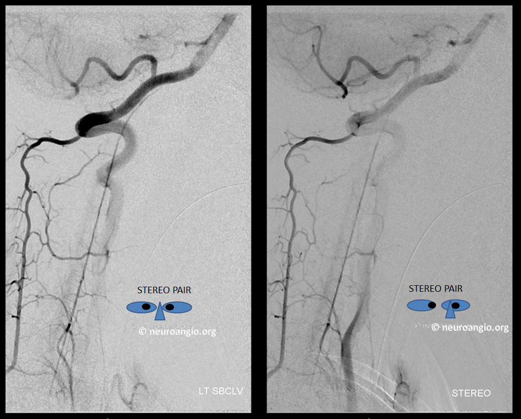
In case you think this is only seen as part of pathology, here is an incidental encounter, with portions of duplicated cervical anterior spinal artery in a child who underwent angiography for an unrelated reason. Frontal and lateral stereo views. Case Courtesy Dr. Eytan Raz
Lateral views — the duplication is not seen well here. However a nice illustration of deep cervical – vertebral anastomoses (so common in children) is present
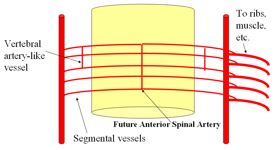
Below are diagrams from “Spinal Arterial Anatomy” page showing how development of the ASA would give rise to the above-shown variant.
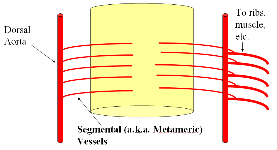
Below is the idea behind the duplicated anterior spinal
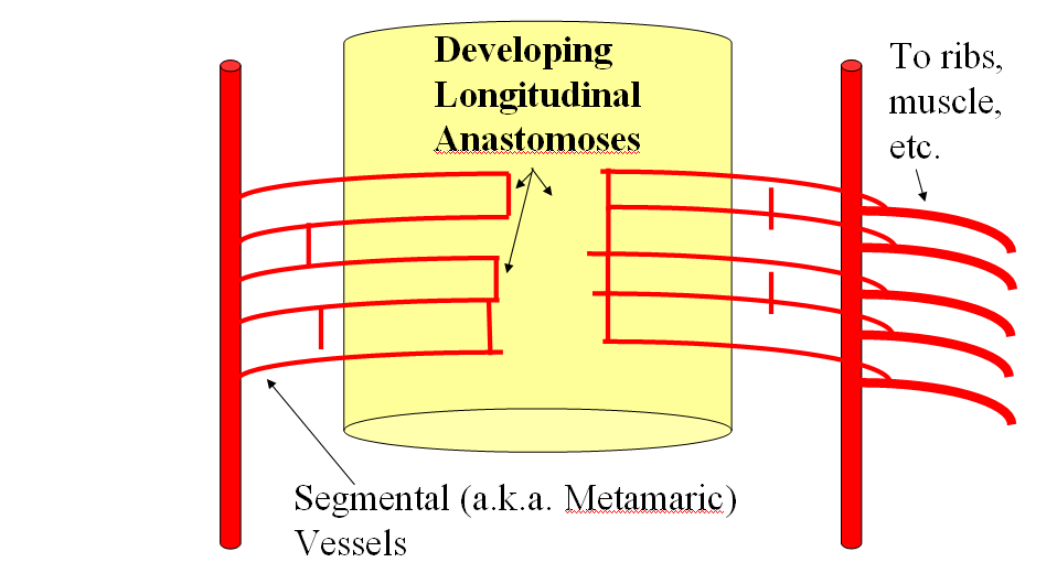
Usually, the ASA ends up being a single vessel, as below
See companion cases of Anterior Spinal Reconstitution of PICA and Lateral Spinal Reconstitution of PICA as well as the Lateral Spinal Artery page