
Another example of dural fistula that does not directly involve a venous sinus. This one is located in the paramedian tentorium cerebelli, adjacent to the torcular. However it does not drain into a sinus — instead congesting cerebellar veins
Sometimes, dural fistulas will parasitize supply from a brain artery — for example arteries of Davidoff-Schechter can supply falcotentorial junction fistulas. Another good example is when posterior meningeal artery arising from PICA supplies a regional dural fistula. In this case, there is prominent contribution from the vermian branch of PICA . However, this does not make this lesion a brain AVM. Why? Because predominant supply is coming from dural branches. Vermian AVMs have a very different appearance. It is key to appreciate this and not mistake this lesion for an AVM. In our opinion, there is no such thing as a dural AVM. It is either a dural fistula or a brain AVM, not both.
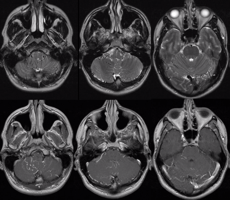
MRI
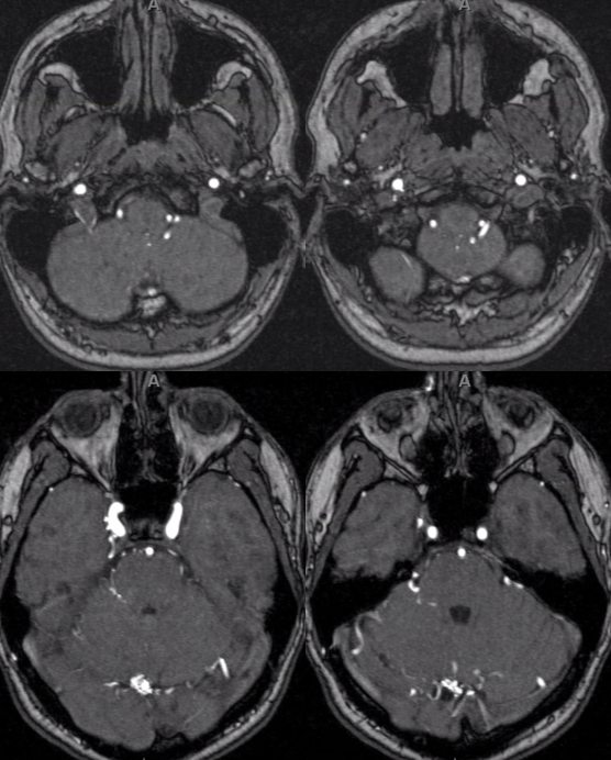
MRA
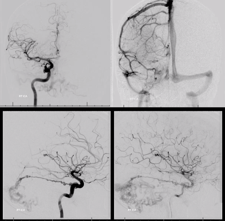
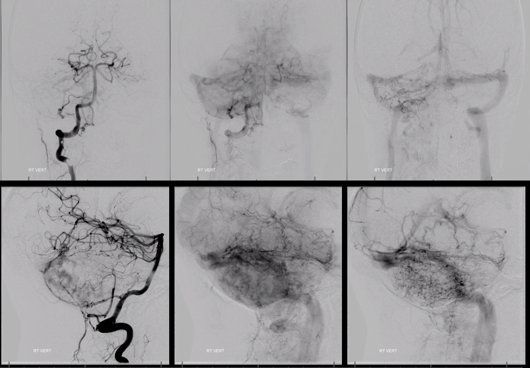
Angio — prominent Bernasconi-Cassinari (medial tentorial arcade) supply — branch of MHT with ILT contribution via recurrent branch. Drainage into cerebellar veins directly
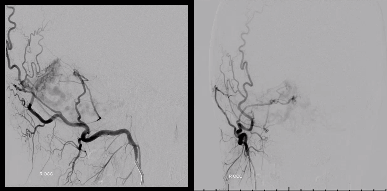
Ascending Pharyngeal — jugular division contribution also seen on MRA
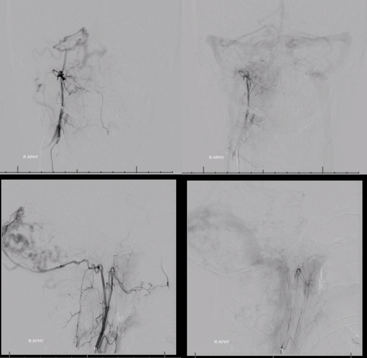
Occipital — contribution is via the same tentorial branch supplied by the Ascending Pharyngeal
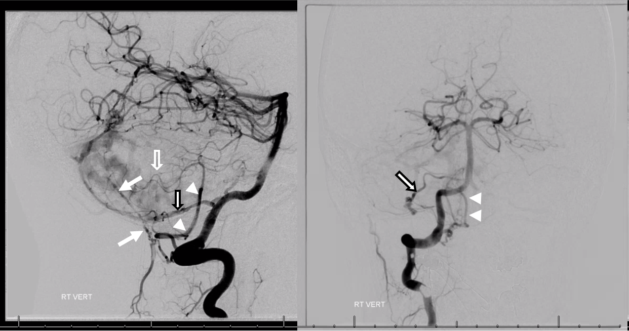
Right vert.
Lets look at it a bit closer. Its an interesting situation. There is an extradural artery of tentorium cerebelli (white arrow). Also there is a C1 origin PICA (arrowheads) which predominantly supplies vermian territory (open white arrow). A separate intradural PICA (black and white arrow) supplies the lateral hemisphere and also contributes to dural fistula.
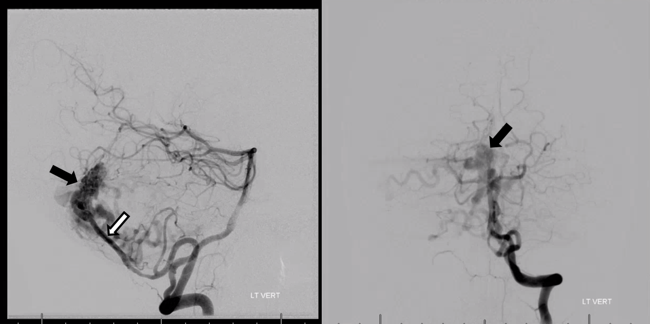
Left vert — contributing a lot to the fistula. Dont mistake this for a brain AVM — the major supply is still dural and dural parasitization of PICA branches is a known entity
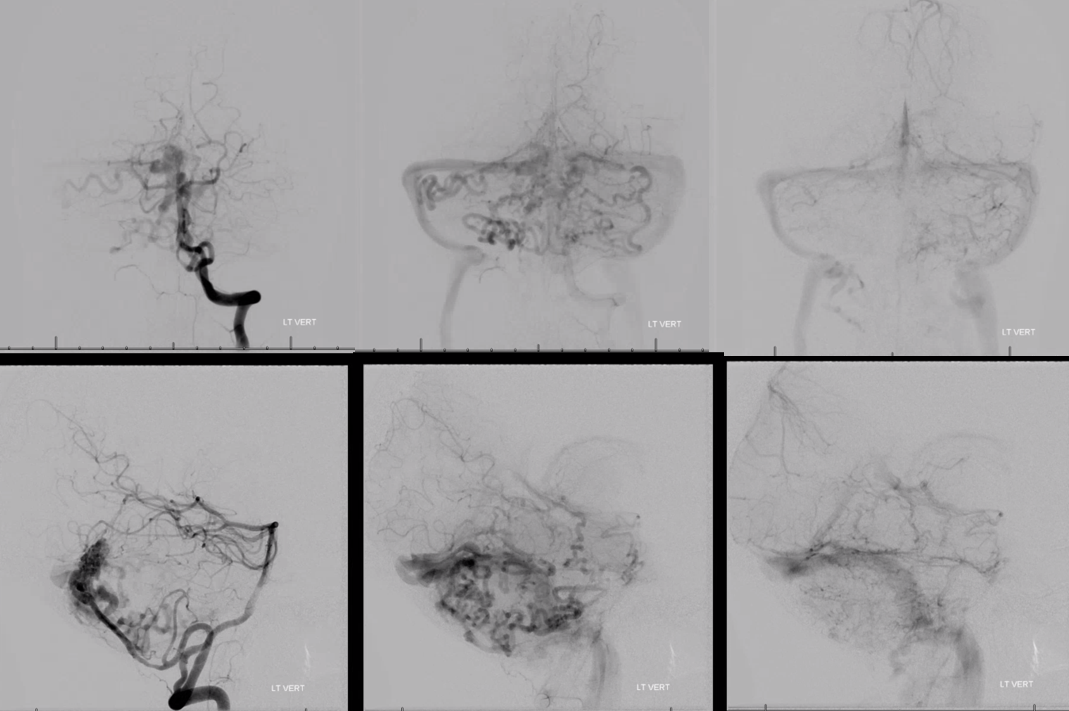
Closer look — vermian branch of PICA (black and white arrow) supplies the shunt — black arrow points to fistula. it is not an AVM.
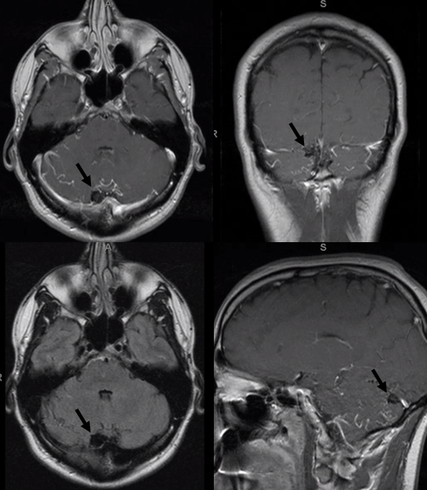
MRI black arrows point to the black void ball of the dural shunt — just next to the torcular.
For other dural venous channel cases see here