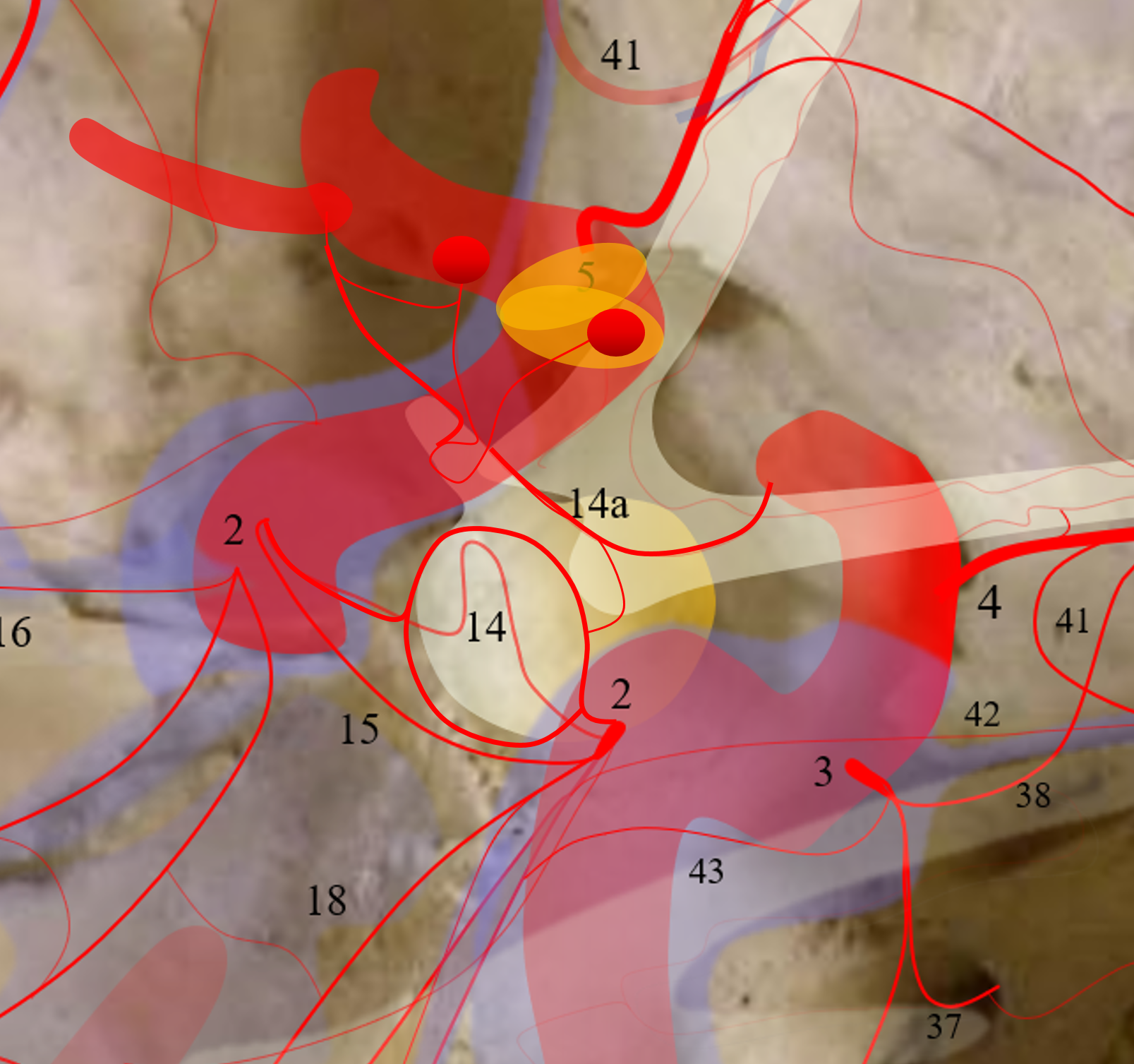
Number 14 above. Branch of the MHT. Unlike the posterior clival arteries (15) which are dural and located behind the clinoid, the inferior hypophyseal supplies the pituitary, and is thus anterior to the posterior clinoid. More specifically, it usually supplies the posterior pituitary. A characteristic anastomotic ring curcumscribing the posterior pituitary links it to its the contralateral twin. The inferior hypophyseals are in balance with each other (left/right), and with the superior hypophyseal arteries (14a). The latter nearly always supply the stalk, with descending network vascularizing the anterior pituitary. The inferior and superior hypophyseals are in balance (see superior hypophyseal artery page also).
Everyting you ever want to know about pituitary blood supply is in the original manuscript by McConnell (after whom the capsular arteries are named — they are NOT the same thing as superior hypophyseals). There is still nothing better.
The balance and specrum approach to anatomy applies here. The superior and inferior hypophyseals are in balance. The right and left ones also. Small branch on one side nearly always means big one on the other.
Supply of the Hypophysis and Balance
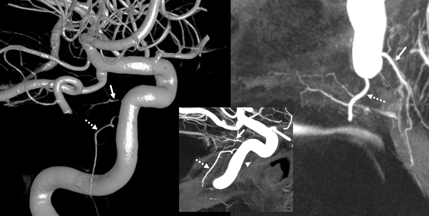
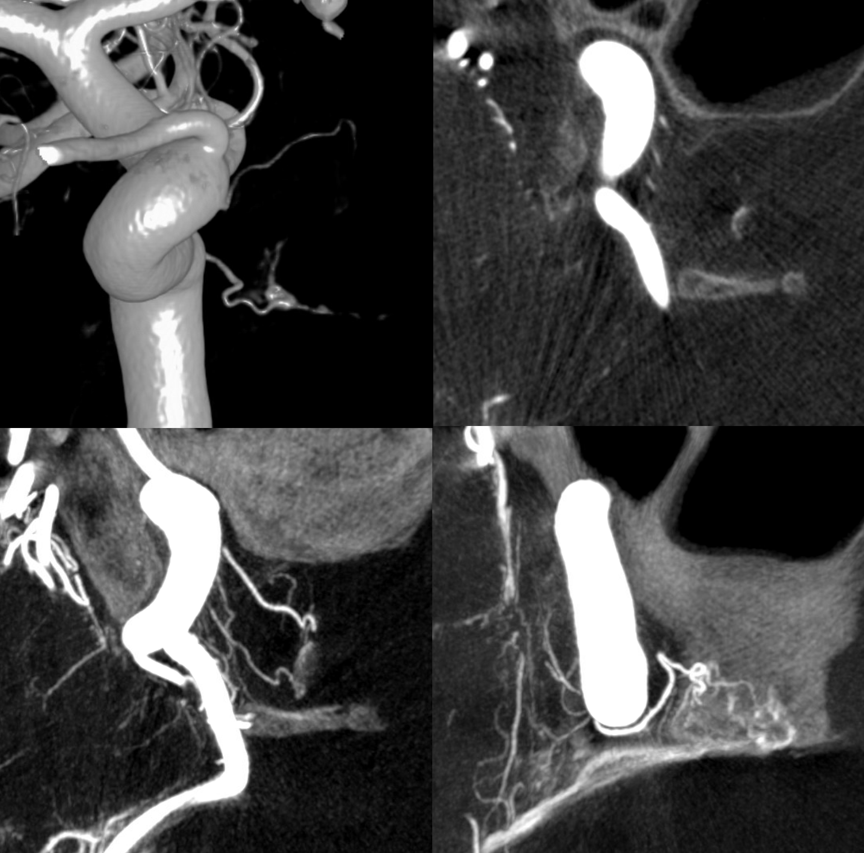
MIP images of different case. Large MHT origin (arrow) inferior hypophyseal supplies the posterior pituitary. A superior hypophyseal artery is shown by arrowhead. The ILT is hypoplastic.
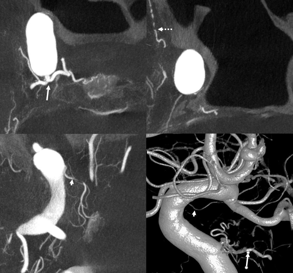
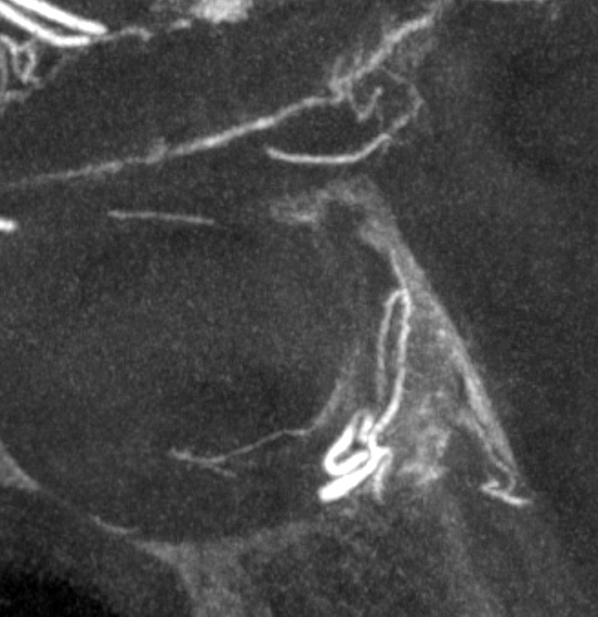
Coronal MIP of the inferior hypophyseal branch with a textbook loop of the posterior pituitary
Sagittal view — loop at the back
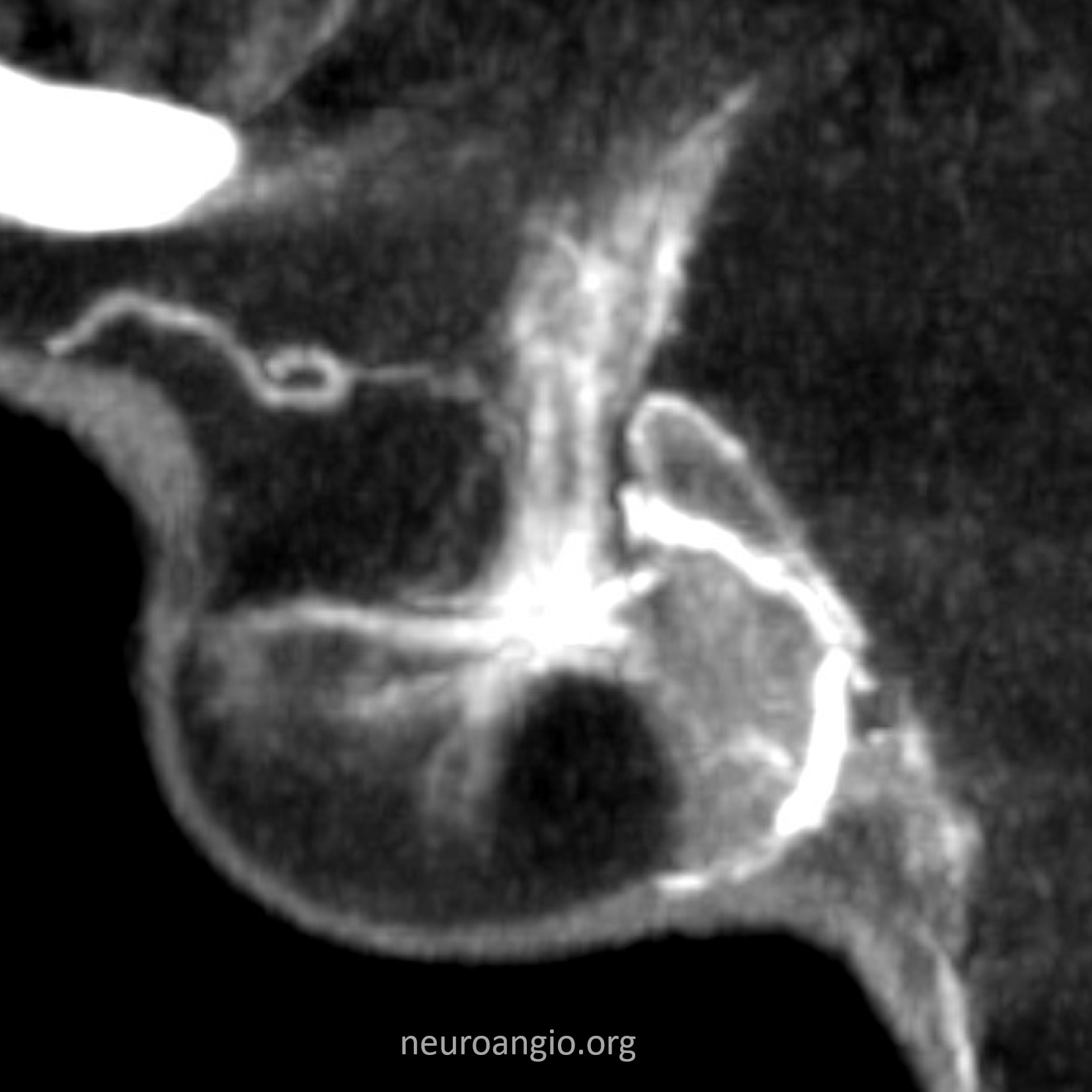
The whole hypophysis can be beautifully seen sometimes
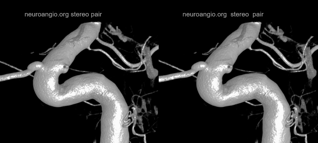
Stereo views — beautiful depth of superior and inferior hypophyseal
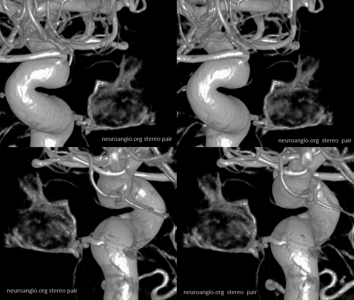
Arrows — superior hypophyseal solid, inferior dashed
More Supply of the Hypophysis and Balance
As always neuroangio is about balance. There may be one or more superior hypophyseal arteries. Usually one is visible by cone beam/flat panel CT. Sometimes more than two. Here there are 3. Nicely shown is supply of the stalk (open arrow). There is also likely contribution to the optic chiasm, which is of course very important. The inferior hypophyseal branches (dashed arrow) from the MHT support the posterior pituitary. The ILT is hypoplastic, with lateral branches of the MHT (white arrowhead) heading towards the meckel cave and the clinoid tentorial arcade branch along the tentorial edge (black arrowhead) supplying territory normally done by the ILT

Without labels
More superior / inferior hypophyseal balance
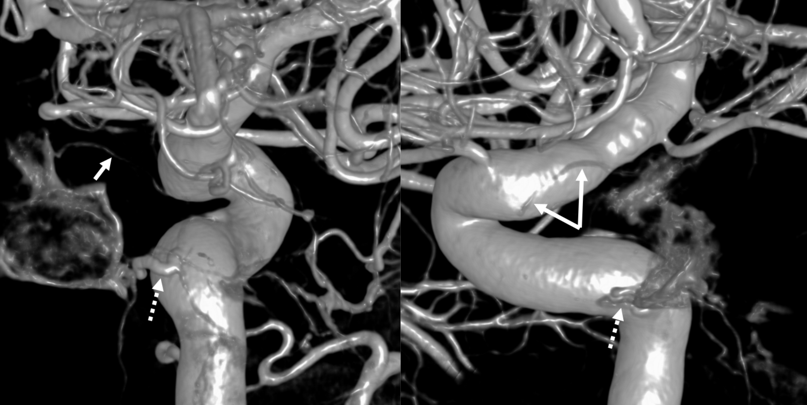
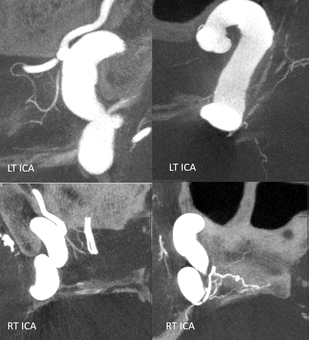
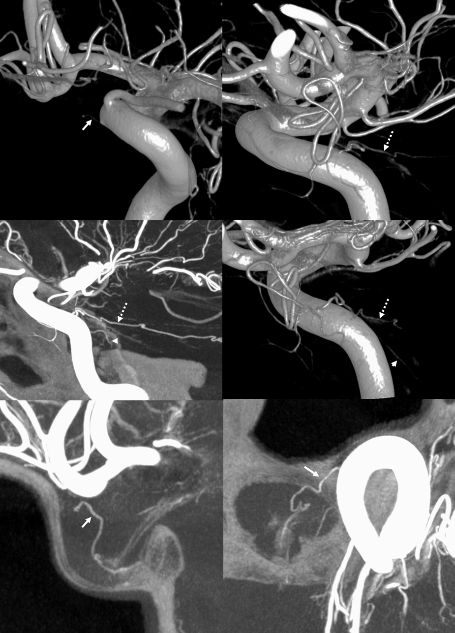
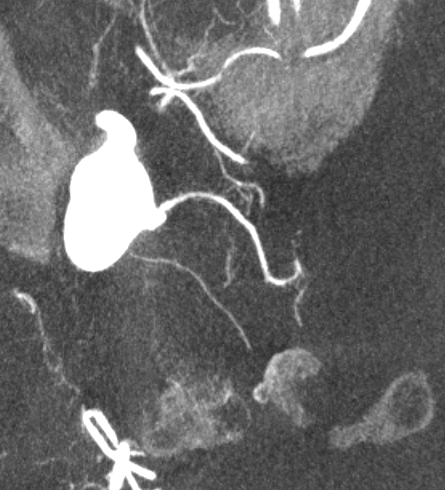
Excellent example of contralateral balance. On the left, there is a hypoplastic posterior hypophyseal network (part of MHT, arrowhead), and a prominent superior hypophyseal artery (associated with a small aneurysm). On the right, the opposite is true — large inferior hypophyseal arteries (dashed arrows) extending past midline due to contralateral hypoplasia, and a smaller superior hypophyseal, without aneurysm — not visible on these VRs, but seen on subsequent MIP images
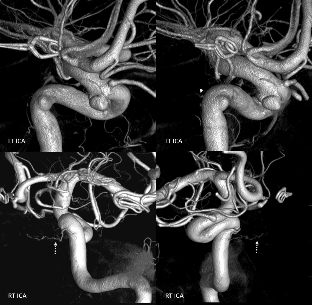
MIP images — there is a small right superior hypophyseal artery present
Another example
Spectrum and balance again. Usually, the inferior hypophyseal branches from the MHT supply the posterior pituitary. Here, the MHT is well-developed, but primarily supplies the marginal tentorial arcade, while the posterior pituitary is fed by the superior hypophyseal branch (arrow).
 Same case MHT — the larger trunk (dashed arrow) is mainly marginal tentorial, smaller one (arrowheads) is clival branches inferiorly towards anastomoses with ascending pharyngeal counterparts
Yet another one — large anterior branch (with infundibulum), no ipsilateral MHT supply
See smaller “middle” hypophyseal as well
MIPS — great anatomy of the MHT, ILT as well. The MHT is somewhat hypoplastic. ILT supplies bulk of its territory via the clinid tentorial arcade. There are 3 superior hypophyseal arteries, supplying large portions of the optic nerve, chiasm, and probably some proximal tract, together with the PCOM
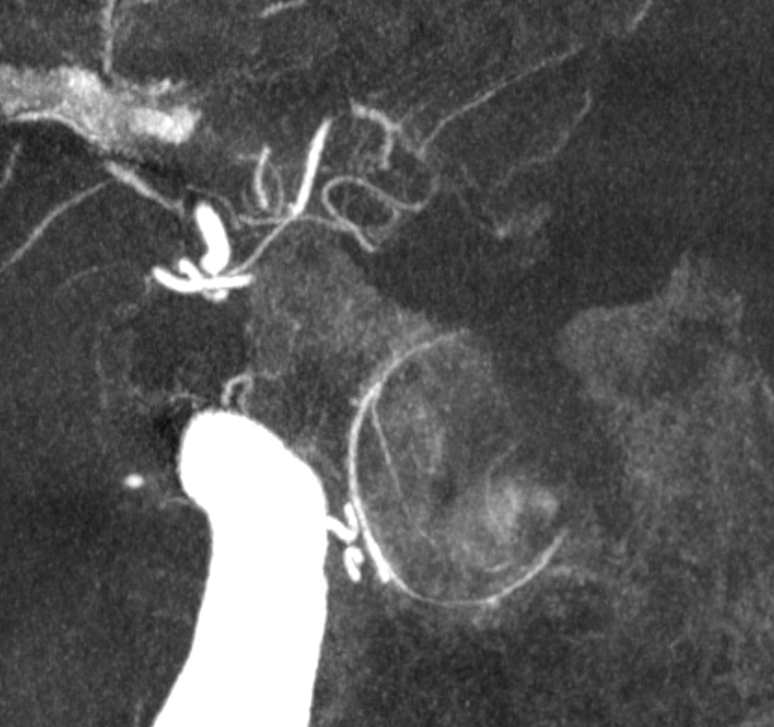
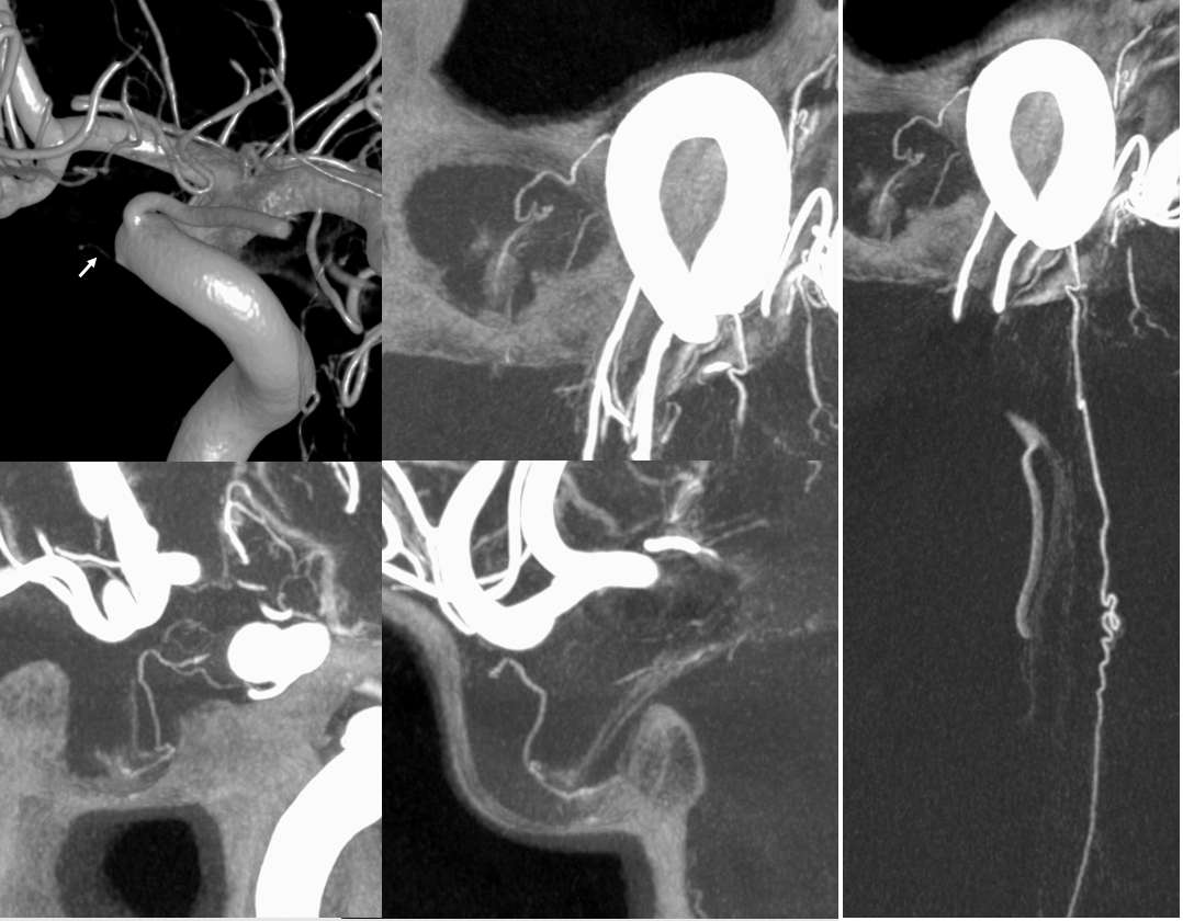
Below is a large inferior hypophyseal branch (arrow), not infrequently separate in origin from the lower clival branches (arrowheads)