
The majority of spinal epidural hematomas are felt to be venous. Here is a case of a man who presented with complete lower extremity plegia following cardiac surgery. A large epidural hematoma (white arrows), to the right of the median raphe, displaces the dura (black arrows) and narrows the spinal canal (yellow)

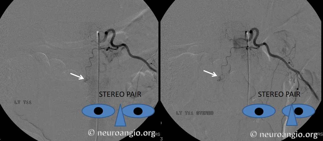
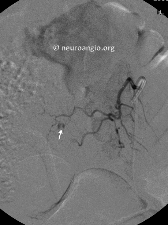
The patient is taken for emergent surgical decompression. Angiogram post-decompression demonstrates contrast extravasation from a dorsal epidural branch of the left T11 segmental artery.
The hemorrhage is slightly to the right of midline
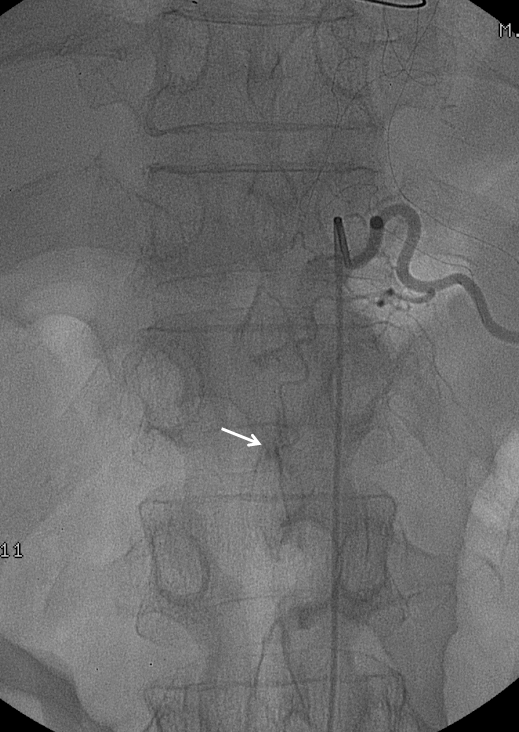
Oblique view
Post-nBCA
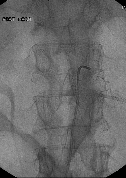
CT post nBCA shows cast in the dorsal epidural space (black arrows) and in the paraspinal musculature (white arrows)

It is impossible to know whether the findings reflect post-surgical arterial injury or primary hemorrhage. But I think it is worth looking at. Certainly, the dorsal epidural venous plexus is usually less vascular than the ventral one, yet here the branches are quite large